Eye of the Snail 1
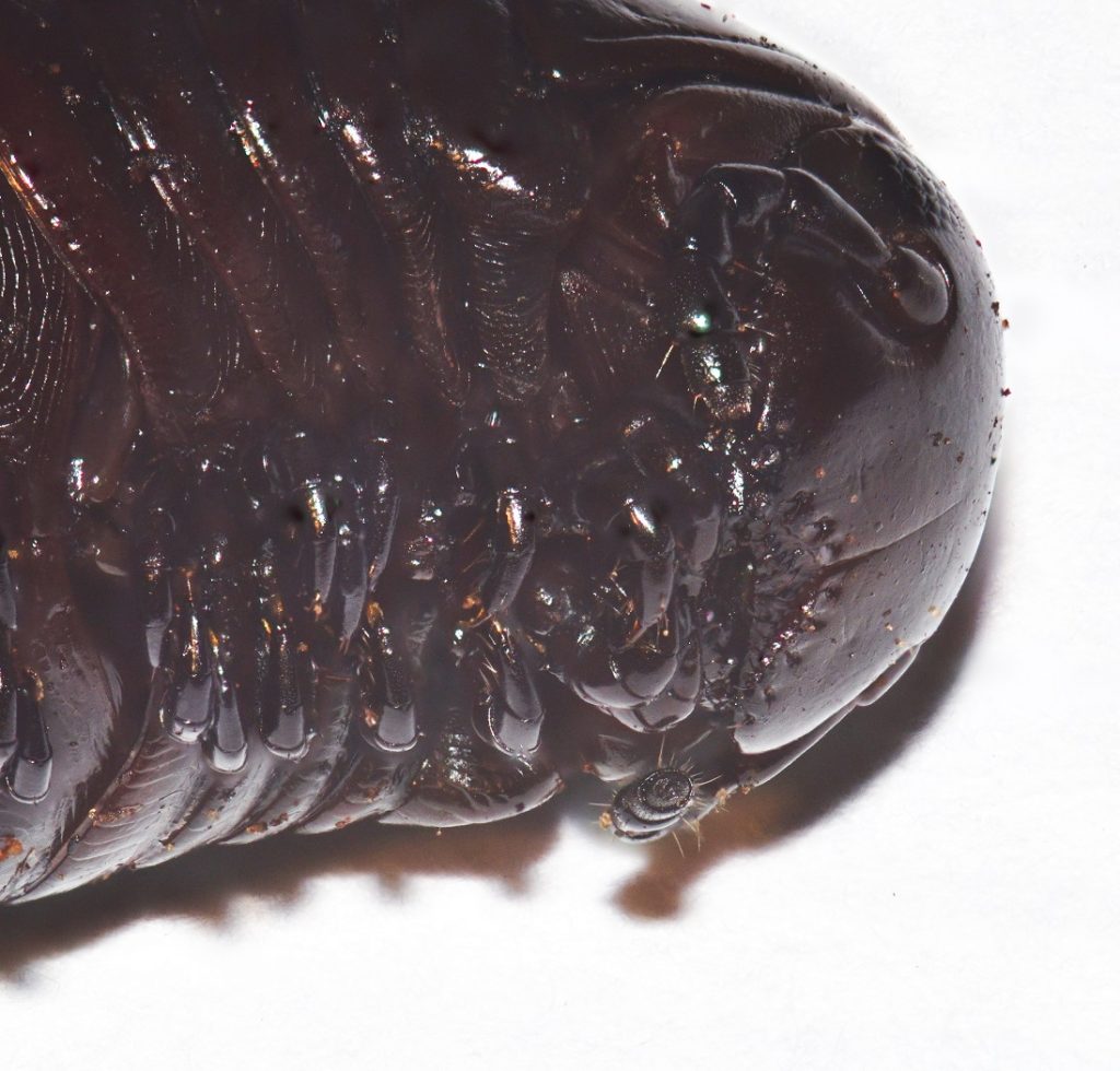
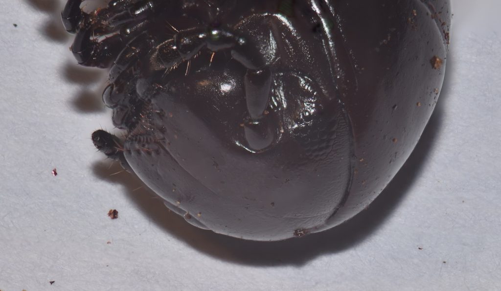
Snails in the group Pulmonata have eyes on the ends of retractable tentacles. These are true eyes with lenses and corneas. The snails have no muscles for focusing the lenses, though, so their vision is probably blurry, at best.
In additional to its eyes, the snail has photoreceptor cells distributed all over its body. These cells enable the snail to sense when a shadow falls over it. Since this usually means a predator, the snail reacts by quickly withdrawing into its shell. This is called the shadow reflex.
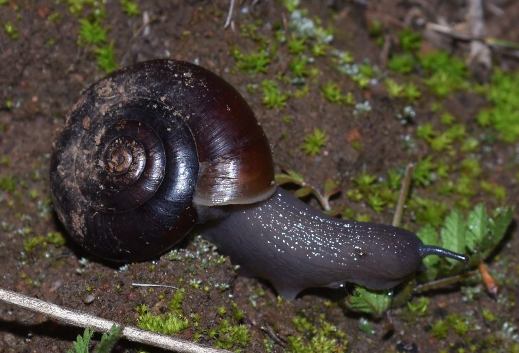
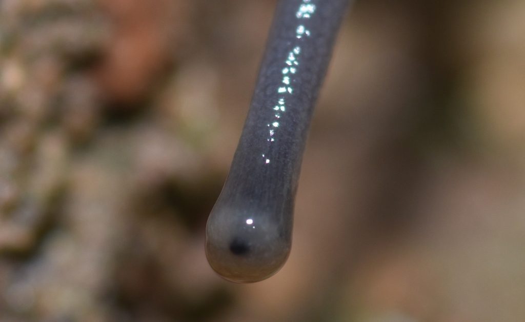
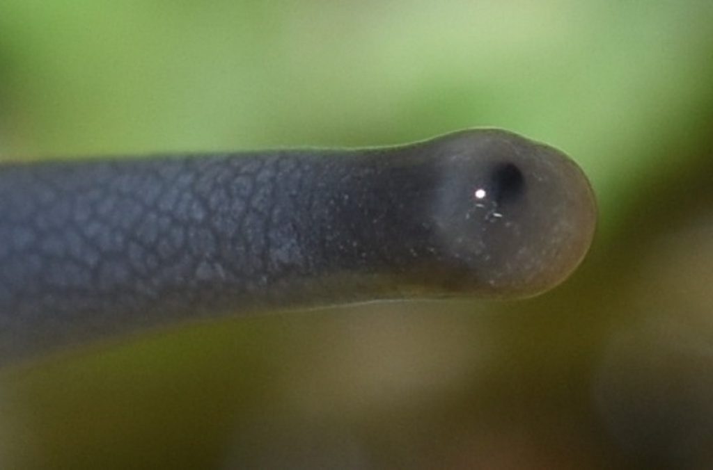
If you watch the following video all the way to the end, you’ll be treated to the weird experience of seeing a snail’s eye disappear into its flesh without a trace!
Eye of the Insect 2
Insects are believed to have evolved from crustaceans and, like crustaceans, many insects have compound eyes. Many insects also have several very simple, single-lens eyes called ocelli (singular ocellus). These are used to detect motion and do not form images. The photo below shows the head of a tarantula hawk wasp. (See my earlier post The Stuff of Nightmares.) In the center of the head, between the two compound eyes, you can see three small ocelli.

Sweetwater River Trail 9/8/17

Sweetwater River Trail 9/8/17
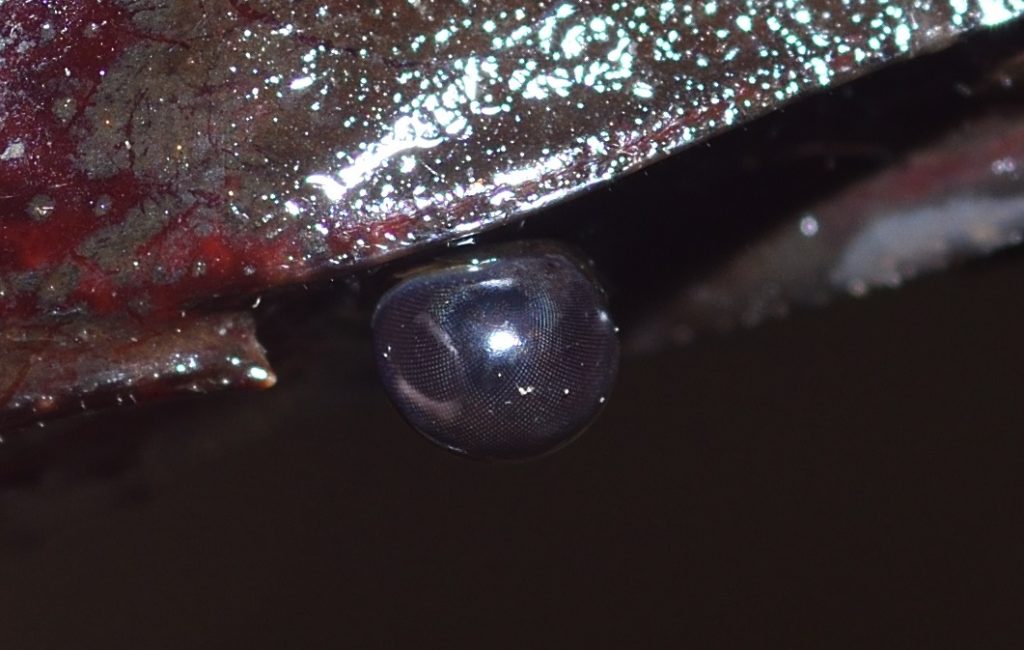
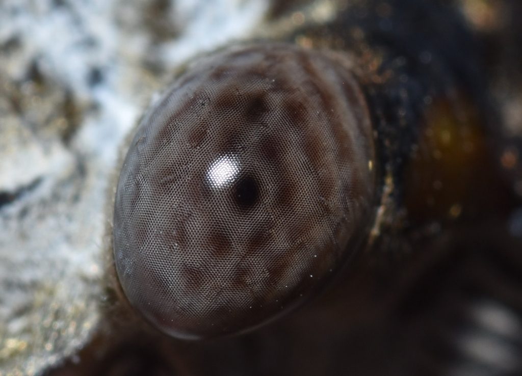
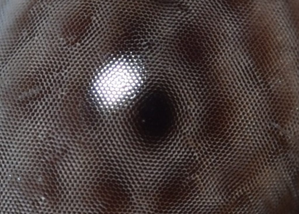
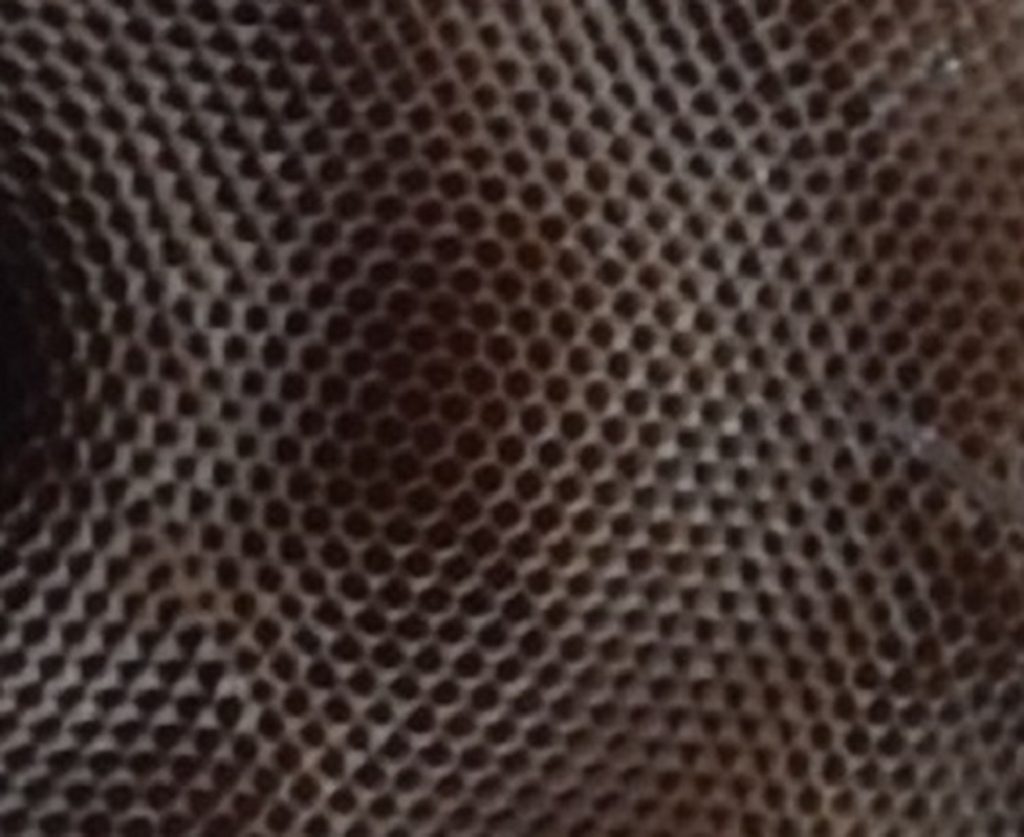
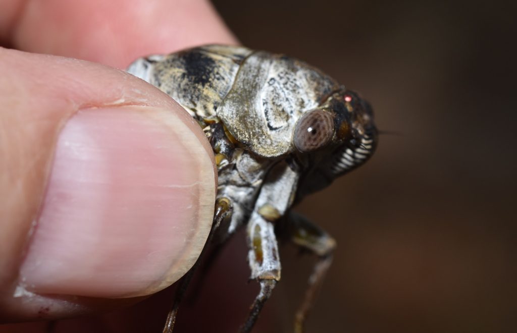
The compound eyes of the tarantula hawk wasp are too dark for you to see the individual facets. Not so with the large compound eyes of the cicada pictured in the following photos. As you can see, this is a very advanced eye with thousands of facets.
Emporia, VA 7/3/18
You may be wondering about the large, dark spot in the middle of the cicada eye. This is a ‘pseudopupil’. As the name implies, it’s not a true pupil. In a sense, it’s not even really there. Each of the tiny facets is the top of a tall columnar structure called an ommatidium. (See figure 3 in part 1.) The ommatidia in the dark spot look black because we’re looking directly down into their dark interiors where the photoreceptors are absorbing the light. We’re viewing the rest of the ommatidia at an angle, so we see the light reflected from their sides. If we could move around the eye and look at it from a different angle, the pseudopupil would appear to move with us.3
Many and Varied Are the Compound Eyes 4
Invertebrates have come up with many variations on the compound eye. These can be divided into two basic types: apposition eyes and superposition eyes.
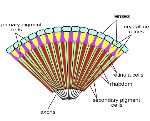
All compound eyes consist of collections of ommatidia. The main parts of an ommatidium are the cornea, the lens, and the rhabdom — a rod-like structure containing photoreceptors. (See figure 3 in Part 1). In an apposition eye, each ommatidium is an independent unit. Light entering the unit is focused only on the rhabdom belonging to that unit. Pigments surrounding the rhabdom prevent light from leaking into neighboring ommatidia. This is the simplest type of compound eye and was almost certainly the first type to evolve. Visual acuity can be very good in an apposition eye, but sensitivity is limited, because each rhabdom receives light from only a small area of the outside world. So organisms with this kind of eye are mostly limited to environments with high light levels. Marine animals with apposition eyes probably live near the surface. Terrestrial ones are probably diurnal.
Figure 4 below shows a typical apposition eye.
Author: Bugboy52.40
Permission is granted to copy, distribute and/or modify this document under the terms of the GNU Free Documentation License, Version 1.2 or any later version published by the Free Software Foundation; with no Invariant Sections, no Front-Cover Texts, and no Back-Cover Texts.
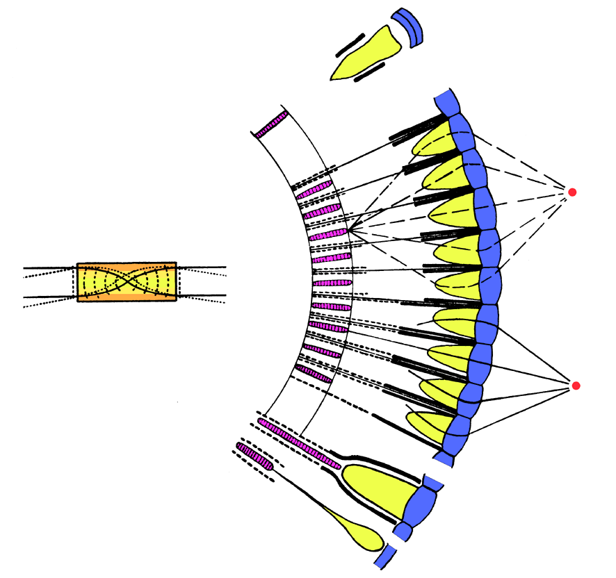
A superposition eye solves the sensitivity problem by focusing light from multiple lenses onto a single receptor. This decreases the acuity, but this can be offset by increasing the size of the eye and adding more ommatidia. Figure 5 illustrates one type of superposition eye.
Author: The original uploader was Kils at English Wikipedia.
This file is licensed under the Creative Commons Attribution-Share Alike 3.0 Unported license.
Each of these two major categories has several subcategories. Apposition eyes are subdivided into focal apposition, afocal apposition, and neural superposition eyes. (The latter is unrelated to true superposition eyes.) I won’t go into these in any detail other than to mention that only butterflies have afocal apposition eyes and neural superposition eyes are limited to true flies (order Diptera).
Superposition eyes are subdivided into refracting, reflecting, and parabolic superposition eyes. Again, I won’t get into the details, but I will mention that reflecting superposition eyes use mirrors to magnify the image, instead of lenses, much like a reflecting telescope! Nature, it seems, never misses a trick! (Actually, it does, but it’s still amazing to see the unexpected innovations it comes up with.)
Eye of the Spider
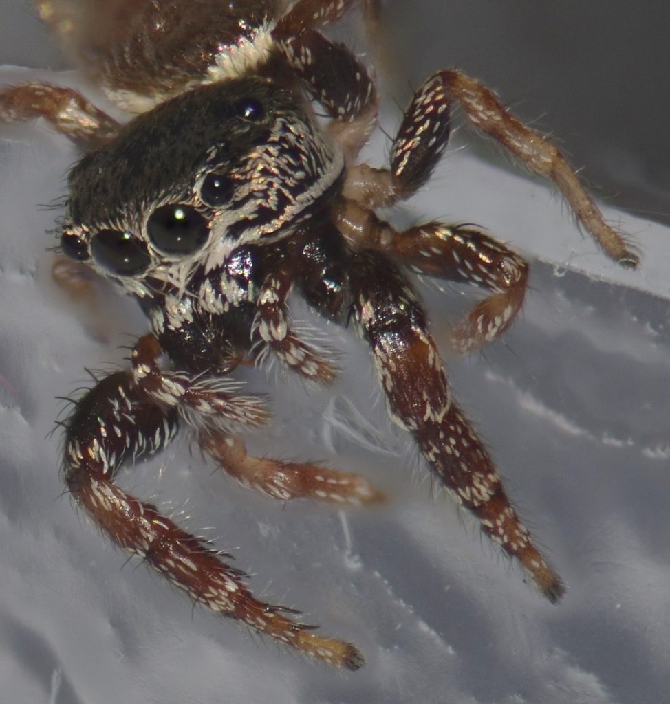
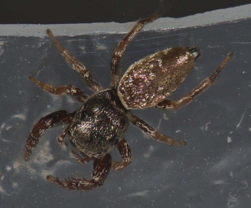
Meet Sassy. Sassy is a buttonhook leaf-beetle jumping spider (Sassacus vitis). She lives in my bathroom. I first encountered her when I was in the shower. Before you freak out, I should mention that Sassy is only a few millimeters long — much too small to be a threat to a human. I caught her in a small plastic container and, after her photo session, I returned her to the bathroom. I still see her now and then.
San Diego County 1/7/20
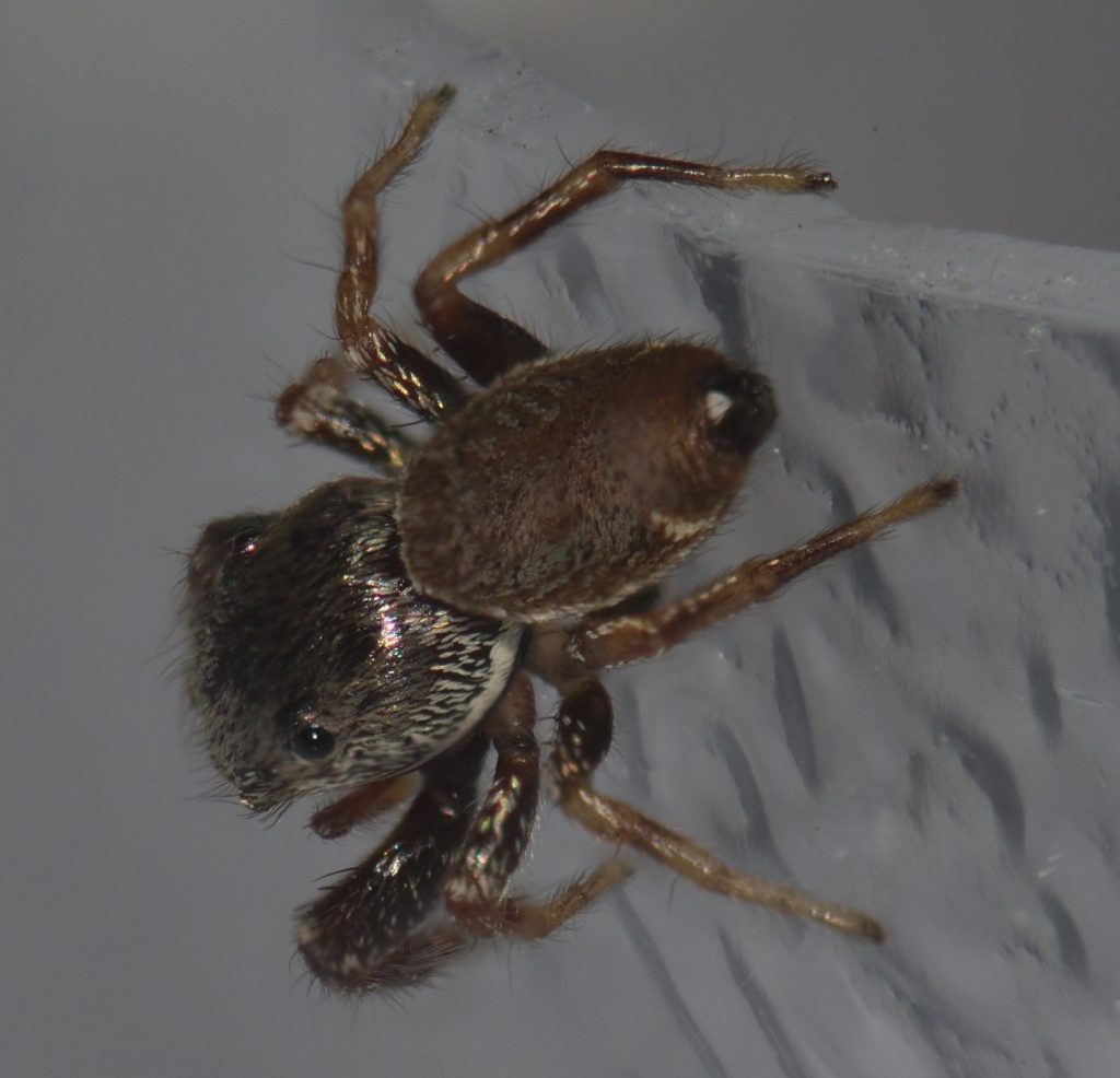
Most spiders have eight eyes. Spiders that build elaborate webs to catch their prey have very small eyes and poor vision. They detect prey by the vibrations they make when they get tangled in the web, so they don’t have much need for good eyesight.
Spiders that actively hunt their prey, like Sassy, are a different matter. Jumping spiders, in particular, have the best eyesight of any spider. The larger of the two pairs of eyes on the sides of the head are for detecting movement. The wide-set pair on the front of the head are for depth perception and the principal eyes — the large pair in the front center — provide a detailed image, probably in color.5 6 As for the remaining pair of eyes — the smaller of the two pairs on the side of the head — their function isn’t clear. They may be vestigial in some species.7
The principle eyes are ‘simple’ eyes — as opposed to the compound eyes of most insects. Like our eyes, they use a single lens to form an image. The remaining pairs of eyes are thought to be derived from the compound eyes of the spider’s ancestors, but they no longer have multiple facets.8
Jumping spiders hunt during daylight hours. Wolf spiders, on the other hand, hunt at night or in twilight. Four of their eyes have a tapetum (plural tapeta) — a reflective layer behind the retina. This is actually a very common feature in many kinds of nocturnal animals. When light hits the retina, not all of it gets absorbed. The light that passes through hits the tapetum and gets reflected back, so the retina has a second chance to absorb it. In this way, the sensitivity of the eye is greatly increased. Oddly, the principal pair of eyes in wolf spiders do not have tapeta.9 This seems counter-intuitive to me — I would expect the principal pair to be the most sensitive — but I haven’t been able to find an explanation for why this is not the case. Maybe having a tapetum would decrease the eye’s acuity somehow, but that’s just a guess. If any of you readers know the answer, please enlighten me in the comments.
Even with a second pass not all of the light gets absorbed. Some of the reflected light escapes the eye entirely. As a result, the eyes of animals with a tapetum, like wolf spiders and cats, glow in the light of a flashlight. If you hike at night in areas with a lot of wolf spiders, you’ll soon get used to spotting the tiny glow of their eyes. Many people say it’s a greenish glow, but it just looks white to me.10
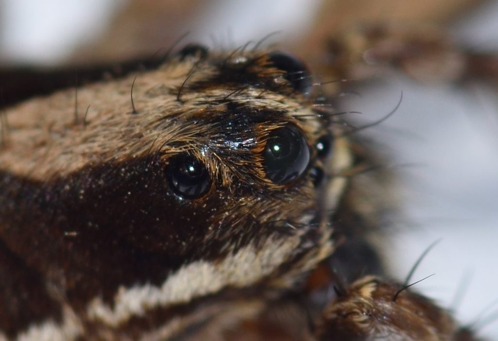
San Diego County. 10/02/19
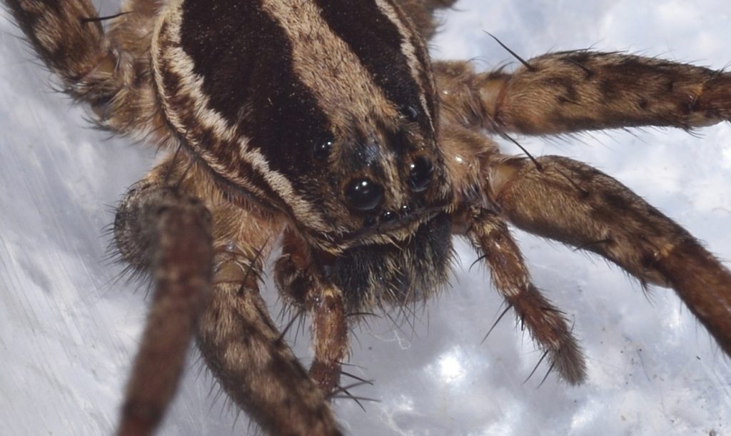
San Diego County. 10/02/19
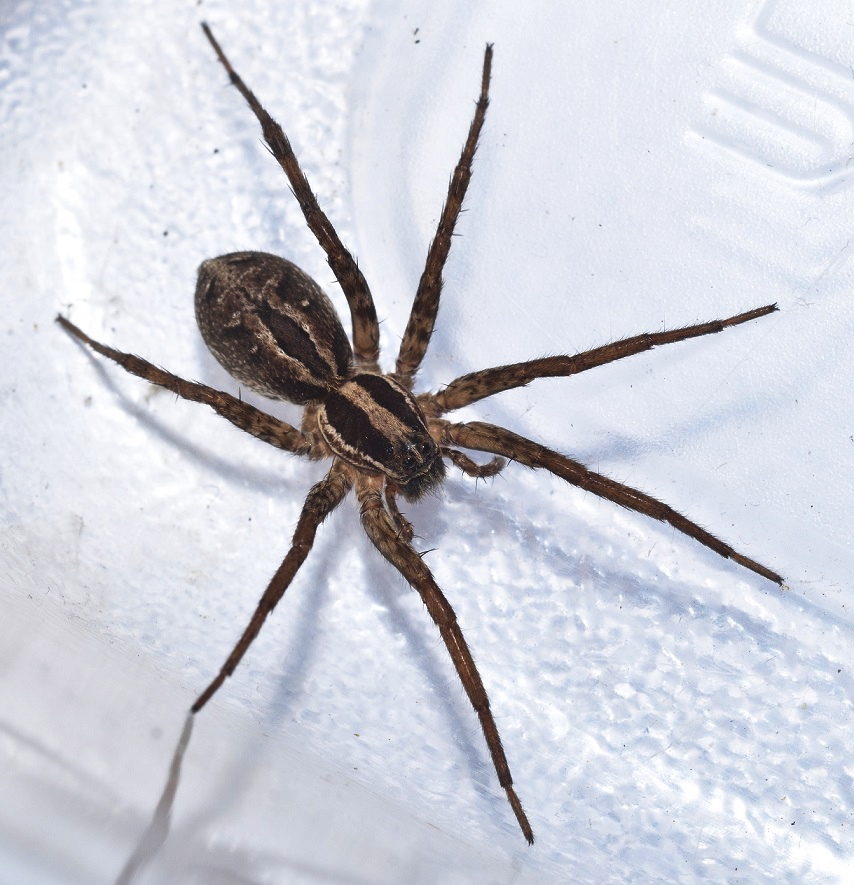
San Diego County. 10/02/19
Seeing the Polarization of Light 11
Some animals can see patterns formed by polarized light. This, of course, begs the question: What is polarized light?
Quantum mechanics tells us that light can behave as either a wave or a particle, depending on the circumstances. I won’t go down that particular rabbit hole. For the purposes of this discussion, we can think of light as a wave. If two people stretch out a rope between them and one of them moves her end rapidly up and down, a vertical wave will travel down the rope. If she moves her end from side to side, a horizontal wave will be produced. She can even create a circular wave by moving it in a quick circle. Think of that rope as a light wave. If the wave is oscillating vertically, then the light wave is vertically polarized. If it’s oscillating horizontally, then it’s horizontally polarized. (It can also be polarized at any angle in between horizontal and vertical.) And, of course, if it’s oscillating in a circle, then it’s circularly polarized.
The rays of light coming from the sun are randomly polarized. (Imagine a whole bunch of ropes oscillating in every direction.) However, when non-polarized light is reflected or refracted, it may become polarized. For instance, light bouncing off the surface of a body of water is polarized.
The sky looks blue because light from the sun is scattered by particles in the atmosphere. Why it’s blue and not some other color isn’t important for our purposes. The important thing is that the scattered light gets at least partially polarized. When the sun is directly overhead, the light is polarized horizontally throughout the sky. At dusk, the light is polarized vertically north to south — that is, perpendicularly to the direction of the sun. If you’re having trouble visualizing this, click here to go to a website that has some good illustrations of what I’m talking about.
The take away from all this is that polarized light forms patterns in nature and some animals are able to see those patterns. How are they able to do this? It turns out that a photoreceptor cell is most sensitive to light that is oscillating in a particular direction, depending on how the cell is rotated. That is, it’s more likely to absorb light that is polarized in that direction. In the retina of most animals, the photoreceptors are rotated randomly, so no matter what direction a ray of light is polarized, some of the receptors are going to be rotated at the correct angle to detect it.
So how does an animal evolve to detect polarized light? It just evolves groups of photoreceptor cells that are all aligned at the same angle. For instance, honey bees use the polarization of the sky (together with an internal clock that tells them what time of day it is) to navigate. If all the ommatidia in a bee’s eye were sensitive to the polarization of light, it would wreak havoc with it’s normal vision — it wouldn’t be able to make out objects in the environment. Instead, only the ommatidia in the upper part of the eye — the ones that are pointing at the sky — are sensitive to polarization.
I’ll be mentioning one or two other animals that can sense polarization later in the article, but before we get to that, it might interest you to know that humans are also able to detect the polarization of light, although we aren’t very good at it…
Haidinger’s Brush 12
Humans did not evolve to see polarization, but because of a quirk in the way the photoreceptors are arranged in our retina, it is possible for us to detect the pattern formed by polarized light in the sky. This pattern appears as kind of a faint cross with two blue or purple arms and two orange ones. Click here to see an illustration. The pattern is called Haidinger’s brush in honor of the scientist who discovered it. It takes some practice to see the pattern and not everyone can do it. If you want to try, click on that last link and follow the directions given there.
Eye of the Mantis Shrimp 13 14
Most animals have at no more than five types of photoreceptors. Many have fewer than that. Humans, for instance, have only four. The mantis shrimp has, depending on the species, between 12 and 16!
Mantis shrimp are not really shrimp — they’re a kind of marine crustacean belonging to the order Stomatopoda. Mantis shrimp are unique in a lot of ways. For instance, some species have clubbed claws with which they are able to smash open shells. These claws move so fast during a strike that they create vapor-filled bubbles called cavitation bubbles in the water. Their prey is hit twice — once by the club and a second time by the shock wave produced by the collapsing cavitation bubble. This wave is powerful enough to stun or kill the prey, even if the actual strike misses.
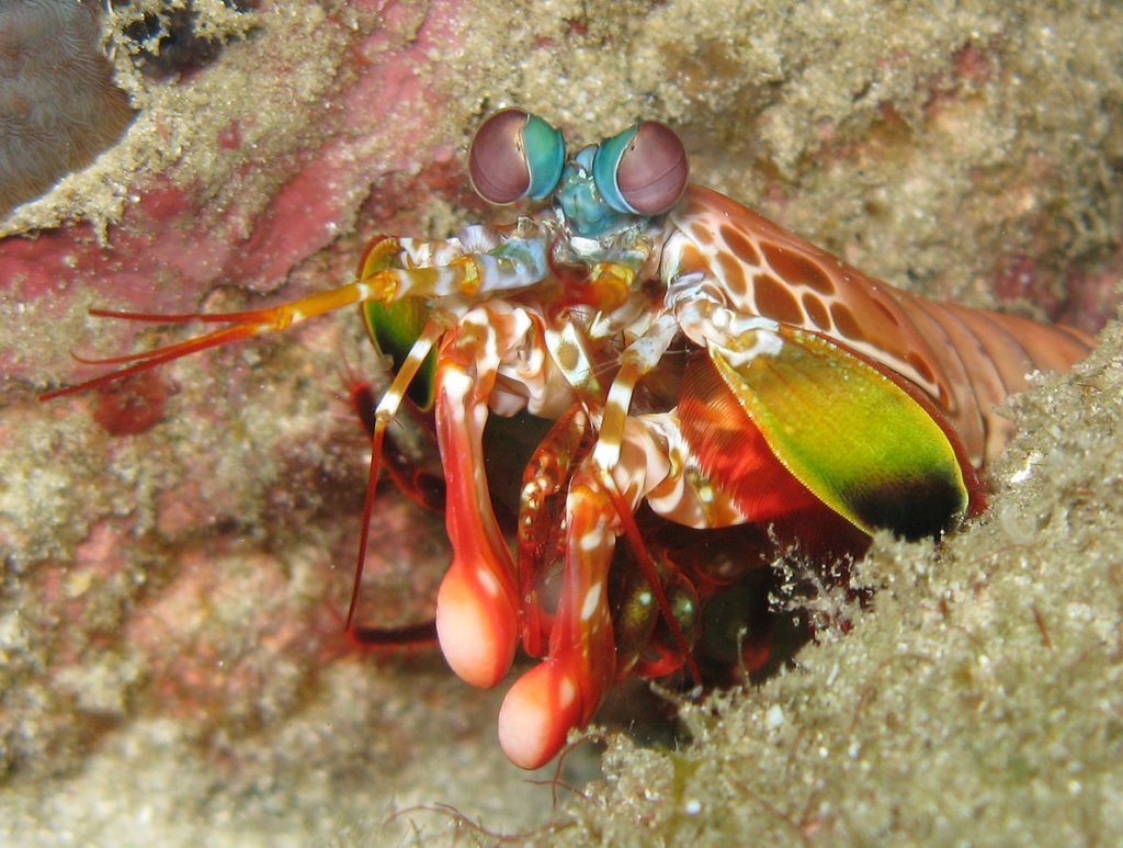
Mantis shrimp have the most complex visual system known to science. If you look at the photo below, you’ll notice a band going across each eye like a belt. Some of the photoreceptors in this band are used to detect polarized light. Most animals, if they can see polarization at all, are limited to detecting linear polarization. Some species of mantis shrimp are unusual in being able to detect both linearly and circularly polarized light. The ability to see polarized light helps the mantis shrimp detect its prey. Animals that blend into the background under ordinary vision often stand out clearly to predators that can see polarization.
The mid-band also contains various photoreceptors for ultraviolet, blue, and a longer wavelength in the green or red range. Other photoreceptor cells contain colored droplets of oil that act like yellow sunglasses, filtering out much of the blue light. Presumably, these are used to see yellow. There are also photoreceptors containing rhodopsin for night vision.
Some of the ommatidia in the rest of the eye (outside the mid-band) are directed toward the same point in space as the ones in the mid-band. This apparently helps with depth perception, but it also means that the animal’s vision is mostly limited to a narrow strip. That is, if the mid-bands of both eyes are held horizontally, then it won’t be able to see what is above or below it very well . The mantis shrimp compensates for this by being able to rotate its stalked eyes in any direction. It can even hold the mid-band of one eye in a vertical position, while the other mid-band is held horizontally!
The description of the mantis shrimp visual system that I’ve given here just scratches the surface. Wikipedia has a much more in-depth treatment. Check it out, if you’re interested.
Author: Silke Baron
This file is licensed under the Creative Commons Attribution 2.0 Generic license.
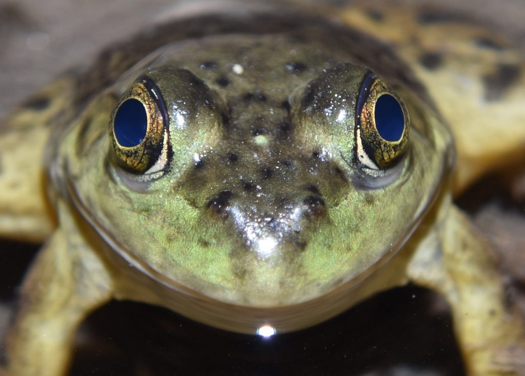
Amphibians represent the transition from an aquatic to a terrestrial environment in vertebrates. They evolved directly from lobe-finned fish and their eyes show it. Like fish, they have round, inflexible lenses and, like fish, they focus their eyes by moving the lens forward and backward. Your typical fish, however, doesn’t have to worry about keeping its eyes moist. Amphibians had to evolve a lacrimal system (tear glands and supporting structures) to keep their eyes from drying out when they’re not in water. They also needed lids to spread the tears. Probably, they evolved tear glands first, closely followed by lids.
Amphibians also evolved a transparent, nictitating membrane that can be drawn across the eye to protect it and to remove dirt. Reptiles, which evolved from amphibians, retained this membrane and improved on it. Mammals, which evolved from early reptiles, sometimes retained it and sometimes didn’t. (Humans, obviously, are in the didn’t category.)
A couple of fossils of lobe-finned fish show evidence of tear ducts that drained into the nasal cavity, so a lacrimal system was probably well on the way before true amphibians evolved. Lids don’t fossilize, though, so it’s not known if these fish had them.
Like most fish, many amphibians are tetrachromats — that is, they have four types of cone cells for detecting color. So some of them, at least, can see more colors than we can.
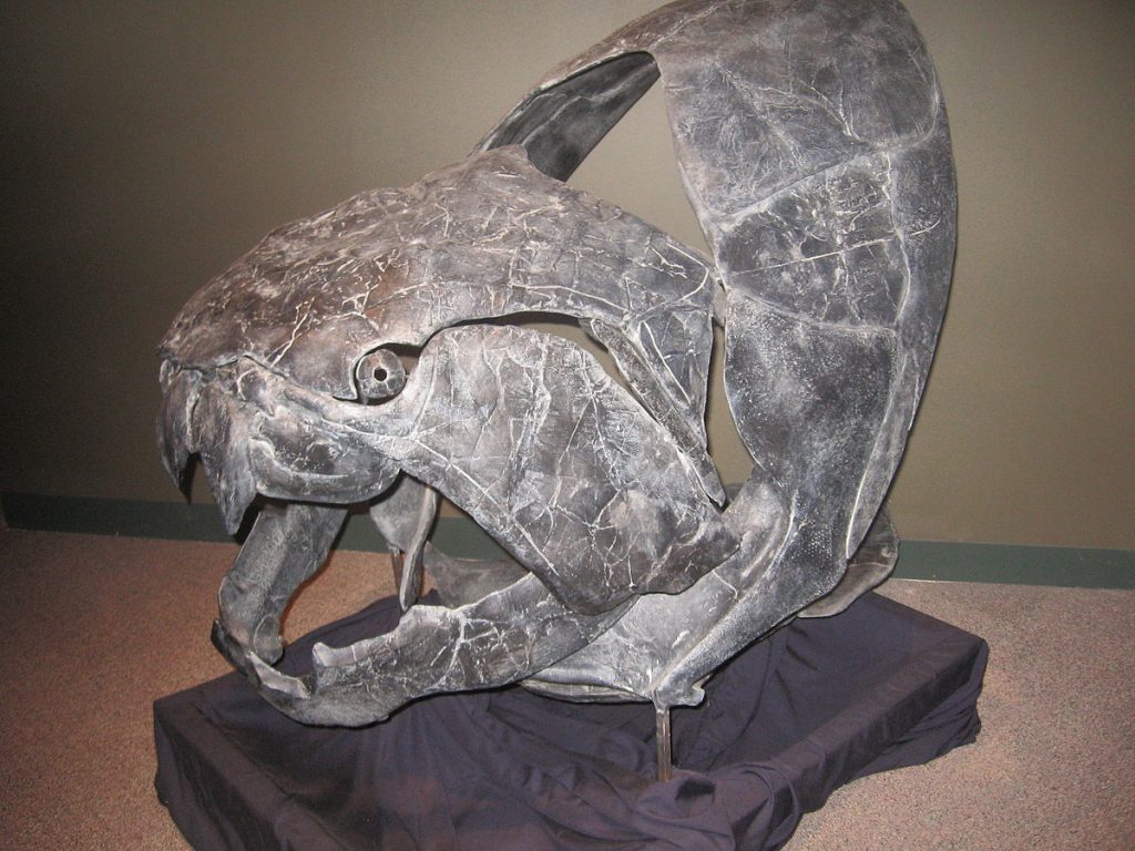
It sounds crazy, but frogs use their eyes to help them swallow. Frogs, salamanders, and newts have a muscle called the retractor bulbi that connects to the back of the eyeball. This muscle is used to pull the eye down and into the roof of the mouth. When a frog swallows a bug, it pulls its eyes in to help push the food down its throat. This is necessary, because the frog’s tongue attaches near the front of the mouth, instead of at the back, so it can’t be used for this purpose. There is evidence that this mechanism goes way back — the last common ancestor of the placoderms and the ray-finned fish probably had a retractor bulbi muscle.
San Diego County, CA 8/19/17
San Diego County, CA 8/19/17
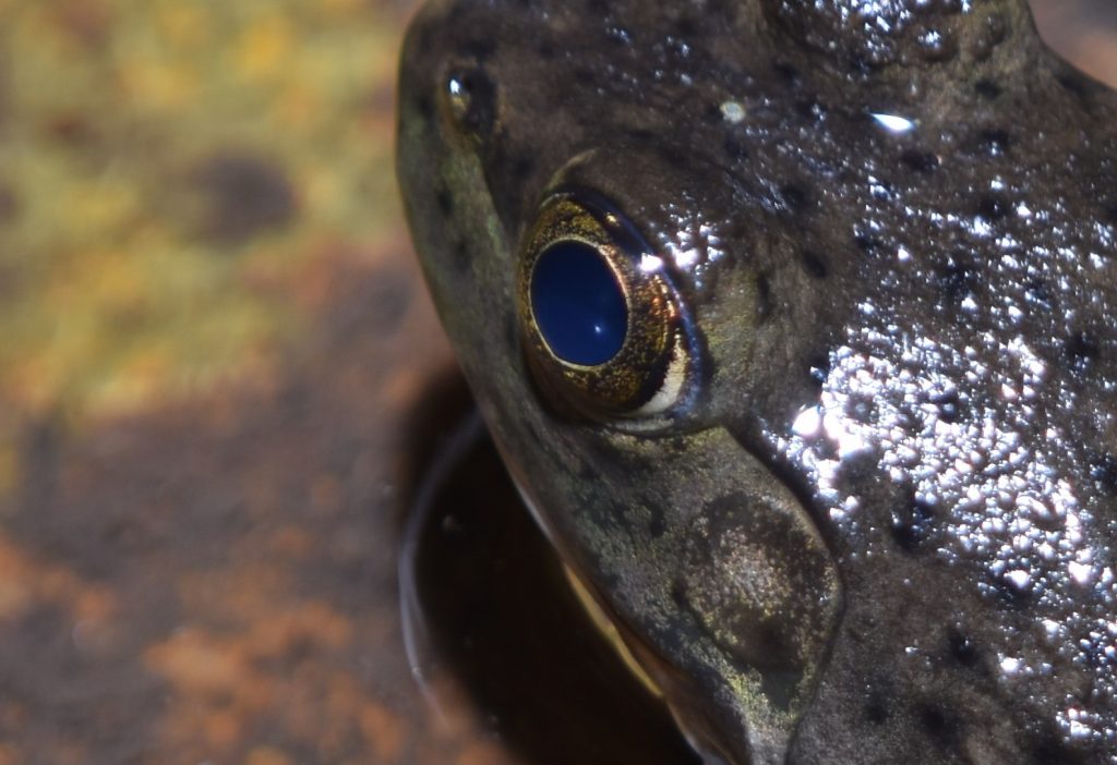
As mentioned in the section on the lens and cornea in part 1, most land vertebrates use the refractive properties of the cornea to supplement the eye’s internal lens. This doesn’t work in water, however. Because a cornea is itself mostly water, it has essentially the same refractive index as water. This means that light passing from the water to the cornea is not bent, so the cornea in fish can’t be used as a lens. Another consequence of this is that any bumps on the cornea will not significantly affect the fish’s vision, so fish corneas tend to be rather roughly textured.
When the lobe-finned fish and, later, the amphibians, started using their eyes in air, a rough cornea became a problem. At the same time, the greater clarity of air opened up the possibility of improved acuity. Amphibians responded by evolving a smoother, more compact, and more transparent cornea. It also became much steeper, so that it could act as a lens. In the frog eye, the cornea has taken over most of the refractive load from the internal lens.
Amphibian vision is specialized for detecting motion. In fact, the only way for an amphibian to see an object is if it is moving across its visual field. An article on the Evergreen website put it as follows:
“…if a frog possessed the mental ability to read, it could only do so if the words were continually passed before it’s eyes. Even then, his visual acuity might not be sufficient to distinguish letters; but if he tried to read a stationary page, it would just appear a gray blur. When you see something in peripheral vision, attention is drawn. You may know that something has moved, but not exactly what. Imagine that your whole visual field is like the peripheral, and that you automatically lunge toward and attempt to eat any moving object that seems a lot smaller than you and that moves erratically enough to seem lively. Imagine that anything static (not moving) in your visual field blurs and fades to obscurity. This is what it would be like if you were an amphibian.” 18
Most amphibians have four types of photoreceptors — two types of cones and two types of rods, including the usual ‘red rod’ that most vertebrates have and also a ‘green rod’ (which is actually attuned to blue rather than green). The green rods are different in form from normal rods and certain of their features suggest that they originated as cones that transformed into rod-like cells as frogs and salamanders transitioned to a nocturnal lifestyle. In some salamanders, these rods have reverted back into cones.
Eye of the Reptile 19
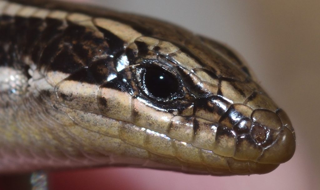
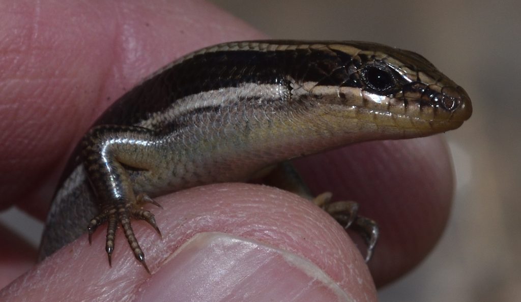
Reptiles evolved from amphibians. The reptile eye began as a diurnal eye. It tends to have a rounded globe shape, especially compared to birds, which evolved from reptiles. The cornea is clear and steep, more so than in amphibians. The uvea or vascular tunic is the eye’s middle coat. It contains the blood vessels that nourish the retina. In reptiles, terrestrial vision is still in the early stages of development and their uveas are quite thin compared to birds. As vision improved, the retina would thicken and the uvea would have to thicken to support it.
A major innovation in reptile eyes is the method of accommodation. Fish and amphibians focus by moving the lens backward and forward. Reptiles, birds, and mammals focus by deforming the lens. The structure that is responsible for this is called the ciliary body. It’s part of the uvea and is located just behind the iris.
This mechanism of accommodation, combined with the use of the cornea to help focus, allows the lens to be much thinner. This, in turn, allows the lens to be enlarged without filling the entire eye. A wider lens captures more light, so the sensitivity of the eye can be increased without having to enlarge the entire eye.
Sheephead Mountain, San Diego County, CA 9/21/19
Sheephead Mountain, San Diego County, CA 9/21/19
Eye of the Bird 20
The theropod lineage of dinosaurs evolved into the birds. In fact, birds are now classified as avian dinosaurs. (See my earlier post, The Coolest Thing About Birds.) The theropods were the top predators of their time and, not surprisingly, they developed good vision. Their bird descendants continued the tradition, eventually evolving the best eyesight of any animals.

Flight requires keeping your weight to a minimum, so birds tend to have small heads. There’s no way to push visual acuity to a maximum without enlarging the eyes, however, so avian eyes evolved to fill their sockets. In most birds, the eyes are bigger than the brain. Bird eyes have grown so large, that they’ve crowded out most of the extraocular muscle. Because of this, birds can generally only move their eyes a small amount in their sockets.
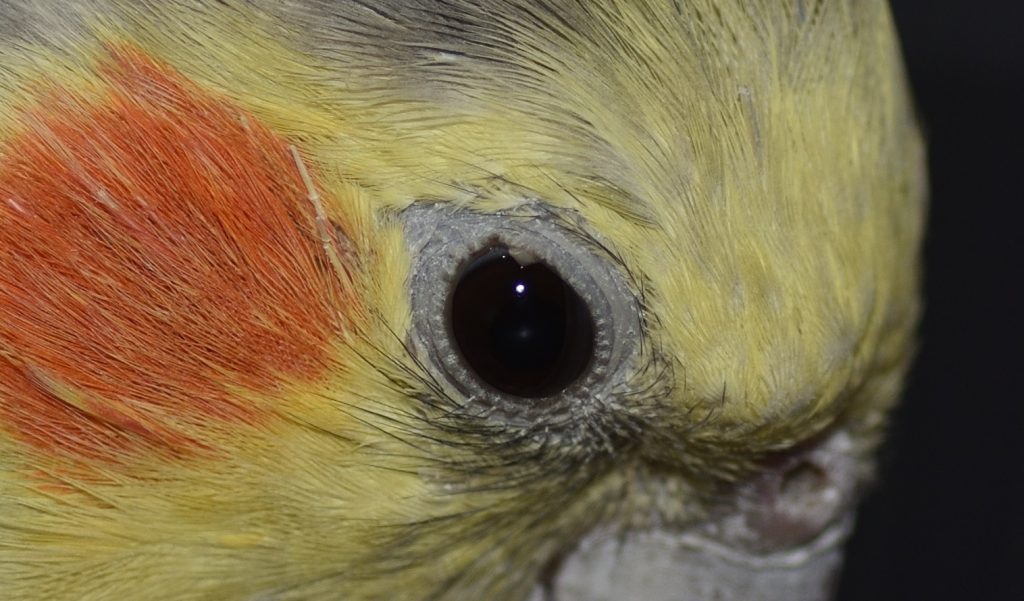
Most birds have roughly spherical, globular eyes like their therapod ancestors, but a couple of other eye shapes have evolved. Aquatic birds that dive for their food, developed flattened globes. As with fish, the cornea cannot help the lens refract light when a bird is underwater, so aquatic birds have evolved flattened corneas, as well. But unlike fish, they still have the soft, relatively thin lenses of their reptile ancestors. To compensate, some diving birds have come up with a novel solution. Muscular contractions of the ciliary body force the lens to bulge out through the pupil. This forces the lens into more of a spherical shape, increasing its refractive power.
The other eye shape that has evolved in birds is the tubular eye, which is seen only in owls and some goatsuckers (Caprimulgiformes), both of which are nocturnal. Having a long tube for an eye allows it to act like a telescope, magnifying the image and, thus, increasing the sensitivity of the eye. This is obviously important for nocturnal animals. The tubular eye of an owl is so large that it has even less room for extraocular muscles than is the case in other birds. They’ve compensated by developing the ability to turn their heads at great speeds through almost 270 degrees.
Eye of the Goat 21
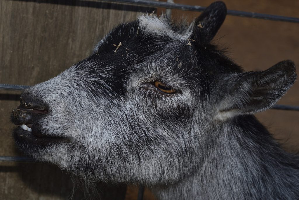
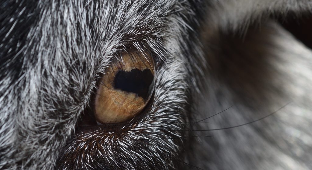
Goats have horizontal pupils. This looks pretty weird if you’re not used to it. Actually, many large herbivores have this kind of pupil. Ambush predators like cats and rattlesnakes, on the other hand, usually have vertical pupils. A study published in 2015 by Martin S. Banks et al. explores the relationship between pupil shape and an animal’s ecological role. The researchers looked at 214 different species of land animals and analyzed how the shape of their pupils affected their vision.
Obviously, prey animals need to be able to spot predators a long way off. A medium-sized herbivore like a goat, which tends to live in open country, probably doesn’t need to worry too much about predators swooping down from the air or jumping from trees. What it needs is wide, panoramic vision. And that’s what horizontal pupils give it. It also helps that its eyes are on the side of its head. This combination of features allows the animal to see a wide swath of its surroundings. (Ambush predators, on the other hand, usually have eyes on the front of the head so that they can focus directly on their prey.)
As important as it is for a prey animal to have a panoramic view of its surroundings, it still needs to be able to see in a forward direction well enough to avoid obstacles when it runs. Bank’s study found that a long, horizontal pupil enhanced the quality of the image both ahead and behind the animal.
Another fascinating thing the study found is that the extraocular muscles of a prey animal automatically rotate the eyes in such a way that the pupils remain in a horizontal position when the animal raises and lowers its head.

Eye of the Rattlesnake
In the previous section, I discussed a study that examined why different animals have differently shaped pupils. This study found that many ambush predators have vertical pupils because it improves their ability to judge the distance to prey.22 One such animal is the rattlesnake. You can clearly see the vertical pupils in the images below.
I’ve already covered rattlesnakes in a previous post (see Rattlesnake!), so I won’t spend a lot of time on them here, but I do want to mention the pit organs. These are also visible in the photos. The snake uses these to detect infrared radiation (a.k.a. heat) given off by prey. The pit organs contain infrared receptors in the form of heat-sensitive proteins. The mouth of the pit is smaller than its diameter, so the pit functions as a crude, infrared version of a pinhole camera. In other words, it actually forms an image! The pit organs don’t contain photoreceptors, so they are not usually thought of as eyes, but maybe they should be — a rattlesnake can see well enough with its pit organs to hunt in complete darkness!
If you haven’t already read my post on rattlesnakes, I urge you to at least read the section on pit organs, where I cover these amazing structures in more detail.

The silvery oval is the pit membrane.
Mission Trails Regional Park, San Diego County 8/14/2017

Mission Trails Regional Park, San Diego County 8/7/2018
Eye of the Human 23
Most mammals have dichromatic vision. That is, they have two different types of cone cells, containing two different photoreceptor proteins. This allows them to see violet, blue, green and yellow. This is a much smaller color range than that of many reptiles and birds, which are often tetrachromatic — having four different types of cone cells.
Why did mammals get short-changed? Back when the dinosaurs ruled, mammals were small, nocturnal, probably burrowing animals. They didn’t have as much need for color vision and, in evolution, if you don’t use it, you tend lose it. I don’t know why mammals didn’t lose color vision completely. Perhaps some color vision was helpful — they just didn’t need the wider range of color vision of birds and reptiles.
Some marsupials have better color vision than placental mammals (the group that most modern mammals belong to). Marsupials evolved before placentals and it’s speculated that some of them never lost the more extended color vision of their ancestors. They still don’t have the range of color sensitivity of birds and reptiles, though.
Most Old World monkeys and primates (including humans), are trichromatic. That is, they have three types of cone cell. Their ancestors, however, were dichromatic, which means that they somehow managed to re-evolve a third cone type. They did this by gene duplication.
When DNA gets copied, occasionally an error occurs that causes a gene to be duplicated. If this happens in a germ cell, it gets passed down to the offspring as a mutation. Depending on the gene, having an extra copy may cause the animal serious problems or it may have no noticeable effect at all. In the latter case, the animal ends up with an extra gene just “lying around” that evolution is free to play with.
So, in our case, the extra gene eventually accumulated mutations that caused it to produce cone cells that were sensitive to a different range of wavelengths and this led to a lineage of mammals with a third kind of color receptors. Specifically, the new receptors allow primates to see red and orange hues.
It’s important to note that this new type of cone cell is not a reappearance of the one that was lost when mammals became nocturnal and burrowing. It’s an independently evolved photoreceptor that extends the color vision of primates and Old World monkeys.
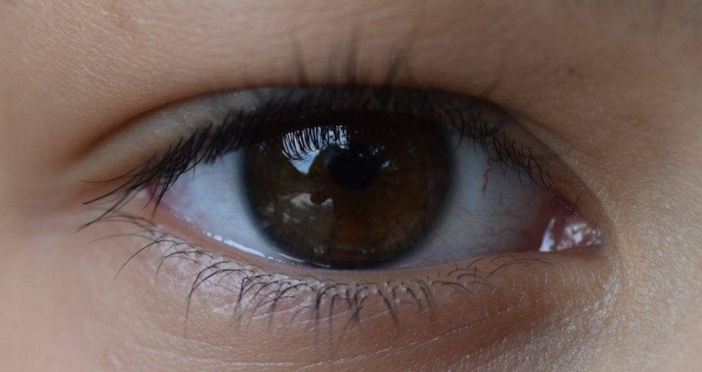
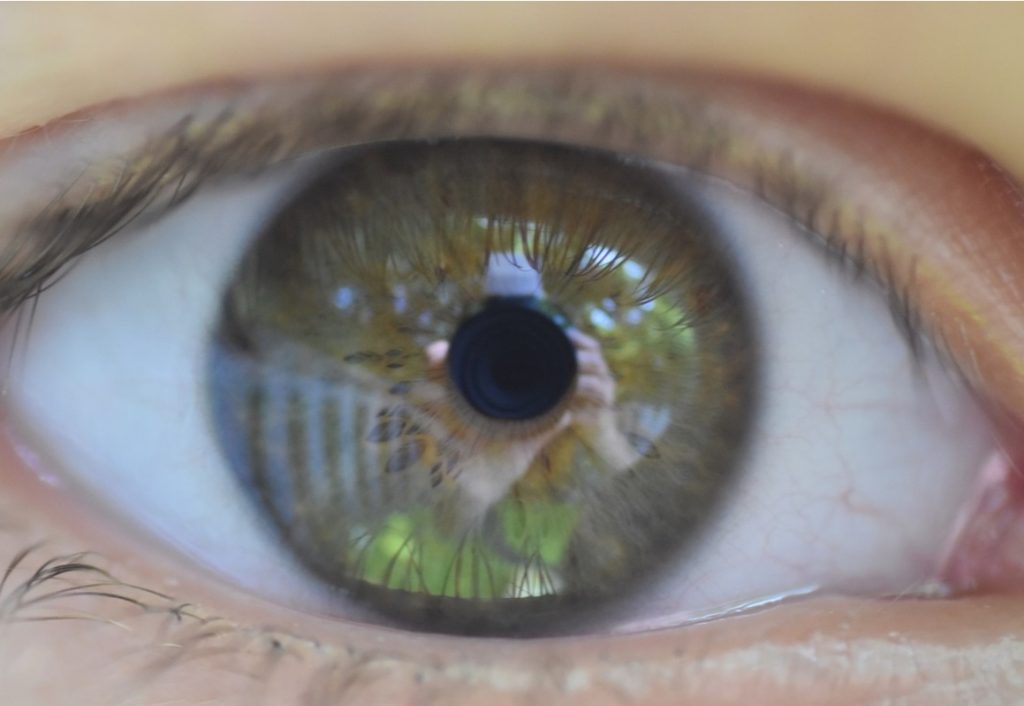
This third cone cell would not have been maintained in the lineage if it didn’t give its owners some advantage. What might that advantage have been?
Color and Climate 24
A howler monkey is a type of Old World monkey that eats mostly leaves. Young leaves tend to be the most nutritious. They also tend to be more reddish or orange, because they haven’t developed enough chlorophyll to mask the colors of the red and orange pigments. (These are the same pigments that make leaves change color in the Fall as they lose their chlorophyll.) In 1999, a team of scientists working in a national park in Uganda spent ten months studying what the various monkeys in the park ate. The researchers found that the howlers were using their extended color vision to selectively eat the most nutritious leaves.
So having trichromatic vision appears to have given howler monkeys an evolutionary advantage. But most monkeys today eat fruit, not leaves. Why would the ancestors of Old World monkeys and primates have evolved an adaptation to eating leaves in the first place?
Molecular studies of the photoreceptor proteins in primates have allowed scientists to estimate when the gene duplication that led to trichromatic vision occurred. It turns out that the lineage of mammals with three color genes goes back to between 40 and 30 million years ago.
It was around this time that the Earth was starting to get significantly cooler. Vast ice sheets pressed down from the poles, pushing back the tropical forests, full of palms and figs, and replacing them with grasslands and deciduous forests with more leafy trees. Gone were the large, abundant fruits, so now our ancestors had to rely much more on eating leaves.
The ice sheets have advanced and receded multiple times in the last 40 million years, but the Earth has never gone back to a climate where tropical forests covered most of the planet. It appears that we have that cooler climate to thank for our ability to experience such a colorful world.