
A while back, I wrote a blog post about the parietal eye of lizards. (See Mysteries of the Third Eye.) In the course of doing research for that article, I read a bit about the evolution of eyes in general and I realized that this would make a fascinating topic for a future post — fascinating and challenging. The first eyes probably evolved around 600 million years ago and natural selection has been diversifying and refining them ever since. The variety of eyes and eye-like structures in modern organisms is truly mind-boggling. In this article and the one that follows, I’m going to cover a broad sampling of visual organs and try to give you some idea how they came to be.
Evolution of the Eye in a Nutshell 1 2
Let’s start with a quick overview. Most eyes consist of soft tissue, which rarely fossilizes, so their fossil record is very sparse. Nevertheless, scientists have a pretty good idea of the major stages in eye evolution. These are shown in the illustration below. An explanation of the various stages follows the figure.

Author: Matticus78 at English Wikipedia
Permission is granted to copy, distribute and/or modify this document under the terms of the GNU Free Documentation License, Version 1.2 or any later version published by the Free Software Foundation; with no Invariant Sections, no Front-Cover Texts, and no Back-Cover Texts.
Stage 1: Sensing the presence of light. (Figure 1-a) One of the earliest ‘proto-eyes’ to evolve was the eyespot. An eyespot consists of a patch of photoreceptive cells. This relatively simple organ is enough to let an organism know whether it’s in light or darkness. This may allow the organism to regulate it’s circadian rhythms. It may also allow it to move around at random until it finds the level of light or darkness that suits it.
Stage 2: Sensing the direction of light. (Figure 1-b) A very small organism, especially a microscopic one, is often transparent, so light can reach its eyespot either by shining directly on it or by first passing through the body. Eventually, some organisms evolved a layer of opaque pigment underneath the eyespot. Now, only light coming from outside the body could reach it. This enabled organisms to get some sense of the direction from which the light was coming.
Eventually, in some organisms, the eyespot became depressed, forming an eyecup. If light shone on the cup at a sharp angle, the rim of the cup would block part of it, so that only some of the photoreceptors in the eyecup were exposed. This allowed the animal to get a better sense of the light’s direction. Over many generations, the depression deepened into a pit and the organism’s ability to sense the direction of light steadily improved.
Stage 3: The pinhole camera eye. (Figure 1-c) Eventually, a limit was reached beyond which increasing the depth of the pit no longer helped. Now, however, evolution was in a position to take a very simple, but incredibly important step — the opening of the pit started to narrow, forming a pinhole camera.
Maybe you played around with one of these in science class back in the day. Take a shoe box and punch a small hole in one side. Light entering the box through this hole will form an image on the opposite wall of the box. (You can cut a larger hole in the top to peek at the image.)
So, as the opening got smaller, the pit started to act as a pinhole camera and a crude image began to form on the layer of photoreceptors lining the pit. The image got sharper as the opening got smaller, but, unfortunately, it also got dimmer. This is a serious limitation of pinhole cameras. Nevertheless, we now have our first true eye.
Step 4: Lenses and other refinements. (Figure 1 d-f) Covering the opening with a transparent layer of cells, displacing the water with a transparent ‘humor’, and adding a lens to collect more light (and thus improve sensitivity) led to the first ‘lens eyes’. Many other enhancements followed, some of which we’ll be covering later.
How Long Did All This Take?
So how long does it take to go from eyespots to eyes? Not as long as you might think, according to a paper by Dan-E. Nilsson and Susanne Pelger.3 These researchers estimated the time required to evolve an eye based on things like the rate of mutation and the relative advantage to the organism. They came up with a conservative estimate that the vertebrate eye could have evolved from a patch of photoreceptor cells in just 364,000 years — so, not even half a million years! This is a remarkably short period of time from an evolutionary standpoint and it helps to explain why there are so many varieties of eyes in modern animals.
Eyespot of the Planarian
Planeria are small, flat, worm-like animals, commonly called flatworms. What looks rather ridiculously like two crossed eyes, are actually eyespots. The dark areas are pigment cells that shade the photosensory neurons from one side, allowing the planarian to detect the direction of light.4 The eyespots are also slightly depressed, forming very shallow eyecups. This further aids the animal in sensing direction.5 I was able to find one site that shows the anatomy of a planarian eye. Click here if you want to check it out.
Phototaxis is the tendency of an organism to move either toward or away from light. Planaria exhibit negative phototaxis, meaning they tend to move away from it.6 7

Kumeyaay Lake, San Diego County 7/17/17
A Quick Note
In the course of these two articles, I’ll be interspersing non-technical sections with slightly more technical ones. The latter will have a cyan background. Feel free to skip over the more technical parts, if you’re not feeling ambitious.
The First Photoreceptors and the Evolution of Multi-cellular Life 8
Bacteria are prokaryotes — single-celled organisms that lack a cell nucleus. Cyanobacteria, as the name suggests, are a type of bacteria. They’ve been around for between 3.75 and 2.6 billion years. Cyanobacteria use the photoreceptive compound chlorophyll for photosynthesis. Chlorophyll may have been the first photoreceptive molecule used by living things, although there are other possibilities.
Another important group of prokaryotes is the Archaea. Archaea are descended from bacteria and look very similar. In fact, they were originally thought to be bacteria. When scientists finally got around to doing molecular studies on them, however, it was found that they are very different genetically. In fact, Archaea are more closely related to eukaryotes — organisms that do have nuclei in their cells. Eukaryotes include single-cell Eukaryoptes called protists (amoebae, paramecia, etc.) and all multi-cellular organisms, including us.
One type of Archaea, halobacteria, uses a photoreceptive molecule called halorhodopsin both as an energy source and to detect light. This could have been the first photoreceptor used for sensory purposes. Halorhodopsin is a member of an important family of photoreceptors called rhodopsins. Various rhodopsins are used in the eyes of all animals.
Eukaryotic cells are much larger and more complex than prokaryotic cells. In addition to the nucleus, they contain various other membrane-enclosed structures called organelles that serve specialized functions within the cell. All eukaryotic cells, for instance, contain mitochondria. These are the ‘powerhouses’ of the cell. They produce the energy that facilitates high-energy activities such as locomotion. Another organelle, the chloroplast, is found only in plant cells. Chloroplasts, like cyanobacteria, contain chlorophyll, so they are used to harvest energy from sunlight.
Molecular studies of mitochondria and chloroplasts have found compelling evidence that both of these organelles originated as free-living bacteria. Chloroplasts, in particular, were originally cyanobacteria. At some point in evolutionary time, a larger single-cell organism must have either engulfed a cyanobacterium in an attempt to eat it or a cyanobacterium tried to parasitize the larger cell. Instead of the cyanobacterium being digested or the larger cell being killed, they somehow formed a ‘truce’. The bacterium reproduced inside the larger organism and, when that organism reproduced by dividing, the descendants of the captured bacterium got distributed to the new cells. Eventually, the cyanobacteria evolved a mutually beneficial relationship with the enclosing cells — they became chloroplasts. This process — of a captured cell being incorporated into a larger cell as an organelle — is called endosymbiosis.
Chloroplasts (and also mitochondria) have their own DNA, separate from the DNA in the nucleus of the parent cell. (This DNA, by the way, is very similar to bacterial DNA, which is what first gave researchers the clue that they originated as bacteria.) They reproduce independently and are passed down to the daughter cells in the cytoplasm. Over time, many of the genes that were originally in the bacterial DNA have been incorporated into the DNA of the parent cell, leaving the chloroplasts and mitochondria with much reduced, but still recognizably bacterial, genomes.
Recent research suggests that it was an Archaean that formed the first endosymbiosis with a bacterium, thus leading to the first eukaryotes and, eventually, to the first multi-cell organisms.
Volvox is a genus of single-cell organisms that forms large colonies. A Volvox colony behaves much like a single organism. It was probably such a colony that evolved to form the first truly multi-cell organism. Volvox contain a photosynthetic rhodopsin compound that is very similar to the rhodopsins in animals. It’s likely that all the rhodopsins used by animals have a common origin.

Note the eyespot attached to the motor apparatus (the flagellum).
Author: Gevictor
This file is made available under the Creative Commons CC0 1.0 Universal Public Domain Dedication.
Eyespot of the Threadsnake 9
Not every animal that uses eyespots is ‘primitive’. Threadsnakes (a.k.a. blindsnakes) spend most of their time underground, so they don’t have much use for full-blown eyes. Their ancestors had eyes, of course, but living underground eliminated the selection pressures on them. What does this mean? Suppose a mutation occurs in one of the genes that’s responsible for the development of part of the eye, resulting in poorer vision. Well, it doesn’t matter, because the snake isn’t using its eyes, anyway. Over thousands of generations, such mutations will tend to accumulate, because natural selection is no longer weeding them out. Eventually, the threadsnake is left with nothing but eyespots.
Does this mean that they will eventually lose the eyespots, too? Threadsnakes do occasionally come to the surface — usually at night or late in the day — so it’s helpful if the snake can at least tell night from day. Eyespots are good for that. So it’s possible that future generations of threadsnakes will keep the eyespots.


and some kind of seed
The Compound Eye 10
Eye evolution has taken two major directions — simple eyes (an eye with one lens) and compound eyes. All vertebrates have simple eyes (if they have any at all). Invertebrates may have either or both. We’ve already seen how simple eyes evolved. Compound eyes evolved by duplication. That is, a simple eye got duplicated multiple times to form a cluster and nerve impulses from all the eyes in the cluster were integrated in the brain to form a single image.
A simple lens eye doesn’t have to be very good. In many animals, the main purpose of a lens is to improve sensitivity by concentrating light. The ability to form an image is secondary. (The first eyes evolved in the depths of the ocean, so just detecting light would have been a priority.) Another way to increase sensitivity is to evolve multiple eyes, so that even more light is collected.
In this way, some animals evolved clusters of simple eyes. Now, suppose that such a cluster happens to form a bulge. Light hitting the bulge at a sharp angle would be blocked by the bulge itself, so that the light couldn’t reach all of the eyes. In this way, the bulge would give the organism a sense of the direction of the light. Increasing the bulge would improve that sense of direction.
So now we have a cluster of lens eyes, with each one oriented in a slightly different direction. In other words, we have the beginnings of a compound eye. In a true compound eye, the individual simple eyes — now just sub-units of the compound eye — are called ommatidia. (See figure 3 for a simplified illustration of an ommatidium.)
As the simple eyes evolved into ommatidia, they stretched out, becoming thin and deep, so that only the rays of light that hit the top of an ommatidium more or less directly were able to penetrate into its depths to reach the photoreceptors. In this way, each ommatidium received light from only a small section of the visual field. Input from all the ommatidia was integrated in the brain to form an image.
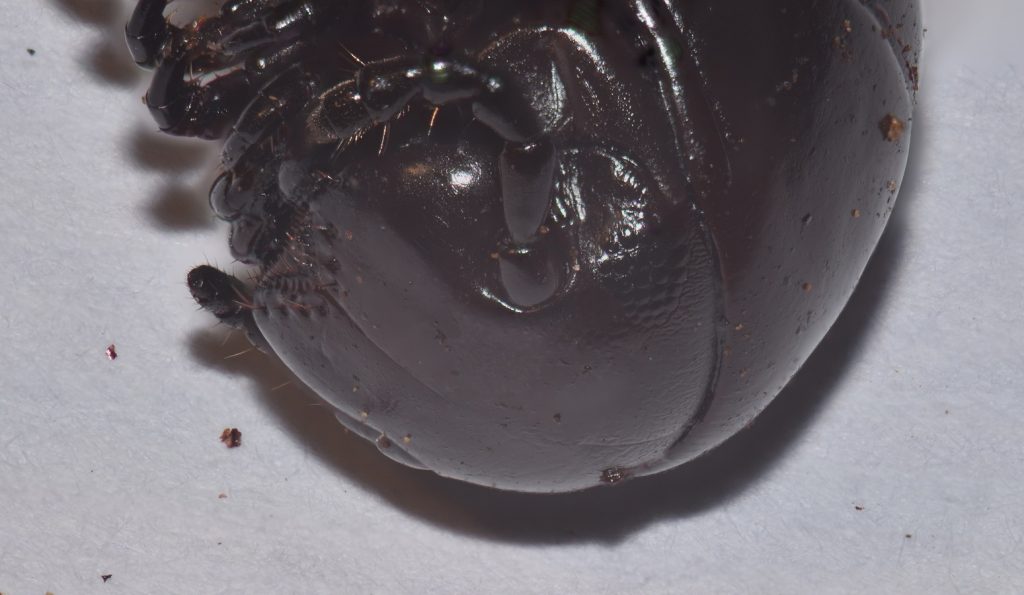
Eye of the Millipede 11
Millipedes are arthropods, like crustaceans, spiders, and insects. Some are eyeless; some have compound eyes; others, like the one pictured here, have patches of simple eyes that are only loosely integrated and maybe don’t quite qualify as fully compound.
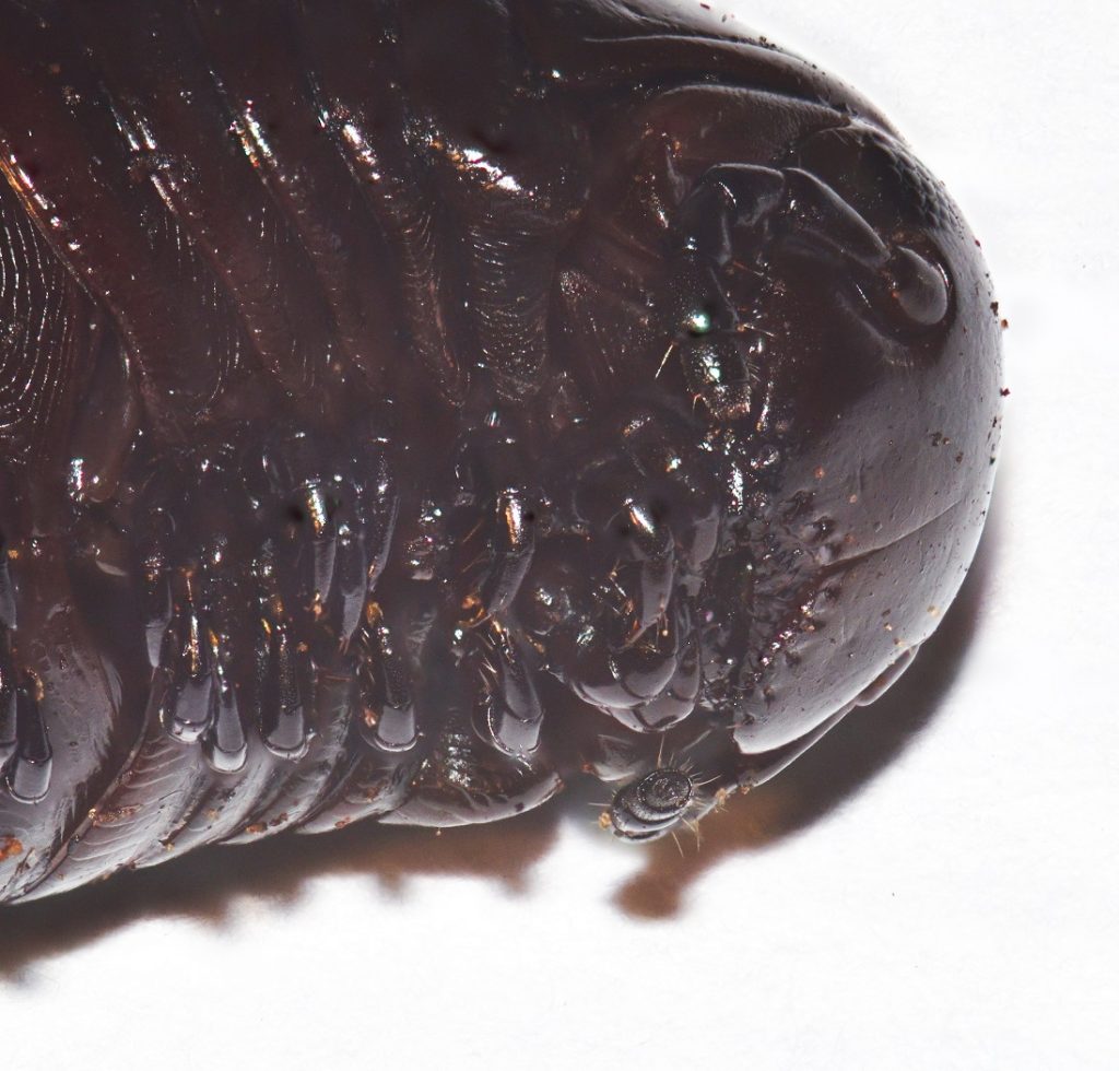
Mission Trails Regional Park, San Diego County 12/03/19
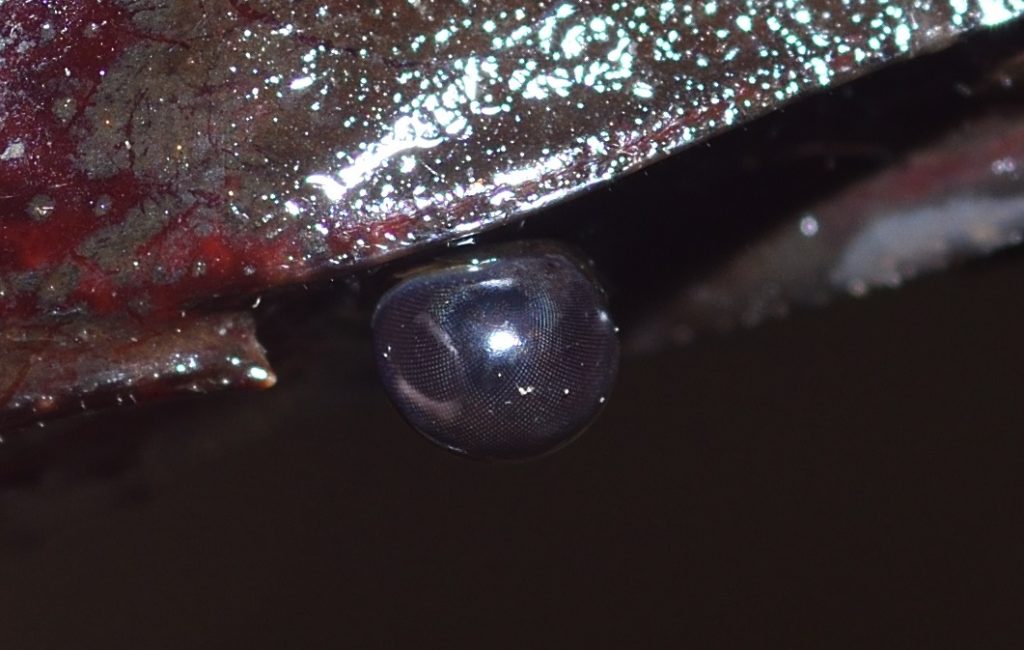
Eye of the Crayfish 12
(Note: Part of this section is taken from my earlier post Crayfish Journeys.)
Crayfish are crustaceans, like crabs, lobsters, and shrimp. Like most crustaceans, they have compound eyes. Each eye consists of thousands of thin, columnar structures called ommatidia. The main parts of an ommatidium are a cornea, a lens, and photoreceptor cells. (Figure 3 shows a generic drawing of an ommatidium.) Each of the tiny, individual facets visible in the following photo is the cornea of an ommatidium. When excited by incoming light, the photoreceptor cells produce electrical impulses representing the intensity and color of the light. These impulses are integrated by the nervous system and brain of the crayfish to form an single image.
Sweetwater River near Jamul, CA

Sweetwater River near Jamul, CA

A and B represent the cornea and lens, respectively. E is the rhabdom, which contains the photosensitive proteins.
This image is in the public domain. It was downloaded from Wiki Commons at this url.

The beginning of the Cambrian explosion is dated to about 541 million years ago and it lasted from around 13 to 25 million years. It was a remarkable period of diversification in which most of the major groups that we see in modern animals appeared. It marks the beginning of the Cambrian Period.
The Ediacaran Period, which immediately preceded the Cambrian, is sparcely represented in the fossil record, probably because most of the organisms at the time had soft bodies that rarely fossilized. In the Cambrian, by comparison, we see the emergence of animals with hard shells or exoskeletons. We also see the emergence of animals with fully developed eyes.
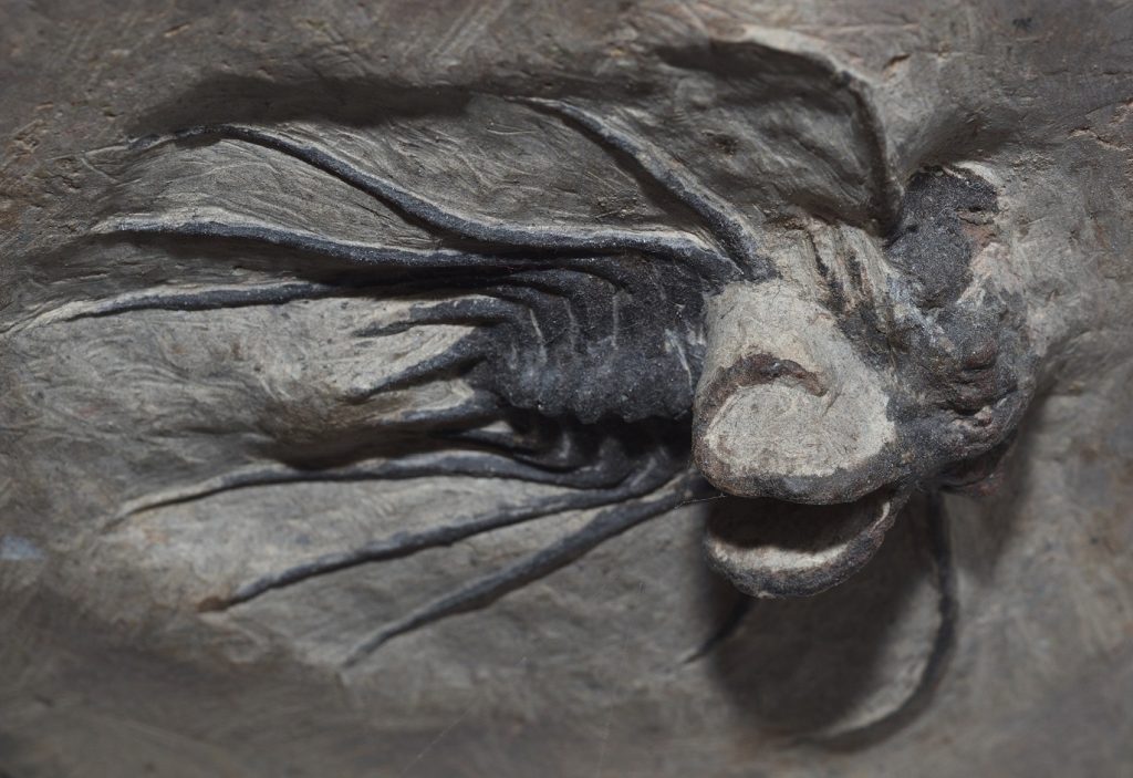
Trilobites, for instance, were a very large, diverse, and highly successful group of arthropods with a wide range of visual organs. Some were blind, but many others had compound eyes with rigid lenses made out of pure, transparent calcite. Each lens was an elongated prism. Some trilobites had only one such lens in each eye (in which case, I suppose it was really a simple eye, not a compound one). Others had thousands of lenses in each eye, rivaling some modern insects. Calcite is a mineral, by the way, so you could argue that trilobite eyes were made of rock (or, at least, the lenses were)!
Trilobites didn’t have corneas. The lenses were fixed to the exoskeleton of the animal. This means it was impossible to focus it, so trilobites were unable to accommodate for distance. Otherwise, though, some of them appear to have had surprisingly good vision. Studies have shown that some types of trilobite eyes had excellent depth of field and minimal spherical aberration.
Scientists are still debating what led to the proliferation of animal forms during the Cambrian. One likely explanation is the evolution of eyes. Having the ability to see other animals at a distance would have been a game changer. Suddenly, predators were able to see and stalk their prey. Their prey, in turn, evolved hard coverings to protect themselves. Of course, all of this took time. The changes that eventually led to the types of animals we see in the Cambrian explosion must have gotten their start in the Ediacaran. According to some estimates, true eyes may have evolved as early as 600 million years ago.

Author: Kennethgass
This file is licensed under the Creative Commons Attribution-Share Alike 4.0 International license.
Nautilus is a genus of cephalopods — the group that also includes squids and octopuses. They are one of the few surviving members of an ancient lineage that evolved in the late Cambrian. The best known species is the chambered nautilus (Nautilus pompilius), pictured below.
N. pompilius is most famous for its shell, which is usually displayed split down the middle to reveal the beautiful geometry of its interior. What doesn’t get as much attention is its large, pinhole camera eyes. These eyes have no cornea or lens. The pupil takes the form of a circular hole near the center of the eye, with a narrow slit trailing down vertically from it. This slit will close completely as the pupil contracts. In life, the eye is filled with a gelatinous substance that is probably similar to the vitreous body of our eyes, but the pupil has no covering, so seawater can freely pass in and out.

Source: http://opencage.info/pics/large_3628.asp
This file is licensed under the Creative Commons Attribution-Share Alike 2.5 Generic license.
The nautilus lives in the Pacific at depths ranging between 150 and 600 meters. It’s active mostly at night and in twilight, so it’s most important source of light is probably bioluminescence. A nautilus can only see a few meters and it’s vision must be rather blurry, but living as a scavenger and opportunistic predator, that’s apparently good enough. Also, its hard shell offers pretty good protection, so it doesn’t have much need to see predators coming a long way off.
Fish (and Their Eyes) Evolve 18
The earliest fish, the ostracoderms, were jawless fish related to the lampreys. The first fish with jaws were the Acanthodii. They showed up around 410 million years ago during the Silurian. Not much is known about their eyes.
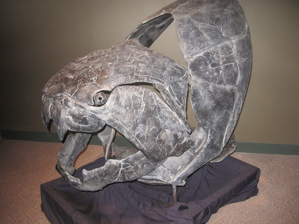
We know a bit more about the eyes of another group of jawed fish, the placoderms, which also appeared during the Silurian. These fish were very heavily armored with bony plates on their heads and bodies. They had more bony plates in their jaws that served as cutting teeth. One of the largest placoderms, Dunkleosteus terrelli, grew up to 6 meters (almost 20 feet) in length. When it opened its jaws, the suction would be strong enough to suck in even large prey.
This work has been released into the public domain by its author, Mitternacht90 at the Wikipedia project. This applies worldwide.
In case this is not legally possible:
Mitternacht90 grants anyone the right to use this work for any purpose, without any conditions, unless such conditions are required by law.
The eyes of some placoderms were encased in bony capsules. The molding of these capsules have allowed paleontologists to study their extraocular muscles (the muscles that attach to the outside of the eye and control its movement). It’s also allowed them to study the choroidal gland — a layer of connective tissue and blood vessels that supplies the outer layers of the retina with oxygen and nourishment. This layer allows an animal to develop a much thicker and more powerful retina than would be possible otherwise.
Although the eyes of modern ray-fin fish (Actinopterygii) are not encased in bone, they are enclosed in a ‘cartilaginous cup’. Many also have scleral ossicles. These are bones that are embedded in the sclera (the white of the eye) and form a ring around it, kind of like an iris of bone (but larger than the actual iris and unrelated in its function).
The scleral bones in fish likely serve as protection for the eye. In reptiles and birds, they serve the additional purpose of preventing distortion of the eyeball during focusing, which the animal does by changing the shape of the lens and cornea. (Fish focus by moving the lens back and forth and the cornea doesn’t change at all, so they don’t have this problem.)
Both the cartilaginous cup and the scleral ossicles are probably homologous to the bony casing of the placoderms. That is, they likely evolved from a similar structure that was present in the common ancestor of the two groups.19
Placoderms, by the way, provide the earliest evidence of a ‘third eye’ or parietal eye. (See Mysteries of the Third Eye.) So this organ must have evolved quite early in vertebrates. The lamprey, which probably evolved earlier than the placoderms (and is still around today), has a pineal gland, which is closely associated with the parietal eye. One source suggests that the parietal eye or something like it could even have evolved before the lateral eyes.20
Eye of the Moray 21
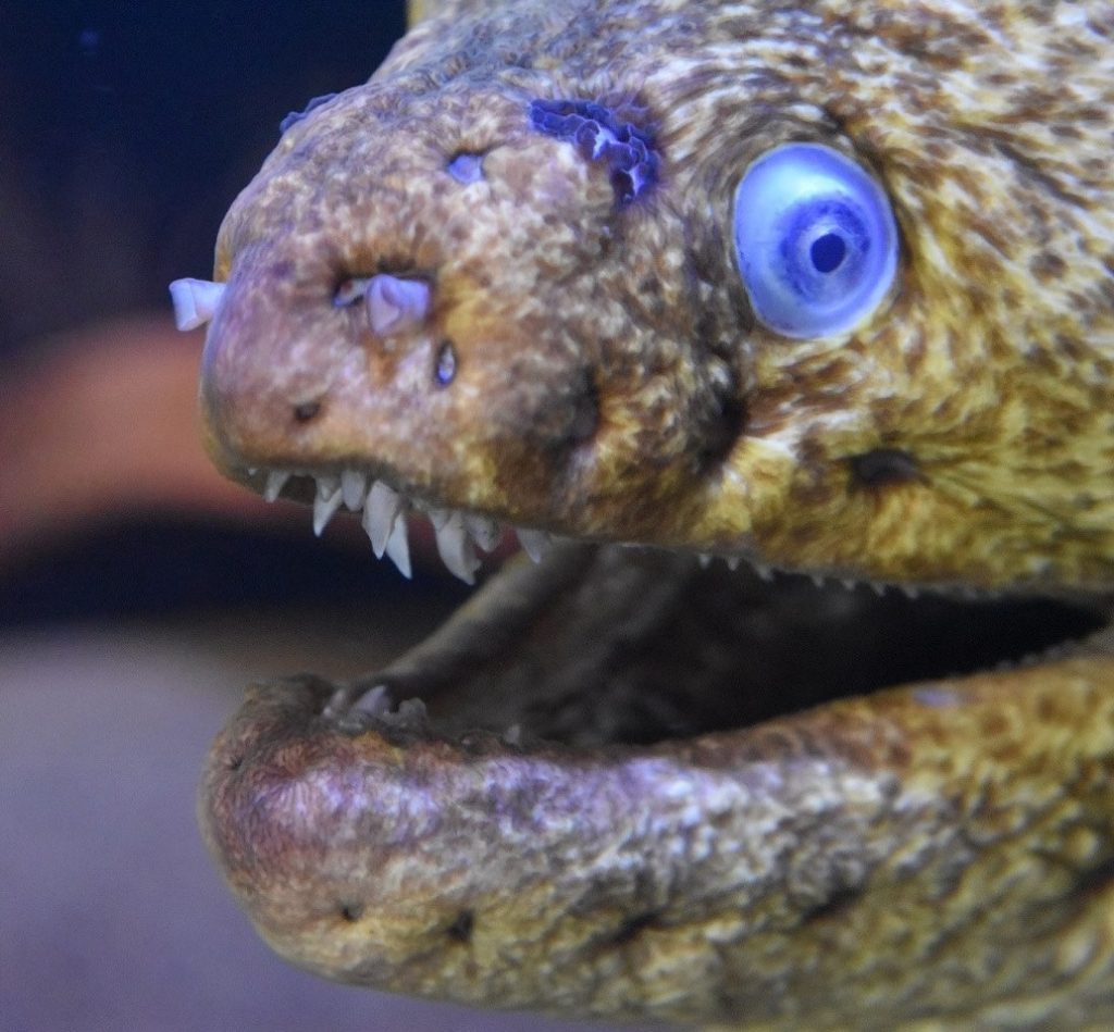
The moray eel, as its name implies, is a type of eel. Despite the unusually elongated and flexible body, eels are ray-finned fish — the most common type of fish. The moray’s eyes are rather small and it probably doesn’t have very good vision. It looks wicked scary, though, so I had to include it. (Weird nostrils, too!)
The Extraocular Muscles 22
Most vertebrates have six or seven ‘extraocular’ muscles that attach to the eye from the outside. Six of these muscles are used for the obvious purpose of changing the direction the eyes are looking. (The seventh, if present, is used to retract the eye. I’ll write more about this in the section on amphibians in part 2.) They serve another purpose that’s even more important, though. If a set of photoreceptors receives continuous stimulation, it becomes “fatiqued” — essentially, it runs out of the chemicals that are responsible for producing electrical impulses and stops working. To compensate for this, extraocular muscles move the eye in microsaccades — fine, fast movements that prevent the light from stimulating the same receptors over and over. This is true of humans, as well. Even when you stare fixedly at an object, your eyes are continually moving in microsaccades. Somehow your brain is wired to ignore these movements, so you’re totally unaware of them.
Eye of the Barreleye 23
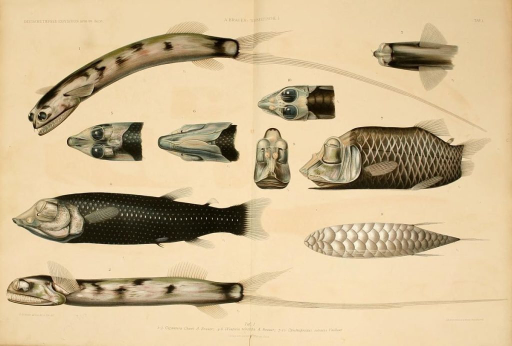
Barreleyes are small, deep-sea fish in the family Opisthoproctidae. The name comes from their bizarre, barrel-shaped eyes. These eyes can be directed straight up when the fish is resting on the bottom or rotated to point forward when it’s hunting.
Source: https://www.flickr.com/photos/biodivlibrary/7413852664
Author: Chun, Carl
This image is in the public domain because it is a mere mechanical scan or photocopy of a public domain original, or – from the available evidence – is so similar to such a scan or photocopy that no copyright protection can be expected to arise.
At least one species of barreleye, Micropinna microstoma, has its eyes totally enclosed inside a transparent skull! Supposedly, even its brain is visible (although, in the video that I link to below, they say something about two large scales behind the eyes, so maybe they block the view of the brain). I so wish I could show you a photo of this insane creature. Unfortunately, I haven’t been able to find one that’s in the public domain, so all I can do is provide a link to a video. Click here to watch it. I strongly urge you to check it out. You’d be hard put to find a stranger animal.
The Lens and Cornea in Vertebrate Eyes 24
Most land vertebrates, such as us humans, use the cornea in conjunction with the lens to refract light and form an image on the retina. In fact, the cornea often does the bulk of the work. The lens serves mostly for accommodation — that is, changing the focus. The eye accomplishes this by using muscles attached to the lens to distort it — making it thicker or thinner as needed. The lens in land animals also tends to be less thick, since it doesn’t have to do all the imaging by itself.
In aquatic animals, this system doesn’t work, because the index of refraction of the cornea is almost the same as that of the surrounding water, so light is not bent as it passes through. The animal has to rely entirely on the lens to form the image. For this reason, the lens tends to be close to spherical in shape and the cornea is usually rather flat. The lens is also stiffer. Focusing is accomplished, not by distorting the lens, but by moving it forwards and backwards, like the lens in a telescope.
In cartilaginous fish (sharks, rays, etc.), the resting state of the lens is toward the front of the eye, which means that the fish are naturally focused on more distant objects. The fish focuses on near objects by pulling the lens backwards, closer to the retina. Bony fish focus in exactly the opposite way — the resting state of the lens is closer to the retina, meaning that the fish is naturally focused on nearby objects. To focus on distant objects, it has to move the lens away from the retina. This marked difference in focusing mechanisms suggests that the eyes of the common ancestor of cartilaginous and bony fish had little or no ability to accommodate. This ability must have evolved independently in the two lineages.
Eye of the Flatfish
Flatfish are members of the order Pleuronectiformes. They include such familiar fish as sole, halibut, and flounder. As far as I know, there’s nothing especially noteworthy about the anatomy of a flatfish’s eye or how it functions. What is noteworthy is the remarkable journey one of the eyes undergoes as the fish grows from a newborn fry to an adult.
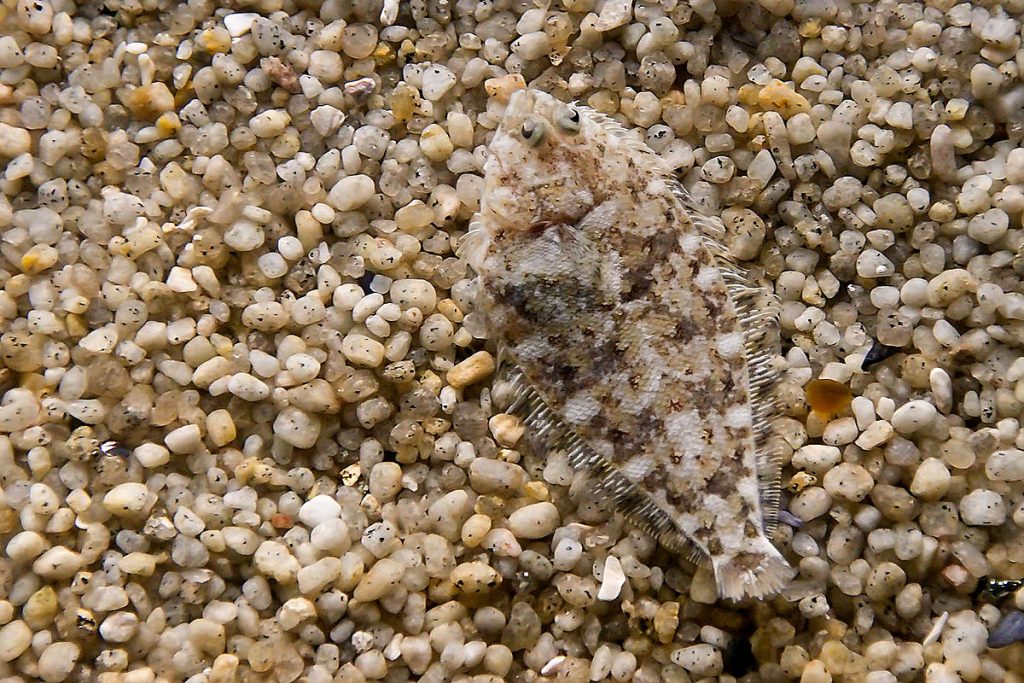
In the adult, both eyes are on the same side of the head. The fish lies on the bottom, where it blends in almost perfectly with the seafloor. It may even cover itself with a thin layer of sediment, so that only its eyes are visible.
It doesn’t start out like this, however. A flatfish begins life as a very tiny and very ordinary looking fry (the standard term for a fish larva) with its eyes in the usual position, swimming around as part of the plankton. After a few weeks, though, a bizarre metamorphosis occurs. The bones of the fish’s skull distort as one of its eyes slowly migrates to the opposite side of the head. The fish’s sense of balance seems to undergo a transformation, as well, and it begins to tip over, swimming at an angle.
The side of the fish that ends up with both eyes develops a speckled coloration, matching the seafloor. The other side, now eyeless, loses pigmentation, turning pallid. Eventually, the fish settles to the bottom as the adult flounder we all know and love. Click here to check out Ikumi Kayama’s excellent illustration of the metamorphosis of a summer flounder (Aralichthys dentatus).
Author: User:Moondigger
This file is licensed under the Creative Commons Attribution-Share Alike 2.5 Generic license.
This is all very interesting, but how on Earth did such a weird fish evolve? Evolution requires intermediate forms and it’s difficult to imagine how any such intermediate form could have been useful. What would be the evolutionary benefit of an eye that was only part-way migrated towards the opposite side of the body?
In a paper published in Nature in 2008, 25 Matt Friedman re-examined two types of fossil fish, Heteronectes and Amphistium, both about 45 million years old, found at a site in Italy. Prior to this, no one had thought that these fossils were related to flatfish, but Friedman used CT scanning to examine the fossils in great detail and found evidence that, not only were they members of this group, but that they represented intermediate steps in flatfish evolution. In both cases, one of the fish’s eyes had migrated towards the top of the head, but had not yet passed over it to the other side. (Various characteristics indicated that the fossils were of adult fish, so these were not fry in an intermediate stage of development.)
Friedman points out that modern flatfish often use their dorsal and anal fins to prop up their bodies above the substrate. Flatfish are ambush predators and such behavior would have allowed their ancestors to “peek” at their surroundings with the bottom eye, increasing their field of vision. A partially migrated eye would have further improved the fish’s view. (I do wonder why this “propping up” behavior persists in modern fish now that the migration has been completed, but maybe it’s still useful for tilting their field of vision towards something that attracts their attention.)
One last thing about flatfish — in most vertebrates, the inner ear contains structures that give the animal a sense of balance. Most fish use their inner ears in combination with their eyes to maintain an upright position. Even in the dark, they can tell which way is up, just as we can. Flatfish seem to be an exception. Flatfish larvae that have been blinded swim on their sides, as if their eyes had already migrated. One researcher used infrared imaging to film fry in complete darkness. Without light, the larvae were incapable of remaining upright. Some bobbed around with their heads pointing down. Older larvae leaned over and drifted to the bottom, landing on the side that would eventually be eyeless.
So in flatfish, the eyes seem to overrule the inner ear. This neatly explains why a fry swims leaning over as one of its eyes is in the process of migrating.
Rods and Cones and More 26
Photoreceptor cells are basically nerve cells that are used to detect light. In most animals, they are located in the retina. Humans and other mammals have three types of photoreceptor cells: rods, cones, and intrinsically photosensitive retinal ganglion cells. (I’m guessing you didn’t cover that last one in science class. In fact, they were only discovered in the 1990s.)
Cone cells are so called because they are roughly cone-shaped. The three types of cone cells in humans are referred to as S-cones, M-cones, and L-cones because the photoreceptor proteins they contain respond more readily to short, medium, and long wavelength light, respectively. Any of the cone types can respond to any of the visible wavelengths of light, but they are much more likely to respond to light in their particular category. In other words, it’s a statistical thing.
As you’ve probably guessed, Rod cells are roughly rod-shaped. While cone cells require a relatively high level of light exposure before they fire, a rod cell can be triggered by a single photon of light. When I was a kid, the numerals of alarm clocks were often painted with a substance that contained radium-226. The radioactive decay of radium produced a faint glow that made the numerals visible in the dark. (So I was probably getting a tiny amount of radiation poisoning, but I guess it wasn’t enough to have any long-term consequences. Knock on wood.) Lying in bed at night, if I stared at the numbers on the clock, I could see the tiny flash of a photon produced by a single decaying atom! 27
The human retina contains around 120 million rods and 6 million cones. Rods and cones are intermingled on the retina, but the cones are concentrated more towards the center — the part directly opposite the lens. At the direct center of the retina is a small pit called the fovea, which contains only cone cells. This is the area of highest visual acuity. When we look directly at an object, we’re positioning it in our visual field so that light from that object falls directly on the fovea. Because cones are much less sensitive than rods, a very dim star may disappear completely when you look at it directly, but reappear when you look off to the side.
A ganglion cell is usually defined as any nerve cell that’s located outside of the brain and spinal cord.28 The impulses produced by the rods and cones feed into bipolar cells (which I won’t get into) and then into ganglion cells, which do some preprocessing before the signals are sent on their way to the brain. Around 1% of the ganglion cells in the retina are intrinsically photosensitive retinal ganglion cells (ipRGCs). The first evidence that there was a third type of photoreceptor cell in the eye was discovered in 1923 when it was found that the pupils of mutant mice born without rods and cones still responded to light by contracting. Recent research suggests that ipRGCs support both the pupillary reflex and circadian rhythms.
As far as I can tell from my reading, most animals possess rods and cones of some sort. The ratio of rods to cones can vary greatly, however. A nocturnal animal will probably have very few cones compared to rods. The cone types can vary greatly, as well. Many birds and reptiles have four types of cones, while most mammals have only two. Some mammals, including humans, have independently evolved a third type. (I’ll cover this in more detail in part 2.)
Eye of the Four-eyed Fish 29
The four-eyed fish (genus Anableps) is a fish with… wait for it… two eyes. They only appear to have four because each eye contains two pupils, one above the other. Four-eyed fish live in Mexico and Central and South America. They inhabit mostly fresh and brackish water. The fish usually holds its eyes right at the surface of the water, so that one pupil is above the water line and the other is below it. Internally, different parts of the eye handle the two views separately. In this way, the fish can ‘keep an eye on’ things going on in both environments.
Among insects, whirligig beetles (family Gyrinidae) independently came up with the same trick. Their compound eyes are each divided into two parts — one for looking above the surface and one for looking below.30
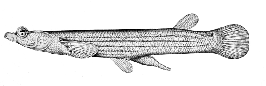
Source: Reptiles, amphibia, fishes and lower chordata
Author: unknown
This work is in the public domain in the United States because it was published (or registered with the U.S. Copyright Office) before January 1, 1925.
End of Part 1
The Story of Eyes will be continued in the next post…
- “Eye to Eye – How Animals See the World” by Steve Jenkins
- “Eye evolution and its functional basis” DAN-E. NILSSON Visual Neuroscience (2013), 30, 5–20. https://www.cambridge.org/core/services/aop-cambridge-core/content/view/E632F655150C8D0E7367566CC99F4717/S0952523813000035a.pdf/eye_evolution_and_its_functional_basis.pdf
- “A Pessimistic Estimate of the Time Required for an Eye to Evolve” Author(s): Dan-E. Nilsson and Susanne Pelger. Reviewed work(s): Source: Proceedings: Biological Sciences, Vol. 256, No. 1345 (Apr. 22, 1994), pp. 53-58 Published by: The Royal Society Stable URL: http://www.jstor.org/stable/49593.
- http://www.marcperkins.net/?p=1553
- https://en.wikipedia.org/wiki/Evolution_of_the_eye#Early_eyes
- https://www.georgefox.edu/academics/undergrad/departments/psychology/ckoch/research/planimal.html
- https://www.biology-online.org/dictionary/Phototaxis
- The Tangled Tree: A Radical New History of Life by David Quammen
- http://theevolvingnaturalist.com/snakes-in-anthills/
- Evolution’s Witness: How Eyes Evolved by Ivan R Schwab.,pg. 23
- http://theevolvingnaturalist.com/a-meandering-of-millipedes/
- http://biology-pages.info/C/CompoundEye.html
- https://en.wikipedia.org/wiki/Cambrian_explosion#Evolution_of_eyes
- https://en.wikipedia.org/wiki/Cambrian#Oceanic_life
- Evolution’s Witness: How Eyes Evolved by Ivan R Schwab.,p. 74-75
- https://en.wikipedia.org/wiki/Nautiloid
- https://en.wikipedia.org/wiki/Nautilus
- Evolution’s Witness: How Eyes Evolved by Ivan R Schwab., p. 77-108
- https://anatomypubs.onlinelibrary.wiley.com/doi/full/10.1002/ar.20639
- Evolution’s Witness: How Eyes Evolved by Ivan R Schwab.,pg. 77
- https://en.wikipedia.org/wiki/Moray_eel
- Evolution’s Witness: How Eyes Evolved by Ivan R Schwab., pgs. 92-93
- https://en.wikipedia.org/wiki/Barreleye
- Evolution’s Witness: How Eyes Evolved by Ivan R Schwab., pg. 82
- https://www.researchgate.net/publication/5237857_The_evolutionary_origin_of_flatfish_asymmetry
- https://en.wikipedia.org/wiki/Photoreceptor_cell
- https://en.wikipedia.org/wiki/Radium_dials
- https://medical-dictionary.thefreedictionary.com/ganglion+cell
- https://en.wikipedia.org/wiki/Four-eyed_fish
- https://en.wikipedia.org/wiki/Whirligig_beetle