Category Archives: Miscellaneous

Strange Fruit and Other Anachronisms

When a bird eats a berry, it digests the pulp and poops out the seed. But not right away. It takes a while for the seed to work its way through the bird’s digestive tract, and by the time it reaches the exit, it’s likely to have traveled a long way from the plant that produced it. In this way, the plant is dispersed.

Every species must have some means of dispersal. Animals can walk or swim or fly to new locations, but plants don’t have that luxury. They have to find more indirect methods of dispersal, and one of the most common is reliance on animals. Usually, this means enclosing the seed in a tough coating that can withstand acidic digestive juices, grinding gizzards, and mashing molars, then wrapping the whole thing in tasty, nutritious pulp that some animal will find irresistible. If the resulting fruit is a small berry, it’ll probably be a bird that’s recruited to disperse it. But many fruits are much larger than your typical berry,1 and larger fruit likely evolved to attract larger, more earth-bound animals.

This leads to a mystery. Some plants produce very large fruit that have very tough rinds. And sometimes the seed or seeds inside are also intimidatingly large. Think of an avocado. I’ll help you out — here’s a photo of one.

Avocado (Persea americana)

And here’s a photo showing the pit inside.2
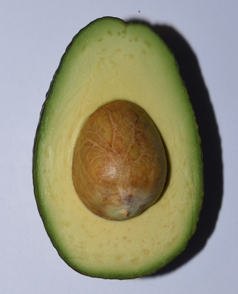
An avocado sliced open to reveal the pit

That is a big pit. What kind of animal could possibly swallow an avocado pit whole? Sure, lot’s of small animals could just eat around it, but that doesn’t help the plant disperse its seed.

Now, I’ll grant you, the avocados of today are the result of thousands of years of domestication, so it’s quite possible that larger fruit have been selected for. But why would the seed also increase in size? In fact, there is evidence that the original version of the fruit had less flesh than today’s avocado. But the pit was actually larger. A larger pit and less pulp — the mystery deepens.

Avocado plants are native to South America, and there is one South American animal that may sometimes eat avocados whole. Surprisingly, that animal is the jaguar! Yes, jaguars are carnivores. Nevertheless, they do occasionally eat avocados, but probably not that often. Also, being carnivores, jaguars are very uncommon. Why? Simple math. A carnivore has to eat lots of other animals during its lifetime, so its prey animals have to greatly outnumber it. For this reason, it’s unlikely that avocados evolved to be dispersed by jaguars — there just aren’t enough of them.3

Maybe you’ve already guessed where I’m going with this. If not, I won’t keep you in suspense any longer. The reason you won’t find any animals in South America large enough to be the dispersal agents for avocados is that they’re all extinct, and they went extinct thousands of years ago!4

The Americas in the Pleistocene

According to current estimates, humans first colonized the Americas around 15,000 years ago. This was toward the end of the Pleistocene epoch, which lasted from 2,580,000 to 11,700 years ago.5 The Americas in the Pleistocene were very, very different from the Americas we know today. And I don’t just mean no cars or roads or white picket fences. I mean, there were some seriously large animals roaming the landscape. 15,000 years ago, North America was home to mammoths, mastodons, giant ground sloths, lions, camels, wild horses, short-faced bears, saber-toothed cats, a beaver the size of a bear, and lots of other beasts you’ve probably never heard of.
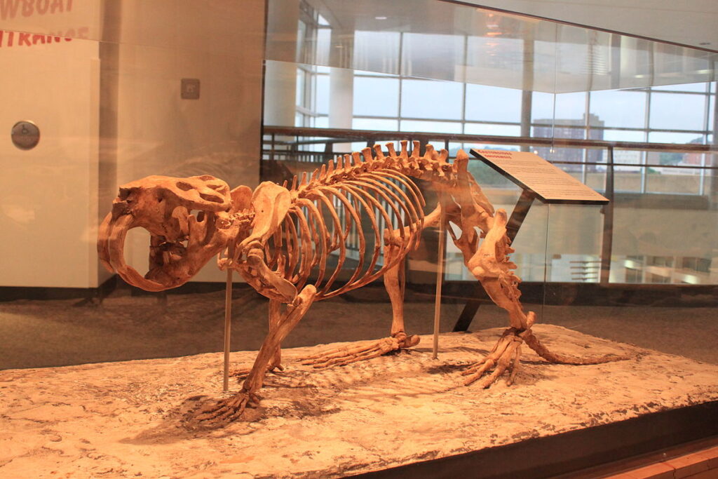
Giant beaver (Castoroides ohioensis)
Minnesota Science Museum: Mississippi River Gallery
Author: Ryan Somma from Occoquan, USA
This file is licensed under the Creative Commons Attribution 2.0 Generic license.

In South America, the situation was much the same, although the selection of large mammals was different. North and South America were separated until around 2.8 million years ago when the Isthmus of Panama formed, so the southern continent evolved its own set of mammals.6 Giant ground sloths actually evolved in South America before crossing the isthmus into the north, so South America did have them.

If you’re not familiar with giant ground sloths, you should look them up. The largest one, Megatherium americanum, was around 20 feet long, 4 tons, and about the size of a modern elephant. On its hind legs, M. americanum would have stood around 12 feet (3.5 meters) tall!7 8
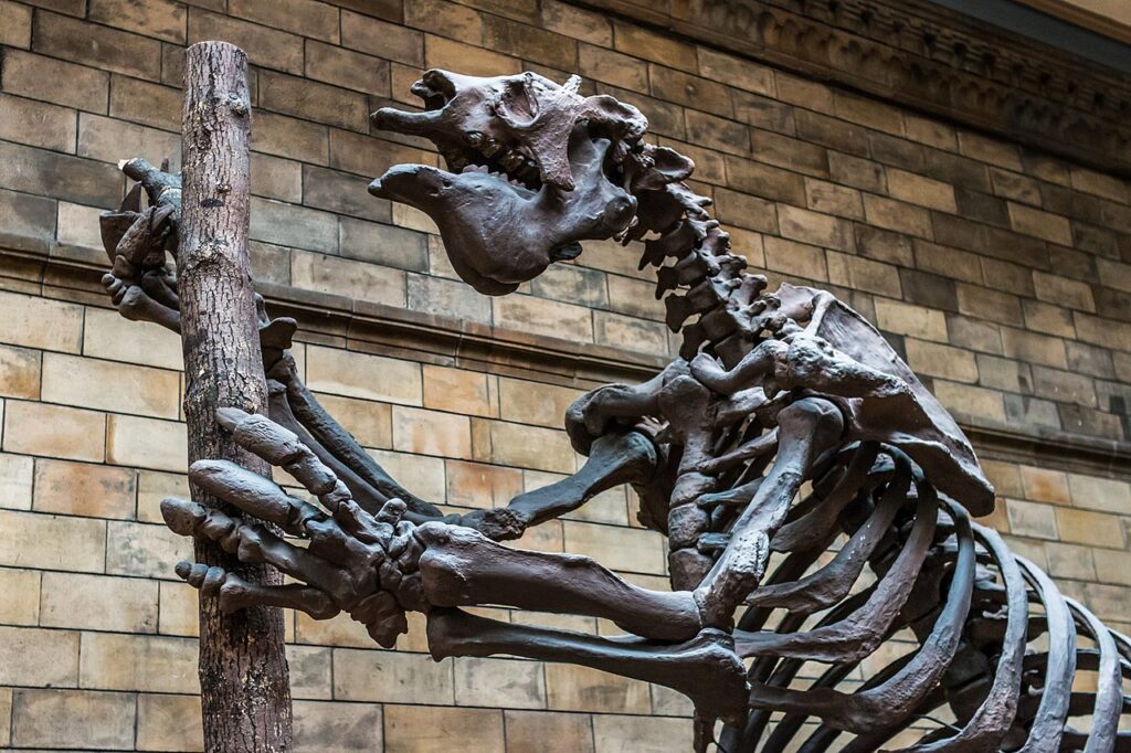
Skeleton of a giant ground sloth (Megatherium sp.)
Natural History Museum, London
Author: Markus Trienke
This file is licensed under the Creative Commons Attribution-Share Alike 2.0 Generic license.

South America didn’t have any mammoths or mastodons, but it had gomphotheres, a family of elephant-relatives, many of which easily held their own in the behemoth department.9
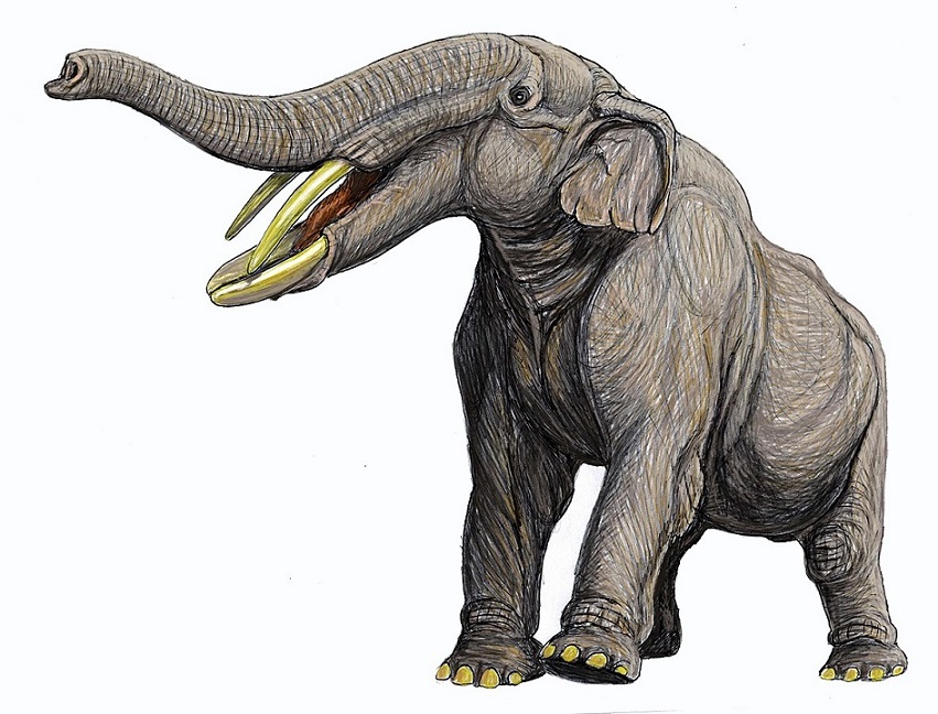
A gomphothere, Amebelodon britti, from the Late Miocene to Early Pliocene of North America
Author: DiBgd
This file is licensed under the Creative Commons Attribution-Share Alike 4.0 International license.

Another large South American mammal of the Pleistocene was the toxodon. Actually, Toxodon is an entire genus nested within an order of extinct hoofed animals called Notoungulata. The most well-known species of toxodon is Toxodon platensis. It’s estimated to have weighed over a ton and was around the size of an American bison or an African black rhino.10
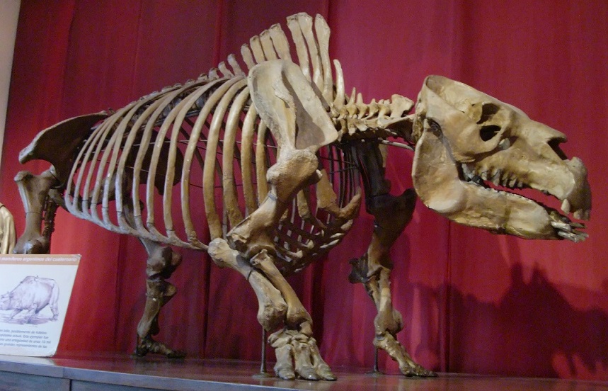
A toxodon skeleton on display in the Bernardino Rivadavia Natural Sciences Museum
Author: WereSpielChequers
This file is licensed under the Creative Commons Attribution-Share Alike 3.0 Unported license.

Yet another group of large extinct mammals from South America are the glyptodonts, members of the suborder Glyptodontinae. They were closely related to modern armadillos and had a similar armored body plan. In fact, molecular studies have found that the glyptodonts actually were armadillos — Glyptodontinae is nested within the family Chlamyphoridae, which includes most modern armadillos.

The best-known of the glyptodonts belong to the genus Glyptodon. These were about the size and weight of a Volkswagon Beetle.11 12 13 Here’s a very old (probably 1920) drawing of a glyptodon.
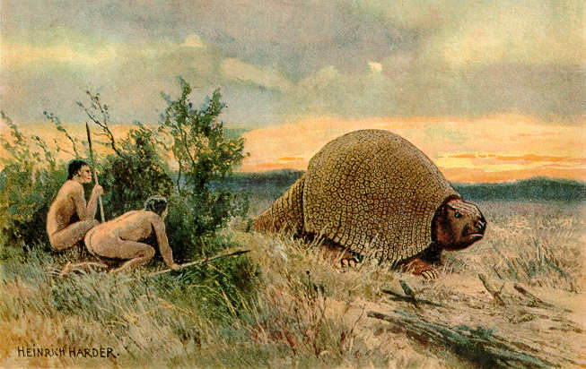
Glyptodon sp.
Source: The Wonderful Paleo Art of Heinrich Harder
Author: Heinrich Harder
This work is in the public domain in the United States because it was published (or registered with the U.S. Copyright Office) before January 1, 1926.

This is just a small sampling of the large mammals that inhabited the Americas of the Pleistocene epoch. Note that all the mammals mentioned here are actually genera or even families. In other words, there wasn’t just one type of gomphothere or one type of glyptodont. These were groups of animals that contained many, many variations. The New World had an incredibly diverse collection of very large mammals .

Scientists use the term megafauna to mean very large animals. How large varies somewhat — the term is a bit loose. Some people define megafauna as including any animal with a mass as great or greater than a human being. Others apply the term only to really big animals — those weighing in over a ton. For the purposes of this article, I’ll stick with the first definition.14

Today, most of the megafauna are gone from North and South America. In fact, most of the megafauna are gone from everywhere except Africa (and we’re working on that). As mentioned, the megafauna were mostly extinct in the New World by 13,000 years ago. (A few literal die-hards, like the giant sloth, managed to hold on until about 11,000 years ago). So what happened to them? Well, humans showed up in North America around 15,000 years ago. 2,000 years later, most of the big mammals were extinct. Seems like a no-brainer.

Actually, some pretty good arguments have been made that humans couldn’t have driven all those animals to extinction in such a short period of time just by over-hunting. It’s possible that other factors, such as climate change, were involved. Still, it’s highly suspicious that most of the megafauna went extinct so soon after humans appeared. Maybe it was a combination of human over-hunting and some other factors. I don’t know. The important thing is, by about 13,000 years ago, most of the really big mammals were gone.

The Concept of Evolutionary Anachronisms15

In modern Africa, large herbivores such as elephants routinely take large fruit into their mouths whole, mash the soft pulp, and swallow the seeds whole, along with the pulp. Later, they poop out the seeds in another location, along with a generous helping of natural fertilizer. Many African plants have evolved large fruit specifically in response to the presence of large megafauna such as elephants, rhinos, giraffes, and so on.16 17

There’s no reason to think that things were any different in the Pleistocene of North and South America. Many plants, including the avocado, must have evolved to rely on the large megafauna for dispersal. When these animals went extinct, the plants were left without their primary dispersal agents, which means it suddenly became much harder for them to spread their seeds. Now, 13,000 years later, these plants are still producing ridiculously large fruit — as if they were still expecting a giant sloth or gomphothere to show up. And, in a sense, they are. 13,000 years is a blip in evolutionary time, especially for plants, which tend to evolve more slowly than animals. It’s as if the plants haven’t yet “noticed” that the megafauna are gone!

When a fruit evolved to be eaten by an animal or group of animals that are no longer around, botanists refer to such a fruit as an evolutionary anachronism. An anachronism, in the general sense, is something that is out of place in time. If you’re reading a story that’s supposed to be taking place in the Middle Ages, and one of the characters uses a slang term from the 21st Century, that’s an example of a literary anachronism. Botanists and evolutionary scientists have adopted the term to describe these strangely out of step fruit.

But strange fruit are not the only examples of evolutionary anachronisms. In general, anytime a species preserves intact a physical or behavioral trait that evolved in response to evolutionary pressures that no longer exist, the trait is said to be an example of an evolutionary anachronism.

So, maybe you’d like to see an example of an evolutionary anachronism that doesn’t involve fruit…

The American Pronghorn18 19

The American pronghorn is sometimes called the American antelope. It’s not a true antelope, although it does resemble one. In scientific terms, it’s a member of the order Artiodactyla, the even-toed ungulates. This is a large order, which includes deer, pigs, true antelopes, giraffes, and (surprisingly) hippos.

The pronghorn is the fastest land mammal in the New World. It’s been clocked at 55 mph for 0.5 mi (88.5 km/h for 0.8 km). The interesting thing about that figure is that it’s too fast! Nothing alive today in North America is anywhere near fast enough to catch a pronghorn. Why would an animal evolve to be so much faster than it needs to be?

The answer, of course, is that it wouldn’t. There’s nothing alive in North America today that can catch a pronghorn, but what about back in the Pleistocene? When I was listing the various megafauna that lived in North America during the Pleistocene, I forgot to mention cheetahs. That’s right, cheetahs used to live in what’s now the United States! Other predators, such as hyenas, short-faced bears, and lions, might also have posed a threat to pronghorns.

So the American pronghorn is likely an example of an evolutionary anachronism — a prey animal that’s still ready to run super-fast at the first glimpse of a cheetah or whatever other scarily-fast predator might have been around all those years ago.

Anachronistic vs. Vestigial20

When I defined the term ‘evolutionary anachronism’, I specified that it had to be an ‘intact’ trait. In other words, the trait cannot have noticeably devolved. Let me give you an example of what I mean.

Many animals that live their entire lives in caves have lost their eyesight. In some cases, they’ve lost their eyes entirely. In other cases, they still have eyes, but those eyes no longer function. In other words, they’re vestigial. If, for whatever reason, a species with vestigial eyes were to migrate out of its cave and re-colonize the surrounding area, the environmental condition that caused animals to evolve eyes in the first place — namely, the presence of light — would reappear. But its vestigial eyes would not start functioning again. By this time, the species has presumably been blind for many thousands of years. So, it’s too late — its eyes are permanently broken.

On the other hand, the pulp and seed of an avocado are still functional. If an animal were to come along tomorrow that liked the taste of the pulp and was big enough to swallow the fruit whole, it could easily fill the role of disperser for the plant. Ergo, the avocado is an anachronistic fruit, not a vestigial one.

Holding On21

So, it’s likely that the fruit of avocados evolved to be swallowed whole by animals like gomphotheres and giant sloths. Such animals were very common for most of the Pleistocene, and, sure enough, avocados had a much larger natural range back then than they do today. But, if the animals that were responsible for dispersing them have been extinct for all this time, why haven’t avocados also gone extinct?

Well, note my use of the phrase ‘natural range’ in the last paragraph. Most of the avocado’s megafaunal dispersers went extinct towards the end of the Pleistocene, but not quite all of them. There was one survivor: humans. Fortunately for the avocado, people like how they taste. And people can be very good dispersers when they want to be. So, it may be that the avocados would have gone extinct if humans hadn’t decided to fill in for the sloths and gomphotheres.

Then again, maybe avocados could have held on for a long time without humans. Just because their primary dispersers went extinct, that doesn’t mean that nothing would ever disperse them. I already mentioned the possibility that jaguars might occasionally disperse an avocado seed. It’s very unlikely that jaguars could have kept the avocados going indefinitely, but they might have enabled them to hold on for a little while, if only by the skin of their non-existent teeth.

Most fruits float. They didn’t evolve for floating. It’s merely a side-effect of the way they’re constructed — most fruits are light enough and waterproof enough to float for a while. For that reason, many plants that appear to be anachronistic can still be found alongside rivers. When the river floods, some of the fruit get washed downstream, and, with luck, some of them may come to rest in a place where they can take root.

Even animals that don’t routinely disperse a plant’s seeds may occasionally disperse them. For example, an animal may be in the habit of eating the pulp of a fruit and spitting out the seeds or eating around them. This doesn’t help the plant because it doesn’t disperse the seeds. Unless, of course, the animal decides to carry the fruit someplace else before it eats it. In that case, the discarded seeds may still be able to find a suitable new location in which to germinate.

If a seed has evolved to survive passage through the digestive tract of an animal, it has necessarily evolved a thick coating. This could make it very hard for the seed to absorb water and germinate. For this reason, many plants have evolved seed coatings that are just barely tough enough to make it through the target animal’s guts. In fact, many won’t make it through, but those that do should be scarified enough by the passage that water will be able to penetrate the coating. The seed absorbs the water, swells up, and germinates. Thus, many plants have evolved seeds that almost have to pass through an animal’s gut if they are ever to germinate.

It’s a tricky balancing act. A seed that hasn’t passed through a digestive tract may eventually germinate, but it may take months or even years. So, if a plant’s primary dispersers are gone, it may still be able to hold on for a long time thanks to that very occasional seed that doesn’t pass though an animal’s gut, but manages to germinate anyway.

But many plants may not be able to hold on without their primary dispersers. Plants rarely fossilize, so we have no way of knowing how many plants went extinct along with their megafaunal dispersers. It seems likely that at least some did, especially the ones for which humans didn’t have any use.

More Strange Fruit

I’d like to spend the rest of the article showcasing a few interesting, and very strange, fruit that are likely anachronistic. It turns out that grocery stores are great places to go hunting for the “ghosts” of extinct megafauna.

“…the most delicious fruit known to men”22

That’s what Mark Twain called the fruit of cherimoya (Annona cherimola), a plant that grows from central Mexico, through Central America, and down the west coast of South America, almost to its very tip. I won’t argue with Twain’s assessment. The one I tried was exquisite, delectable, scrumptious… just seriously good. Its taste is complemented by the texture of the flesh, which is similar to custard and gives it the other common name it goes by — custard apple.

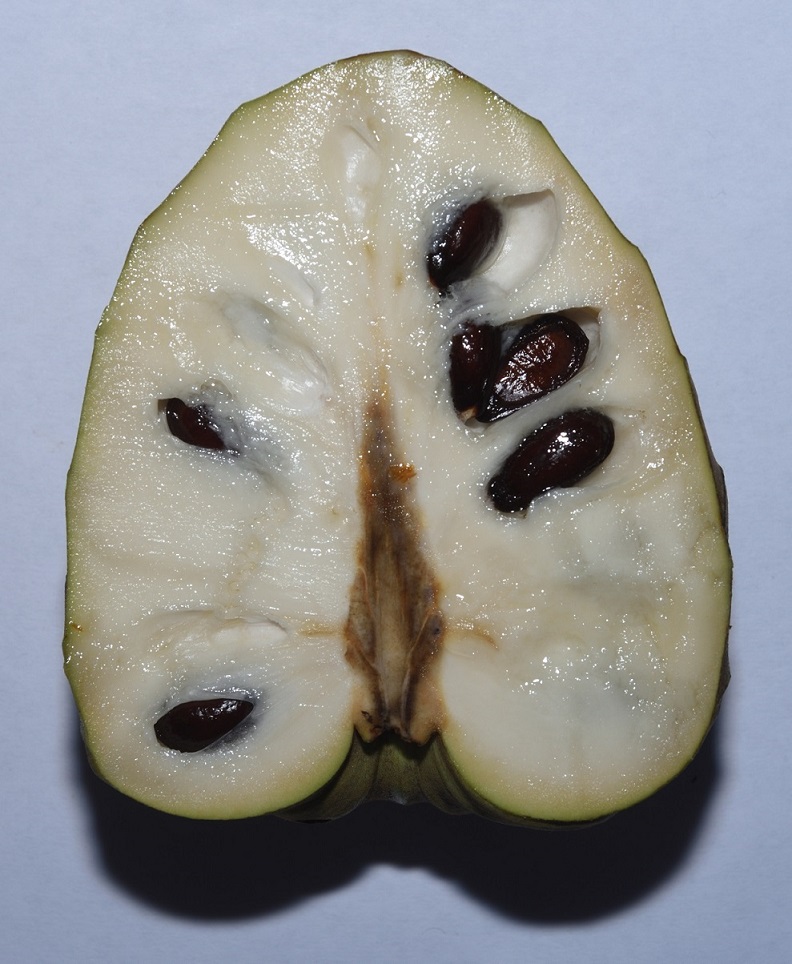
My only complaint is that the seeds are numerous and evenly distributed throughout the pulp, so that you have to spit them out with every mouthful. This makes eating one a bit of a hassle. If someone could come up with a seedless variety, I expect it would take the world by storm.
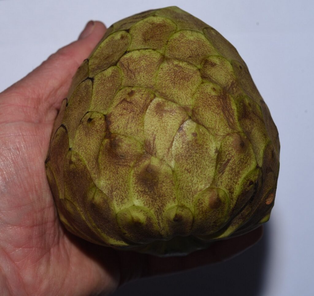
Cherimoya

I googled “seedless cherimoya” just to see if, by chance, anyone had come up with one. I found a number of articles referencing work done at UC Davis that isolated a mutant gene in a closely related fruit — the sugar apple — that caused it to be seedless. It seems like it would be a simple matter to knock out the same gene in cherimoya, thereby producing a seedless variety. But the articles I saw were from 2011, and I haven’t found any follow up articles, so I guess it’s not that easy.23
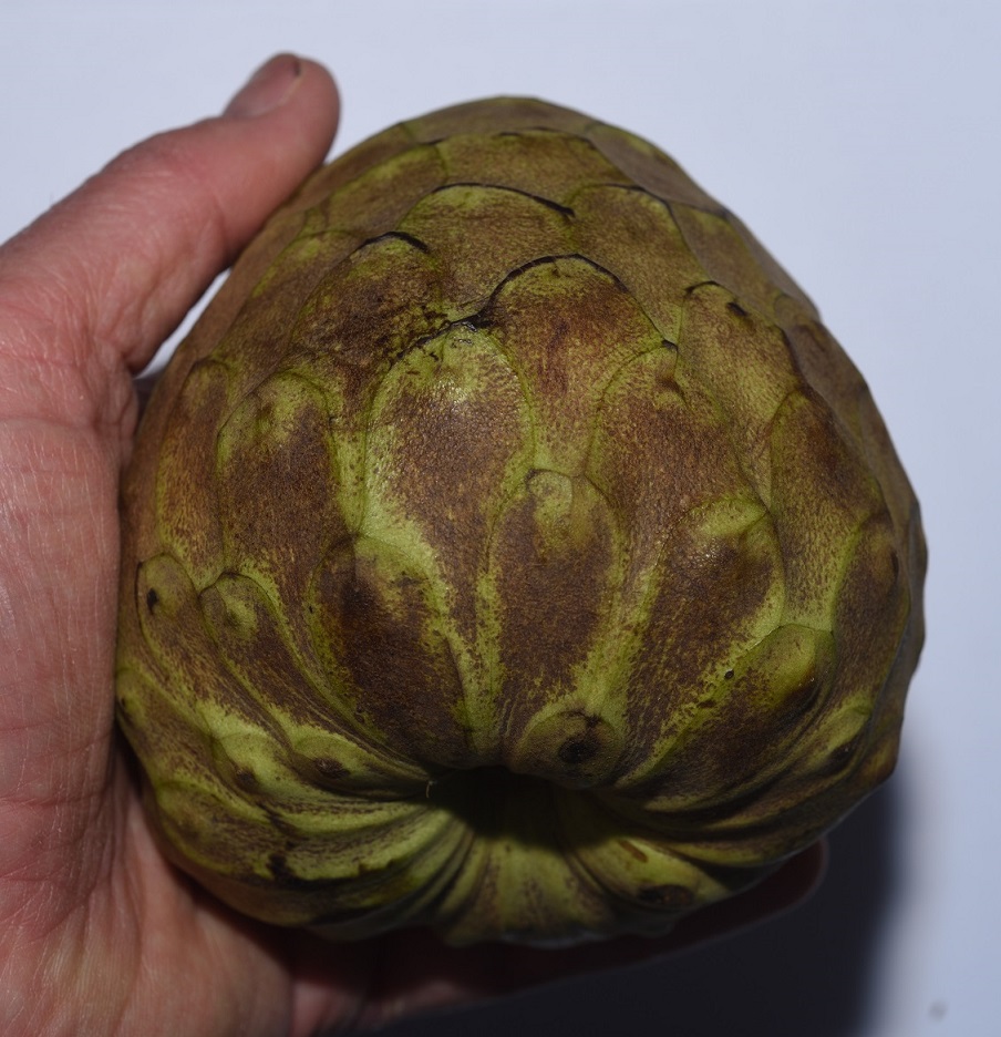
Cherimoya
A cherimoya fruit sliced down the middle

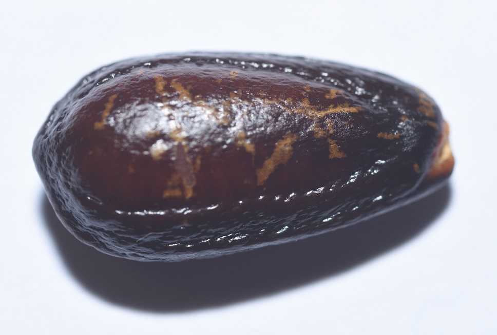
The best evidence that cherimoya is anachronistic is its large size. The seeds are also fairly large and not super easy to separate from the pulp, so it seems likely that the fruit evolved to be eaten by large mammals that would mash the fruit and swallow the pulp along with the seeds.

Sliced for eating
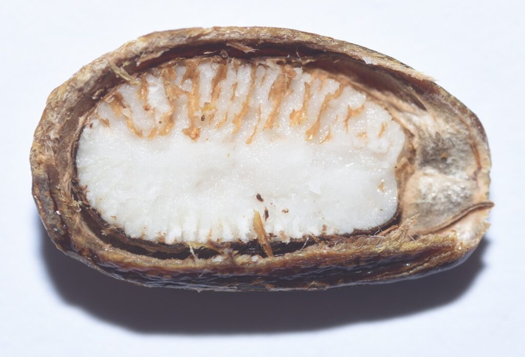
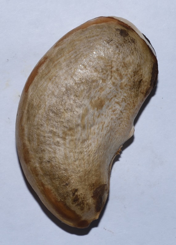
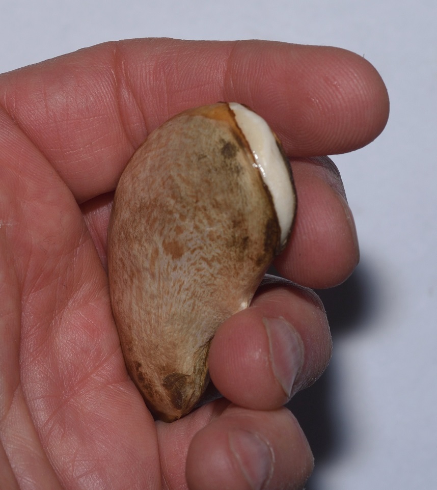
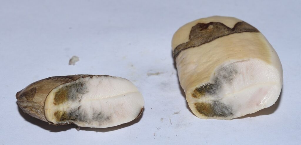
A cherimoya seed
A cherimoya seed dissected

There are at least two other edible fruits produced by plants in the Annona genus that may be anachronistic: soursop (Annona muricata)24 and sweetsop or sugar apple (Annona squamosa).25. Both are similar in appearance to cherimoya, but I haven’t tried them, so I don’t know how they compare taste-wise.

Way down yonder in the pawpaw patch26 27

I’ve never eaten a pawpaw (Asimina triloba). Chances are, you haven’t either. In fact, it’s possible your only clue to the existence of this fruit is the lyrics of that old folksong. (If you’re not familiar with the song, well now… you should just click here and have a listen.)

The plant is a small deciduous tree that’s native to the eastern United States and Canada. It’s in the same family, Annonaceae, as the aforementioned cherimoya, and is quite closely related. Supposedly, the fruit has a “sweet, custardish flavor somewhat similar to banana, mango, and pineapple”.28 The fruit has a shelf life of only about three days, which explains why you don’t find them in supermarkets. If you’re lucky, you might find some in a roadside stand. Otherwise, unless you happen to live way down yonder near a pawpaw patch, you probably won’t run across them.
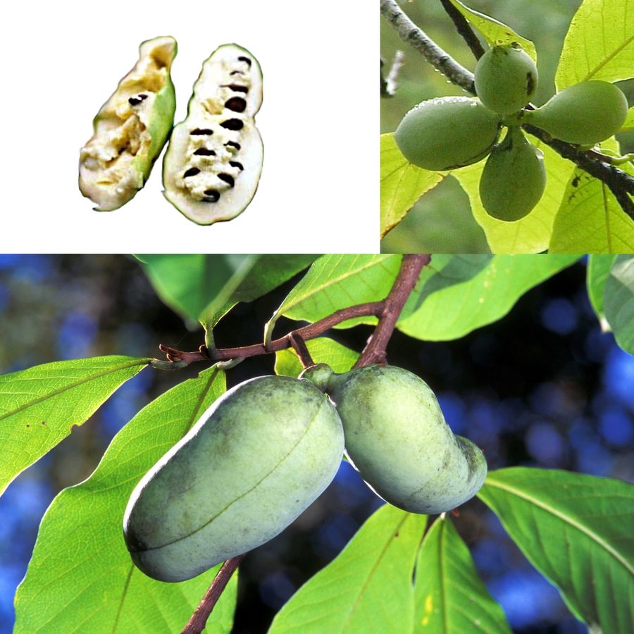
Pawpaws (Asimina triloba)
Author: Agnieszka Kwiecień (Nova)
Permission is granted to copy, distribute and/or modify this document under the terms of the GNU Free Documentation License, Version 1.2 or any later version published by the Free Software Foundation; with no Invariant Sections, no Front-Cover Texts, and no Back-Cover Texts. A copy of the license is included in the section entitled GNU Free Documentation License.

Several things point to pawpaws being anachronistic. First, of course, is their large size. The seeds are also fairly large, although not ridiculously so. Second, the fruits tend to fall from the tree within a day of ripening, and then they rot very quickly. This suggests that they are targeted towards animals large enough to pick them directly from the tree.

Third, pawpaw patches are rare, except where indigenous people routinely eat them. This suggests that their only remaining primary dispersers may be humans. Finally, wild pawpaw trees usually only grow in floodplains, and yet, they grow just fine when they’re planted in uplands. This, again, suggests that people are their only remaining primary dispersers.

…the largest fruit of all trees29 30

The jackfruit (Artocarpus heterophyllus) has the distinction of being the largest fruit of all trees. Here’s a photo of a jackfruit that was found in Kerala, India.

A jackfruit found in Kerala, India
Author: Suniltg at English Wikipedia
Permission is granted to copy, distribute and/or modify this document under the terms of the GNU Free Documentation License, Version 1.2 or any later version published by the Free Software Foundation; with no Invariant Sections, no Front-Cover Texts, and no Back-Cover Texts. A copy of the license is included in the section entitled GNU Free Documentation License. Subject to disclaimers.

The one I bought in an Asian market wasn’t quite that big, but it wasn’t exactly small, either. Here it is on the cutting board.
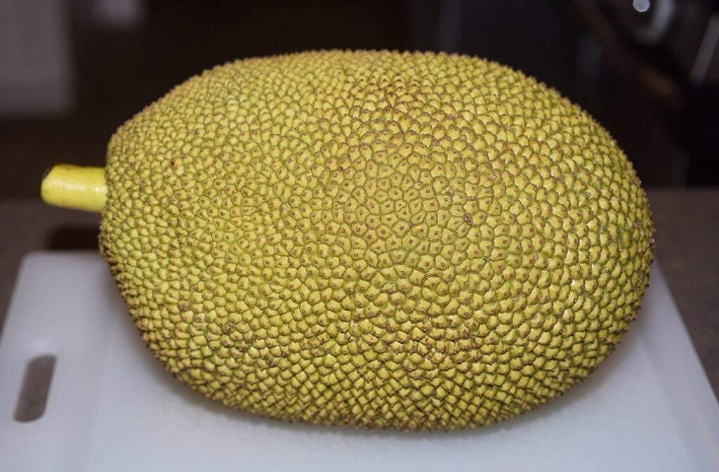
Jackfruit

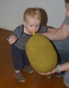
And here’s one of my grandnephews for scale.

Mother, son, and jackfruit

I don’t know if anyone has ever been killed by a jackfruit falling from a tree, but it wouldn’t surprise me. Then again, maybe people just instinctively avoid walking under something that looks like this.
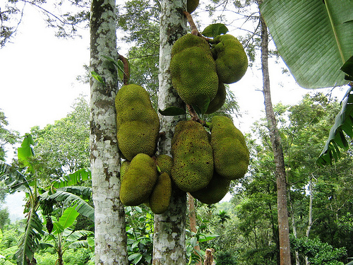
Jackfruit on a jackfruit tree
Kerala Agricultural University, Ambalavayal, Kerala, India
Author: Prof. Mohamed Shareef from Mysore
This file is licensed under the Creative Commons Attribution-Share Alike 2.0 Generic license.

When I cut my specimen open, I was surprised how tough the rind was. It took some effort to cut through it. I was also surprised by how sticky the insides were. The pulp stuck to my hands like glue. It took a lot of soap and water to get it off. It was also kind of slimy. My grandnephew (the older one, who’s four) refused to eat it because it was “too slimy”. (This is the same kid who routinely handles earthworms and thinks they’re “cute”!)

A jackfruit cut open

The fruit is very good. According to Wikipedia, it has a “subtle pineapple- or banana-like flavor”.31 Personally, I thought it tasted more like cantaloupe.
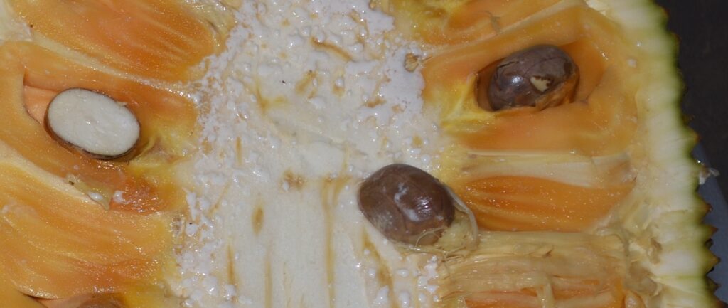
A closeup of the pulp and a few seeds
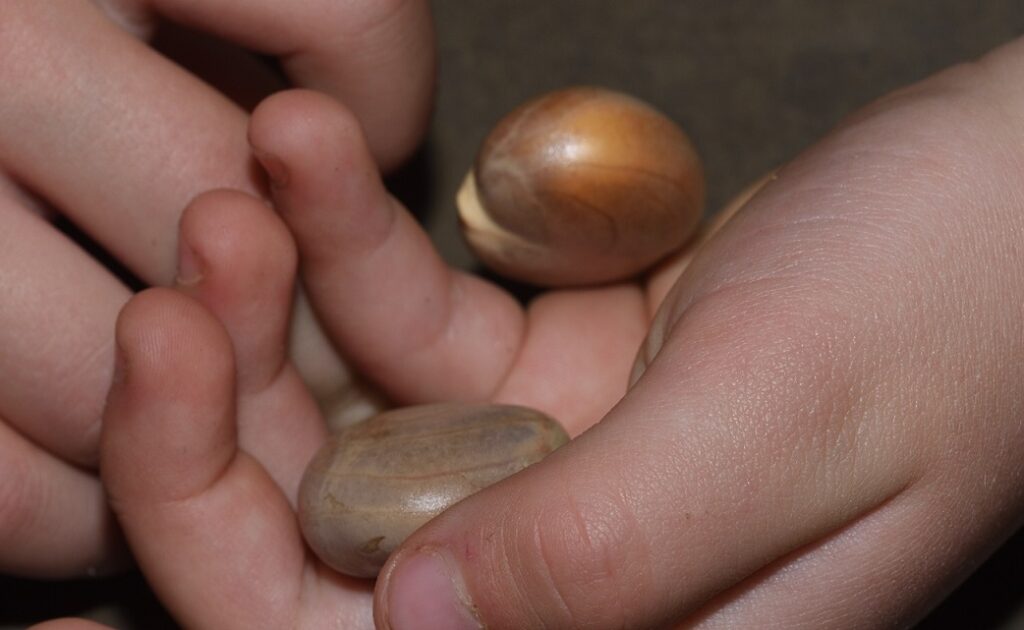
My grandnephew holding a couple of seeds

The seeds can also be eaten. I boiled and ate a bunch of them. After 10 minutes of boiling, the water was stained a dark reddish-brown from all the tannins. After boiling, the seeds have to be peeled, which is no fun, but they do have a nice, sweet taste to them.


A closeup of a seed
Dangsuriya Jackfruit ready to eat
Thailand
Author: Tris T7
This file is licensed under the Creative Commons Attribution-Share Alike 4.0 International license.

So is jackfruit anachronistic? It’s important to realize that, like the avocado, and like most fruit consumed by people, the jackfruit has been domesticated. It’s probably quite a bit larger than the original, wild variety. But also like the avocado, the original fruit was probably still pretty big, and the seeds likely haven’t changed significantly.

So the huge size, tough rind, and large seeds strongly suggest a fruit that evolved to be mashed in the mouths of megafauna and the seeds swallowed whole without grinding. The presence of high levels of tannins in the seeds would be a deterrent to insects, but it would also make them distasteful to any large animal that bit into them or ground them between its molars.

So what kind of megafauna are we talking about? It’s not known exactly where jackfruit originated, but they’re believed to be indigenous to the rainforests of the Western Ghats in southern India. This area is a hotspot of biodiversity. Among the many large animals found there is the Indian elephant (Elephas maximus indicus) — an obvious candidate seed disperser for the ancestral jackfruit. Turn the clock back around 50,000 years, and India was home to, not one, but two giant elephants — Palaeoloxodon namadicus and Stegodon namadicus. P. namadicus, in particular, may have been the largest land mammal that ever lived. According to one estimate, it weighed around 24 tons (21,772 kg) and stood 16 feet (4.8 m) tall at the shoulder.32 The largest mammal and the largest fruit? Seems like a good match.33

Mango

The mango (Mangifera sp.) is another fruit that probably originated on the Indian subcontinent. The most common variety, appropriately named the common mango, is Mangifera indica. I’m not sure what variety I got when I visited the local grocery, but here it is.

A mango (Mangifera sp.)

And here it is cut in half. Notice how the pulp clings to the pit.
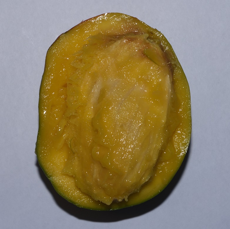
A mango sliced in half

In the next photo, I’ve extracted the pit. Notice how large it is compared to the fruit as a whole. I actually used a scouring pad to remove most of the pulp from the pit and I still couldn’t get it all off — that’s how tightly the pulp is bound to it. This is no accident. A large animal eating the fruit isn’t going to spit out the pit — that would be a waste of pulp — it’s going to mash the fruit and swallow both pulp and pit.

…and with the pit extracted.

I continued my dissection, extracting the seed from the pit.
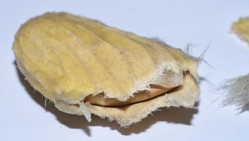
Extracting the seed…
The final cut

Papaya34

Papayas (Carica papaya) are sometimes called pawpaws, but they are unrelated to the pawpaw (Asimina triloba) discussed earlier. They have the usual large size, suggestive of an affinity for megafauna, but the seeds are a surprise.
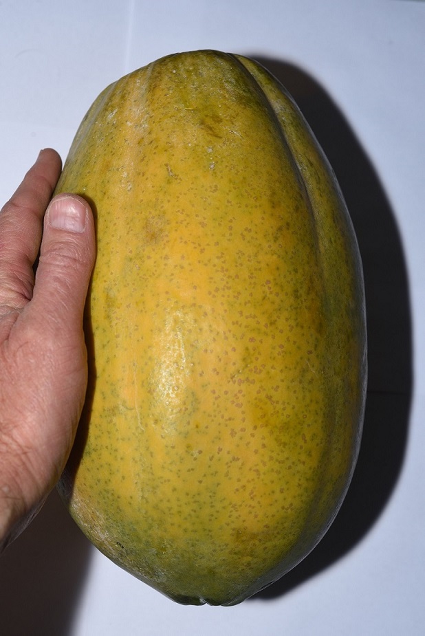
A papaya (Carica papaya)

Instead of one huge seed or a few dozen moderately large ones, the papaya has lots of very small seeds concentrated in a large central cavity. This is an interesting variation. The large cavity reduces the weight of the fruit, so the stalk attaching it to the tree doesn’t have to be quite as thick. The large number of seeds increases the chances that one of them will successfully germinate and grow to maturity.
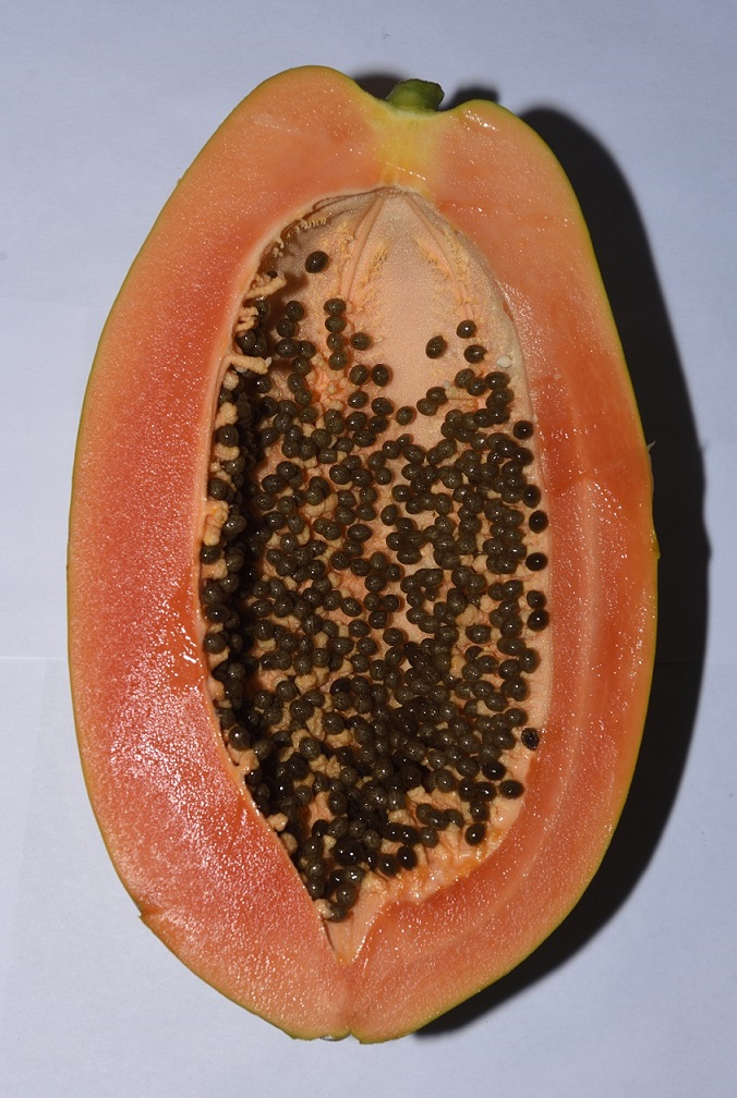
A papaya cut in half

The seeds themselves are fascinating. They’re wrapped in some sort of gelatinous pouch. Here’s a photo of a whole seed (right) and a seed with its pouch removed (left).
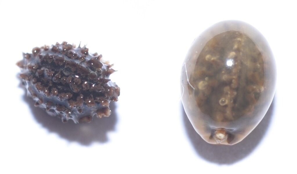
A papaya seed with its covering removed (left) and with its covering intact (right)
I dried this one off as best I could…
…before slicing it open.

Papaya seeds are edible and taste a bit like horseradish. I would love to know more about that gelatinous pouch. I’m not familiar with any other type of seed that has such a covering. I’ve been searching for more info online, but all I’ve found so far is some stuff about papaya seeds’ reputed health benefits and a lot of rather dubious articles extoling their value for detoxing and parasite cleansing.

Papaya is thought to be native to Mexico, Central America, and northern South America. Several members of the papaya family can be found growing wild in the tropical valleys of the Andes. If papaya originated in South America, then, like the avocado, it was probably dispersed by South American giants such as ground sloths, gomphotheres, and toxodons.

Osage-Orange35 36 37

The osage-orange or hedge-apple (Maclura pomifera) is a small tree or large shrub, which has a small natural range in parts of Texas, Oklahoma, and Arkansas. The fruit is neither an orange nor an apple. It’s technically edible, but no humans actually eat it, because it contains an unpalatable milky latex. Even horses don’t much care for it, although one of its many names is horse-apple.
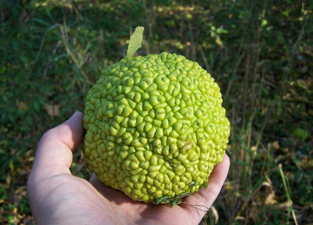
Osage-orange (Maclura pomifera)
Winfield, IL USA
Author: Bruce Marlin
This file is licensed under the Creative Commons Attribution-Share Alike 2.5 Generic license.

In fact, nothing much eats it. When the fruits ripen, they drop to the ground. There, unless they roll downhill or get dispersed by a flood, they rot underneath the tree that produced them. This is, itself, a clear sign of anachronism. Nothing today eats them, but something must have eaten them at one time — something that’s no longer around. Molecular analysis indicates that M. pomifera probably diverged from other members of Maclura during the Oligocene, around the same time that some groups of mammoths, mastodons, and ground sloths first appear in the evolutionary record. This suggests that these mammals could have been the original seed dispersers for the plant.

You might be wondering, if the latex contained in the osage-orange is distasteful to horses and people, why would these larger mammals be any different? The fact is, many plants that people do eat produce chemicals that are toxic to some degree. Peppers, for instance, are eaten by no other animal other than humans. Large mammals, such as us, are able to handle limited amounts of mild toxins, and much larger mammals, such as elephants, are able to eat some fruits that would be toxic or unpalatable to us. Partly, this is because their slower digestion gives them more time to break down the toxins. Bad-taste is often an indication that something is not safe to eat. If something is safe for an animal to eat, chances are it won’t taste bad to that animal. So, to a mammoth or ground sloth, an osage-orange might have tasted pretty good.

Although we don’t eat the fruit, people have managed to find other uses for osage-orange. The wood was prized by Native Americans as the best wood for making bows (from which it gets another one of its common names, bois d’arc, which is French for bow wood.) They prized it so much, in fact, that they would travel hundreds of miles to get it.

The common name ‘hedge-apple’ refers to its use by European settlers as living fences or hedges around their fields to keep out free-range livestock. Pruning the plants would cause them to sprout adventitious roots from their bases. These would interweave, forming a dense, thorny barrier.

The use of osage-orange as hedges allowed it to become naturalized far beyond its limited natural range. The invention of barbed wire eventually ended its use by farmers, but, by that time, it had become well-established in much of the U.S. It’s still occasionally used as an ornamental.

More Anachronisms and More Theory38

I’ve concentrated on fruits in this article because they represent the most obvious examples of anachronisms, and because most of the research has been focused on them. This is probably only scratching the surface of the subject, though. One thing I haven’t mentioned is the presence on some plants of ridiculously large thorns — thorns that seem unnecessarily large to fend off the tiny herbivores the plants have to deal with in their modern environment. In the case of trees, these thorns often go far up the trunk — far beyond the reach of any modern animal — and then stop. Plants don’t waste resources any more than they have to. If nothing can reach beyond a certain height, then there’s no point in growing thorns beyond that height.39 There are many more examples of evolutionary anachronisms that I haven’t covered here, and as more research is done on the subject, many more examples will undoubtedly be found.

One final thing I should point out is that whether or not a particular trait is anachronistic rarely, if ever, has a simple yes or no answer. The person responsible for originating the concept of evolutionary anachronisms is a scientist named Dan Janzen, and as Janzen points out “…each trait is to some degree anachronistic.” The reason for this is simple. Evolution is slow. When the environment changes, it takes time for poorly adapted organisms to die out, leaving better adapted ones to carry on. This means that organisms are not really adapted to their current environment — they are adapted to their past environment.

Another important point is that the fruits I’ve been discussing are probably not adapted to a particular species of megafauna, or even a particular genus or family. As has already been pointed out, plants tend to evolve more slowly than animals. If most of a plant’s original dispersers have gone extinct, chances are, another set of dispersers will have evolved to take their place. (At least, that was the case until humans came along and upset the apple cart.)

All this leads to the concept of degrees of anachronism. Some organisms represent extreme anachronisms and are probably on their way to extinction (unless, of course, humans have stepped in and taken the place of their former dispersers.) Other organisms may have evolved traits in response to environmental conditions that have long since changed, but they still manage to get by pretty well, and they will probably have time to adapt to the new conditions. As in all things biological, it’s complicated.

The Story of Eyes: Part 2

Eye of the Snail 28

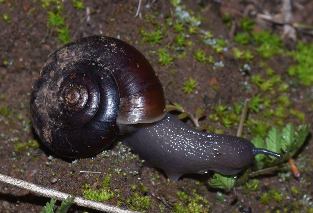
Snails in the group Pulmonata have eyes on the ends of retractable tentacles. These are true eyes with lenses and corneas. The snails have no muscles for focusing the lenses, though, so their vision is probably blurry, at best.

In additional to its eyes, the snail has photoreceptor cells distributed all over its body. These cells enable the snail to sense when a shadow falls over it. Since this usually means a predator, the snail reacts by quickly withdrawing into its shell. This is called the shadow reflex.

An unidentified snail in the taxon Pulmonata

If you watch the following video all the way to the end, you’ll be treated to the weird experience of seeing a snail’s eye disappear into its flesh without a trace!

Eye of the Insect 35

Insects are believed to have evolved from crustaceans and, like crustaceans, many insects have compound eyes. Many insects also have several very simple, single-lens eyes called ocelli (singular ocellus). These are used to detect motion and do not form images. The photo below shows the head of a tarantula hawk wasp. (See my earlier post The Stuff of Nightmares.) In the center of the head, between the two compound eyes, you can see three small ocelli.

Tarantula Hawk Wasp (Pepsis mildei)
Sweetwater River Trail 9/8/17
Tarantula Hawk Wasp (Pepsis mildei)
Sweetwater River Trail 9/8/17

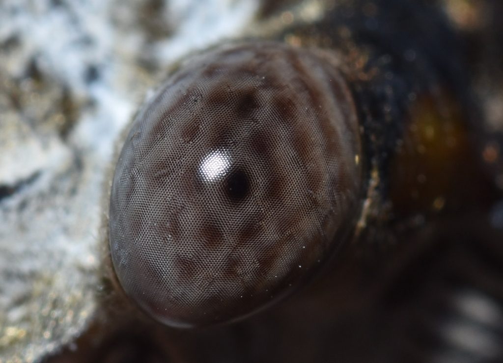
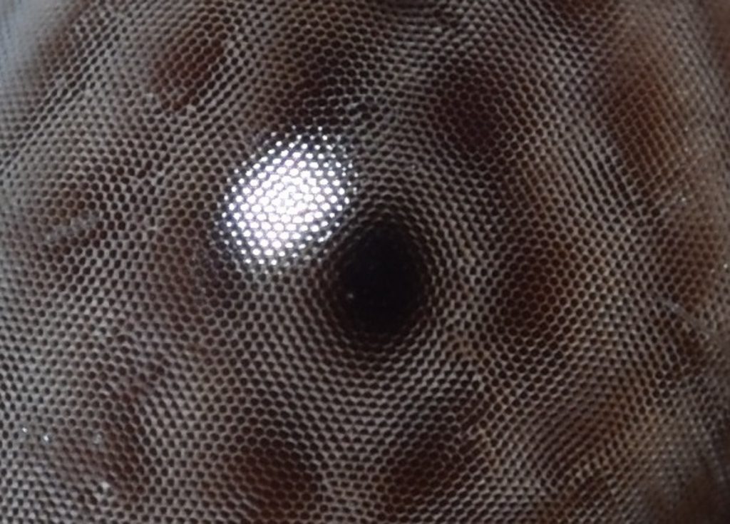
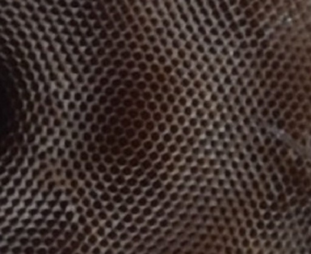
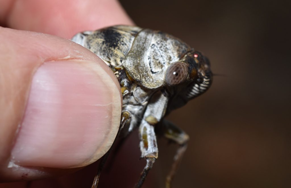
The compound eyes of the tarantula hawk wasp are too dark for you to see the individual facets. Not so with the large compound eyes of the cicada pictured in the following photos. As you can see, this is a very advanced eye with thousands of facets.

Eye of an unidentified cicada
Closer view
Closer still
Unidentified cicada
Emporia, VA 7/3/18

You may be wondering about the large, dark spot in the middle of the cicada eye. This is a ‘pseudopupil’. As the name implies, it’s not a true pupil. In a sense, it’s not even really there. Each of the tiny facets is the top of a tall columnar structure called an ommatidium. (See figure 3 in part 1.) The ommatidia in the dark spot look black because we’re looking directly down into their dark interiors where the photoreceptors are absorbing the light. We’re viewing the rest of the ommatidia at an angle, so we see the light reflected from their sides. If we could move around the eye and look at it from a different angle, the pseudopupil would appear to move with us.40

Many and Varied Are the Compound Eyes 41

Invertebrates have come up with many variations on the compound eye. These can be divided into two basic types: apposition eyes and superposition eyes.

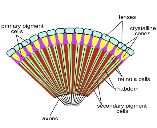
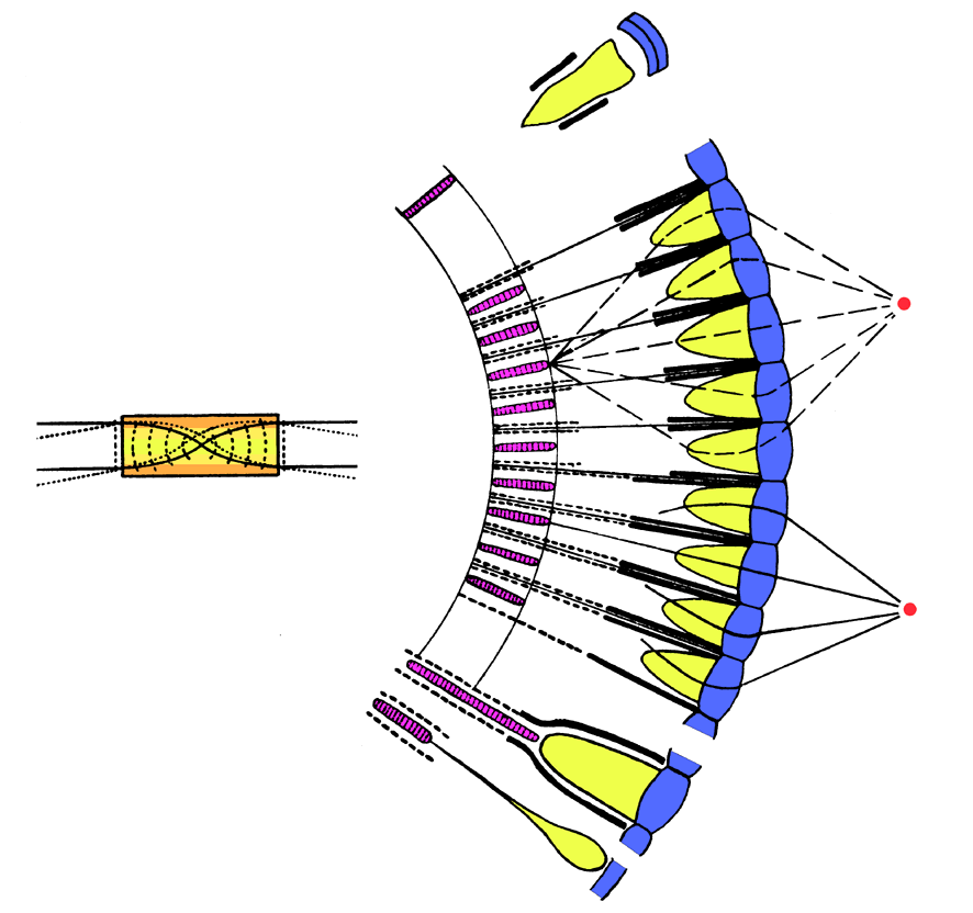
All compound eyes consist of collections of ommatidia. The main parts of an ommatidium are the cornea, the lens, and the rhabdom — a rod-like structure containing photoreceptors. (See figure 3 in Part 1). In an apposition eye, each ommatidium is an independent unit. Light entering the unit is focused only on the rhabdom belonging to that unit. Pigments surrounding the rhabdom prevent light from leaking into neighboring ommatidia. This is the simplest type of compound eye and was almost certainly the first type to evolve. Visual acuity can be very good in an apposition eye, but sensitivity is limited, because each rhabdom receives light from only a small area of the outside world. So organisms with this kind of eye are mostly limited to environments with high light levels. Marine animals with apposition eyes probably live near the surface. Terrestrial ones are probably diurnal.

Figure 4 below shows a typical apposition eye.

Figure 4. Apposition compound eye
Author: Bugboy52.40
Permission is granted to copy, distribute and/or modify this document under the terms of the GNU Free Documentation License, Version 1.2 or any later version published by the Free Software Foundation; with no Invariant Sections, no Front-Cover Texts, and no Back-Cover Texts.

A superposition eye solves the sensitivity problem by focusing light from multiple lenses onto a single receptor. This decreases the acuity, but this can be offset by increasing the size of the eye and adding more ommatidia. Figure 5 illustrates one type of superposition eye.

Figure 5. Superposition compound eye
Author: The original uploader was Kils at English Wikipedia.
This file is licensed under the Creative Commons Attribution-Share Alike 3.0 Unported license.

Each of these two major categories has several subcategories. Apposition eyes are subdivided into focal apposition, afocal apposition, and neural superposition eyes. (The latter is unrelated to true superposition eyes.) I won’t go into these in any detail other than to mention that only butterflies have afocal apposition eyes and neural superposition eyes are limited to true flies (order Diptera).

Superposition eyes are subdivided into refracting, reflecting, and parabolic superposition eyes. Again, I won’t get into the details, but I will mention that reflecting superposition eyes use mirrors to magnify the image, instead of lenses, much like a reflecting telescope! Nature, it seems, never misses a trick! (Actually, it does, but it’s still amazing to see the unexpected innovations it comes up with.)

Eye of the Spider

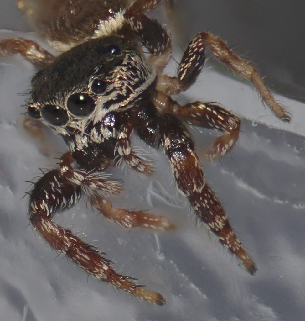
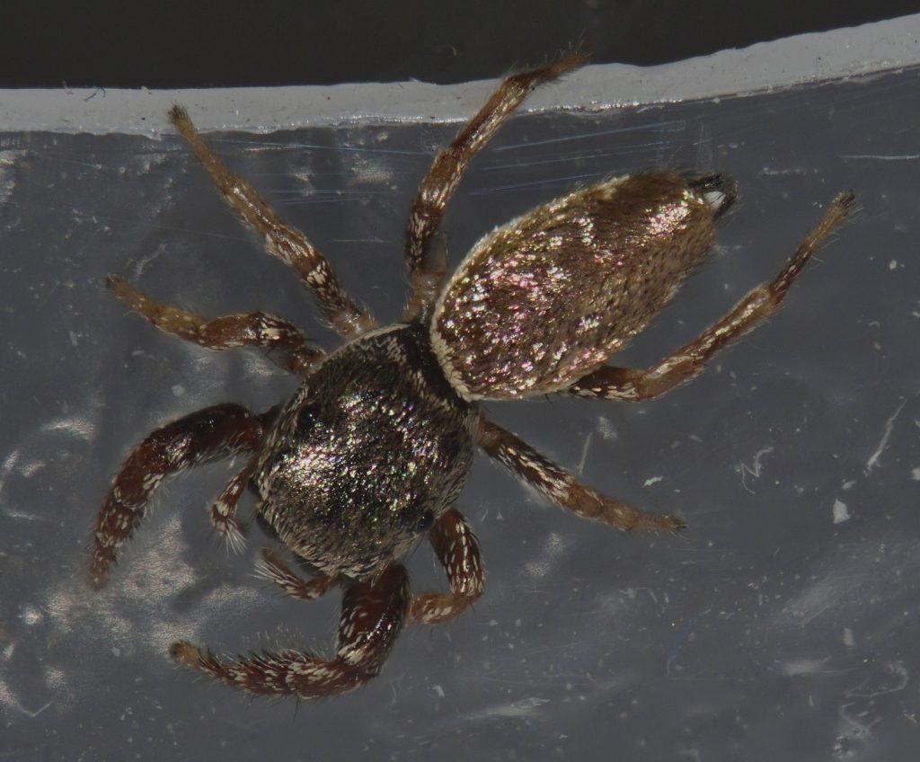
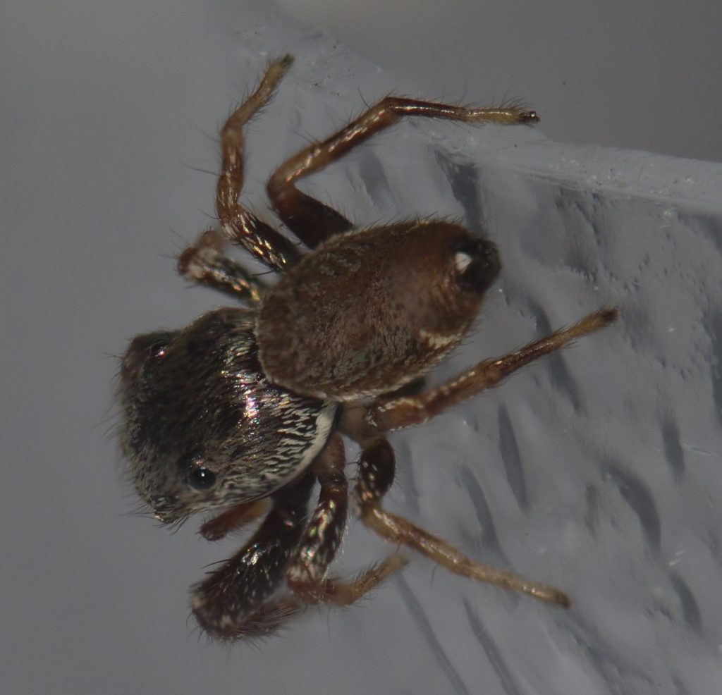
Meet Sassy. Sassy is a buttonhook leaf-beetle jumping spider (Sassacus vitis). She lives in my bathroom. I first encountered her when I was in the shower. Before you freak out, I should mention that Sassy is only a few millimeters long — much too small to be a threat to a human. I caught her in a small plastic container and, after her photo session, I returned her to the bathroom. I still see her now and then.

Buttonhook leaf-beetle jumping spider (Sassacus vitis)
San Diego County 1/7/20

Most spiders have eight eyes. Spiders that build elaborate webs to catch their prey have very small eyes and poor vision. They detect prey by the vibrations they make when they get tangled in the web, so they don’t have much need for good eyesight.

Spiders that actively hunt their prey, like Sassy, are a different matter. Jumping spiders, in particular, have the best eyesight of any spider. The larger of the two pairs of eyes on the sides of the head are for detecting movement. The wide-set pair on the front of the head are for depth perception and the principal eyes — the large pair in the front center — provide a detailed image, probably in color.42 43 As for the remaining pair of eyes — the smaller of the two pairs on the side of the head — their function isn’t clear. They may be vestigial in some species.44

The principle eyes are ‘simple’ eyes — as opposed to the compound eyes of most insects. Like our eyes, they use a single lens to form an image. The remaining pairs of eyes are thought to be derived from the compound eyes of the spider’s ancestors, but they no longer have multiple facets.45

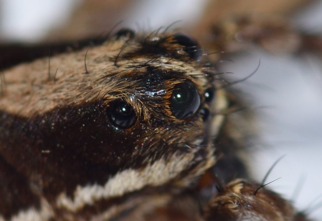
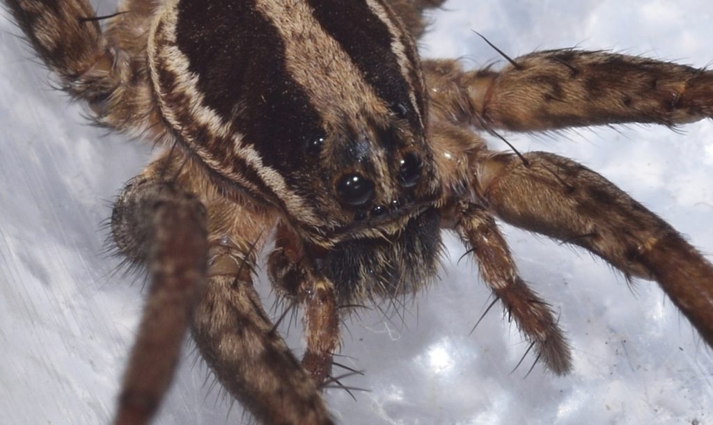
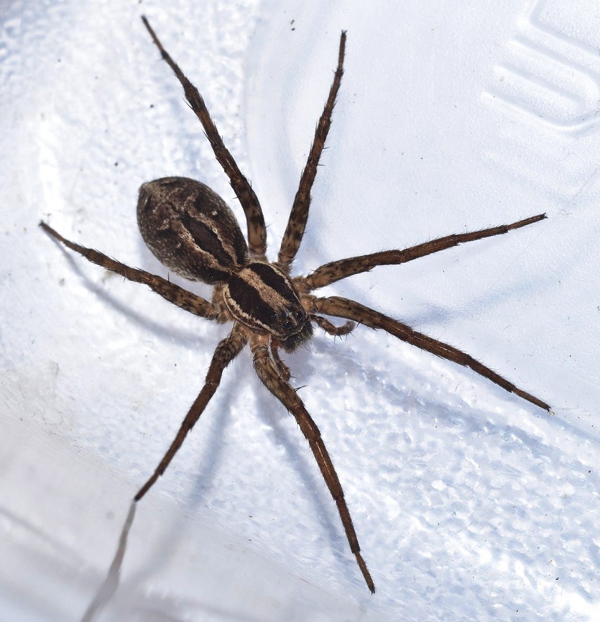
Jumping spiders hunt during daylight hours. Wolf spiders, on the other hand, hunt at night or in twilight. Four of their eyes have a tapetum (plural tapeta) — a reflective layer behind the retina. This is actually a very common feature in many kinds of nocturnal animals. When light hits the retina, not all of it gets absorbed. The light that passes through hits the tapetum and gets reflected back, so the retina has a second chance to absorb it. In this way, the sensitivity of the eye is greatly increased. Oddly, the principal pair of eyes in wolf spiders do not have tapeta.46 This seems counter-intuitive to me — I would expect the principal pair to be the most sensitive — but I haven’t been able to find an explanation for why this is not the case. Maybe having a tapetum would decrease the eye’s acuity somehow, but that’s just a guess. If any of you readers know the answer, please enlighten me in the comments.

Even with a second pass not all of the light gets absorbed. Some of the reflected light escapes the eye entirely. As a result, the eyes of animals with a tapetum, like wolf spiders and cats, glow in the light of a flashlight. If you hike at night in areas with a lot of wolf spiders, you’ll soon get used to spotting the tiny glow of their eyes. Many people say it’s a greenish glow, but it just looks white to me.47

Wolf spider (Schizocosa mccooki)
San Diego County. 10/02/19
Wolf spider (Schizocosa mccooki)
San Diego County. 10/02/19
Wolf spider (Schizocosa mccooki)
San Diego County. 10/02/19

Seeing the Polarization of Light 48

Some animals can see patterns formed by polarized light. This, of course, begs the question: What is polarized light?

Quantum mechanics tells us that light can behave as either a wave or a particle, depending on the circumstances. I won’t go down that particular rabbit hole. For the purposes of this discussion, we can think of light as a wave. If two people stretch out a rope between them and one of them moves her end rapidly up and down, a vertical wave will travel down the rope. If she moves her end from side to side, a horizontal wave will be produced. She can even create a circular wave by moving it in a quick circle. Think of that rope as a light wave. If the wave is oscillating vertically, then the light wave is vertically polarized. If it’s oscillating horizontally, then it’s horizontally polarized. (It can also be polarized at any angle in between horizontal and vertical.) And, of course, if it’s oscillating in a circle, then it’s circularly polarized.

The rays of light coming from the sun are randomly polarized. (Imagine a whole bunch of ropes oscillating in every direction.) However, when non-polarized light is reflected or refracted, it may become polarized. For instance, light bouncing off the surface of a body of water is polarized.

The sky looks blue because light from the sun is scattered by particles in the atmosphere. Why it’s blue and not some other color isn’t important for our purposes. The important thing is that the scattered light gets at least partially polarized. When the sun is directly overhead, the light is polarized horizontally throughout the sky. At dusk, the light is polarized vertically north to south — that is, perpendicularly to the direction of the sun. If you’re having trouble visualizing this, click here to go to a website that has some good illustrations of what I’m talking about.

The take away from all this is that polarized light forms patterns in nature and some animals are able to see those patterns. How are they able to do this? It turns out that a photoreceptor cell is most sensitive to light that is oscillating in a particular direction, depending on how the cell is rotated. That is, it’s more likely to absorb light that is polarized in that direction. In the retina of most animals, the photoreceptors are rotated randomly, so no matter what direction a ray of light is polarized, some of the receptors are going to be rotated at the correct angle to detect it.

So how does an animal evolve to detect polarized light? It just evolves groups of photoreceptor cells that are all aligned at the same angle. For instance, honey bees use the polarization of the sky (together with an internal clock that tells them what time of day it is) to navigate. If all the ommatidia in a bee’s eye were sensitive to the polarization of light, it would wreak havoc with it’s normal vision — it wouldn’t be able to make out objects in the environment. Instead, only the ommatidia in the upper part of the eye — the ones that are pointing at the sky — are sensitive to polarization.

I’ll be mentioning one or two other animals that can sense polarization later in the article, but before we get to that, it might interest you to know that humans are also able to detect the polarization of light, although we aren’t very good at it…

Haidinger’s Brush 49

Humans did not evolve to see polarization, but because of a quirk in the way the photoreceptors are arranged in our retina, it is possible for us to detect the pattern formed by polarized light in the sky. This pattern appears as kind of a faint cross with two blue or purple arms and two orange ones. Click here to see an illustration. The pattern is called Haidinger’s brush in honor of the scientist who discovered it. It takes some practice to see the pattern and not everyone can do it. If you want to try, click on that last link and follow the directions given there.

Eye of the Mantis Shrimp 50 51

Most animals have at no more than five types of photoreceptors. Many have fewer than that. Humans, for instance, have only four. The mantis shrimp has, depending on the species, between 12 and 16!

Mantis shrimp are not really shrimp — they’re a kind of marine crustacean belonging to the order Stomatopoda. Mantis shrimp are unique in a lot of ways. For instance, some species have clubbed claws with which they are able to smash open shells. These claws move so fast during a strike that they create vapor-filled bubbles called cavitation bubbles in the water. Their prey is hit twice — once by the club and a second time by the shock wave produced by the collapsing cavitation bubble. This wave is powerful enough to stun or kill the prey, even if the actual strike misses.

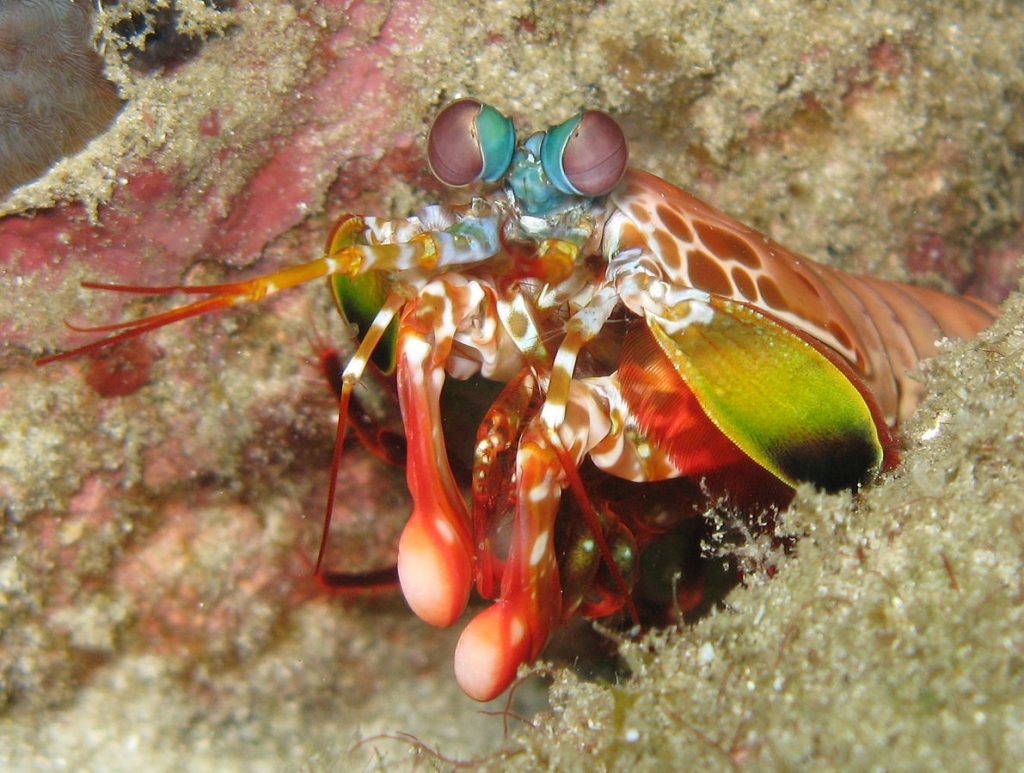
Mantis shrimp have the most complex visual system known to science. If you look at the photo below, you’ll notice a band going across each eye like a belt. Some of the photoreceptors in this band are used to detect polarized light. Most animals, if they can see polarization at all, are limited to detecting linear polarization. Some species of mantis shrimp are unusual in being able to detect both linearly and circularly polarized light. The ability to see polarized light helps the mantis shrimp detect its prey. Animals that blend into the background under ordinary vision often stand out clearly to predators that can see polarization.

The mid-band also contains various photoreceptors for ultraviolet, blue, and a longer wavelength in the green or red range. Other photoreceptor cells contain colored droplets of oil that act like yellow sunglasses, filtering out much of the blue light. Presumably, these are used to see yellow. There are also photoreceptors containing rhodopsin for night vision.

Some of the ommatidia in the rest of the eye (outside the mid-band) are directed toward the same point in space as the ones in the mid-band. This apparently helps with depth perception, but it also means that the animal’s vision is mostly limited to a narrow strip. That is, if the mid-bands of both eyes are held horizontally, then it won’t be able to see what is above or below it very well . The mantis shrimp compensates for this by being able to rotate its stalked eyes in any direction. It can even hold the mid-band of one eye in a vertical position, while the other mid-band is held horizontally!

The description of the mantis shrimp visual system that I’ve given here just scratches the surface. Wikipedia has a much more in-depth treatment. Check it out, if you’re interested.

A mantis shrimp (Odontodactylus scyllarus)
Author: Silke Baron
This file is licensed under the Creative Commons Attribution 2.0 Generic license.

Eye of the Amphibian 52 53 54

Amphibians represent the transition from an aquatic to a terrestrial environment in vertebrates. They evolved directly from lobe-finned fish and their eyes show it. Like fish, they have round, inflexible lenses and, like fish, they focus their eyes by moving the lens forward and backward. Your typical fish, however, doesn’t have to worry about keeping its eyes moist. Amphibians had to evolve a lacrimal system (tear glands and supporting structures) to keep their eyes from drying out when they’re not in water. They also needed lids to spread the tears. Probably, they evolved tear glands first, closely followed by lids.

Amphibians also evolved a transparent, nictitating membrane that can be drawn across the eye to protect it and to remove dirt. Reptiles, which evolved from amphibians, retained this membrane and improved on it. Mammals, which evolved from early reptiles, sometimes retained it and sometimes didn’t. (Humans, obviously, are in the didn’t category.)

A couple of fossils of lobe-finned fish show evidence of tear ducts that drained into the nasal cavity, so a lacrimal system was probably well on the way before true amphibians evolved. Lids don’t fossilize, though, so it’s not known if these fish had them.

Like most fish, many amphibians are tetrachromats — that is, they have four types of cone cells for detecting color. So some of them, at least, can see more colors than we can.

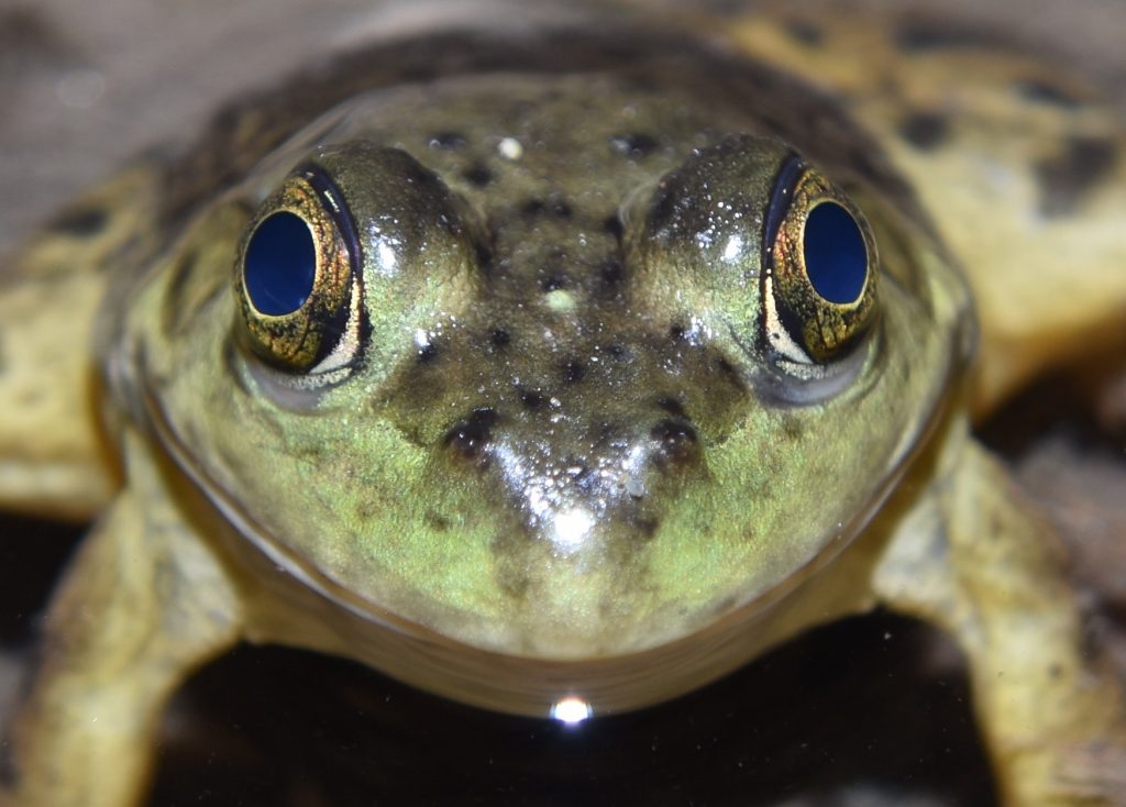
It sounds crazy, but frogs use their eyes to help them swallow. Frogs, salamanders, and newts have a muscle called the retractor bulbi that connects to the back of the eyeball. This muscle is used to pull the eye down and into the roof of the mouth. When a frog swallows a bug, it pulls its eyes in to help push the food down its throat. This is necessary, because the frog’s tongue attaches near the front of the mouth, instead of at the back, so it can’t be used for this purpose. There is evidence that this mechanism goes way back — the last common ancestor of the placoderms and the ray-finned fish probably had a retractor bulbi muscle.

American bullfrog (Lithobates catesbeianus)
San Diego County, CA 8/19/17
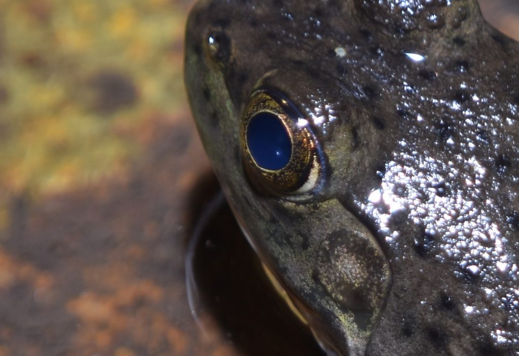
American bullfrog (Lithobates catesbeianus)
San Diego County, CA 8/19/17

As mentioned in the section on the lens and cornea in part 1, most land vertebrates use the refractive properties of the cornea to supplement the eye’s internal lens. This doesn’t work in water, however. Because a cornea is itself mostly water, it has essentially the same refractive index as water. This means that light passing from the water to the cornea is not bent, so the cornea in fish can’t be used as a lens. Another consequence of this is that any bumps on the cornea will not significantly affect the fish’s vision, so fish corneas tend to be rather roughly textured.

When the lobe-finned fish and, later, the amphibians, started using their eyes in air, a rough cornea became a problem. At the same time, the greater clarity of air opened up the possibility of improved acuity. Amphibians responded by evolving a smoother, more compact, and more transparent cornea. It also became much steeper, so that it could act as a lens. In the frog eye, the cornea has taken over most of the refractive load from the internal lens.

Amphibian vision is specialized for detecting motion. In fact, the only way for an amphibian to see an object is if it is moving across its visual field. An article on the Evergreen website put it as follows:

“…if a frog possessed the mental ability to read, it could only do so if the words were continually passed before it’s eyes. Even then, his visual acuity might not be sufficient to distinguish letters; but if he tried to read a stationary page, it would just appear a gray blur. When you see something in peripheral vision, attention is drawn. You may know that something has moved, but not exactly what. Imagine that your whole visual field is like the peripheral, and that you automatically lunge toward and attempt to eat any moving object that seems a lot smaller than you and that moves erratically enough to seem lively. Imagine that anything static (not moving) in your visual field blurs and fades to obscurity. This is what it would be like if you were an amphibian.” 55

Most amphibians have four types of photoreceptors — two types of cones and two types of rods, including the usual ‘red rod’ that most vertebrates have and also a ‘green rod’ (which is actually attuned to blue rather than green). The green rods are different in form from normal rods and certain of their features suggest that they originated as cones that transformed into rod-like cells as frogs and salamanders transitioned to a nocturnal lifestyle. In some salamanders, these rods have reverted back into cones.

Eye of the Reptile 56

Reptiles evolved from amphibians. The reptile eye began as a diurnal eye. It tends to have a rounded globe shape, especially compared to birds, which evolved from reptiles. The cornea is clear and steep, more so than in amphibians. The uvea or vascular tunic is the eye’s middle coat. It contains the blood vessels that nourish the retina. In reptiles, terrestrial vision is still in the early stages of development and their uveas are quite thin compared to birds. As vision improved, the retina would thicken and the uvea would have to thicken to support it.

A major innovation in reptile eyes is the method of accommodation. Fish and amphibians focus by moving the lens backward and forward. Reptiles, birds, and mammals focus by deforming the lens. The structure that is responsible for this is called the ciliary body. It’s part of the uvea and is located just behind the iris.

This mechanism of accommodation, combined with the use of the cornea to help focus, allows the lens to be much thinner. This, in turn, allows the lens to be enlarged without filling the entire eye. A wider lens captures more light, so the sensitivity of the eye can be increased without having to enlarge the entire eye.

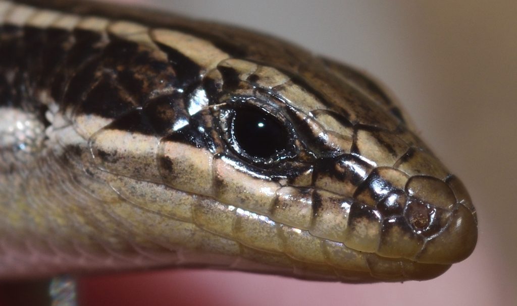
Skink (Plestiodon sp.)
Sheephead Mountain, San Diego County, CA 9/21/19
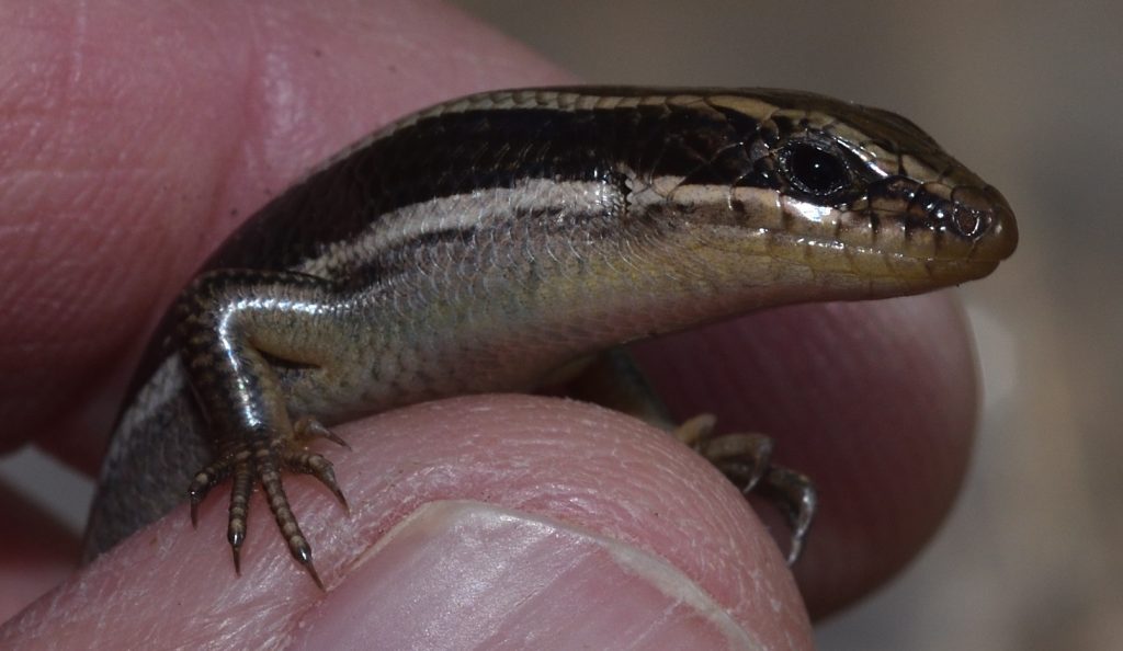
Skink (Plestiodon sp.)
Sheephead Mountain, San Diego County, CA 9/21/19

Eye of the Bird 57

The theropod lineage of dinosaurs evolved into the birds. In fact, birds are now classified as avian dinosaurs. (See my earlier post, The Coolest Thing About Birds.) The theropods were the top predators of their time and, not surprisingly, they developed good vision. Their bird descendants continued the tradition, eventually evolving the best eyesight of any animals.

Burrowing owl

Flight requires keeping your weight to a minimum, so birds tend to have small heads. There’s no way to push visual acuity to a maximum without enlarging the eyes, however, so avian eyes evolved to fill their sockets. In most birds, the eyes are bigger than the brain. Bird eyes have grown so large, that they’ve crowded out most of the extraocular muscle. Because of this, birds can generally only move their eyes a small amount in their sockets.

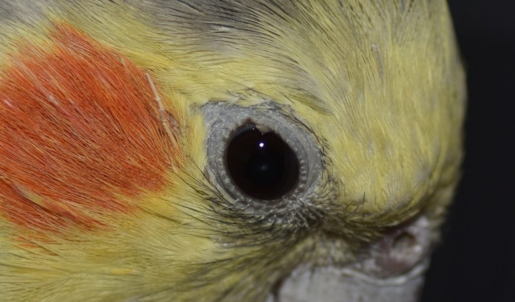
My pet dinosaur, Elvis. A cockatiel.

Most birds have roughly spherical, globular eyes like their therapod ancestors, but a couple of other eye shapes have evolved. Aquatic birds that dive for their food, developed flattened globes. As with fish, the cornea cannot help the lens refract light when a bird is underwater, so aquatic birds have evolved flattened corneas, as well. But unlike fish, they still have the soft, relatively thin lenses of their reptile ancestors. To compensate, some diving birds have come up with a novel solution. Muscular contractions of the ciliary body force the lens to bulge out through the pupil. This forces the lens into more of a spherical shape, increasing its refractive power.

The other eye shape that has evolved in birds is the tubular eye, which is seen only in owls and some goatsuckers (Caprimulgiformes), both of which are nocturnal. Having a long tube for an eye allows it to act like a telescope, magnifying the image and, thus, increasing the sensitivity of the eye. This is obviously important for nocturnal animals. The tubular eye of an owl is so large that it has even less room for extraocular muscles than is the case in other birds. They’ve compensated by developing the ability to turn their heads at great speeds through almost 270 degrees.

Eye of the Goat 58

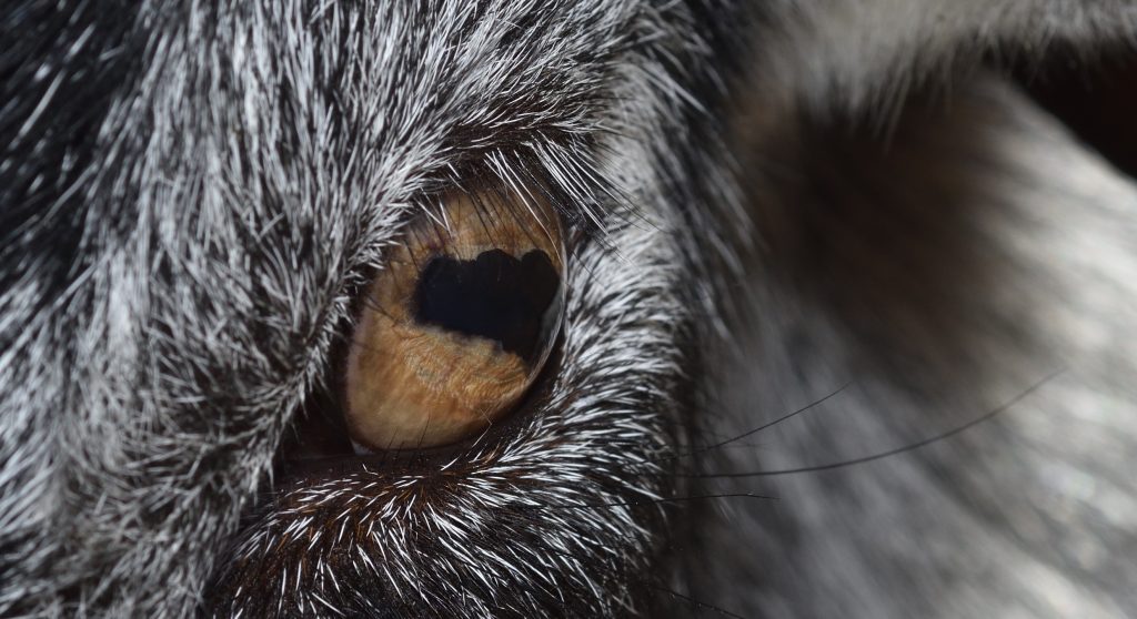
Goats have horizontal pupils. This looks pretty weird if you’re not used to it. Actually, many large herbivores have this kind of pupil. Ambush predators like cats and rattlesnakes, on the other hand, usually have vertical pupils. A study published in 2015 by Martin S. Banks et al. explores the relationship between pupil shape and an animal’s ecological role. The researchers looked at 214 different species of land animals and analyzed how the shape of their pupils affected their vision.

Obviously, prey animals need to be able to spot predators a long way off. A medium-sized herbivore like a goat, which tends to live in open country, probably doesn’t need to worry too much about predators swooping down from the air or jumping from trees. What it needs is wide, panoramic vision. And that’s what horizontal pupils give it. It also helps that its eyes are on the side of its head. This combination of features allows the animal to see a wide swath of its surroundings. (Ambush predators, on the other hand, usually have eyes on the front of the head so that they can focus directly on their prey.)

As important as it is for a prey animal to have a panoramic view of its surroundings, it still needs to be able to see in a forward direction well enough to avoid obstacles when it runs. Bank’s study found that a long, horizontal pupil enhanced the quality of the image both ahead and behind the animal.

Another fascinating thing the study found is that the extraocular muscles of a prey animal automatically rotate the eyes in such a way that the pupils remain in a horizontal position when the animal raises and lowers its head.

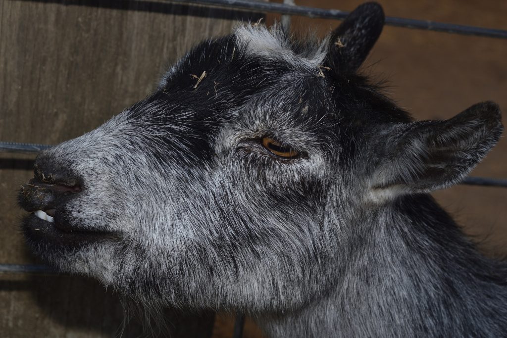
Unidentified goat
Notice how the eye protrudes from the socket, giving the goat a view of what’s in front of it.

Eye of the Rattlesnake

In the previous section, I discussed a study that examined why different animals have differently shaped pupils. This study found that many ambush predators have vertical pupils because it improves their ability to judge the distance to prey.59 One such animal is the rattlesnake. You can clearly see the vertical pupils in the images below.

I’ve already covered rattlesnakes in a previous post (see Rattlesnake!), so I won’t spend a lot of time on them here, but I do want to mention the pit organs. These are also visible in the photos. The snake uses these to detect infrared radiation (a.k.a. heat) given off by prey. The pit organs contain infrared receptors in the form of heat-sensitive proteins. The mouth of the pit is smaller than its diameter, so the pit functions as a crude, infrared version of a pinhole camera. In other words, it actually forms an image! The pit organs don’t contain photoreceptors, so they are not usually thought of as eyes, but maybe they should be — a rattlesnake can see well enough with its pit organs to hunt in complete darkness!

If you haven’t already read my post on rattlesnakes, I urge you to at least read the section on pit organs, where I cover these amazing structures in more detail.

Probably a southern Pacific rattler (Crotalus oreganus)
The silvery oval is the pit membrane.
Mission Trails Regional Park, San Diego County 8/14/2017
Red diamondback (Cotalus ruber)
Mission Trails Regional Park, San Diego County 8/7/2018

Eye of the Human 60

Most mammals have dichromatic vision. That is, they have two different types of cone cells, containing two different photoreceptor proteins. This allows them to see violet, blue, green and yellow. This is a much smaller color range than that of many reptiles and birds, which are often tetrachromatic — having four different types of cone cells.

Why did mammals get short-changed? Back when the dinosaurs ruled, mammals were small, nocturnal, probably burrowing animals. They didn’t have as much need for color vision and, in evolution, if you don’t use it, you tend lose it. I don’t know why mammals didn’t lose color vision completely. Perhaps some color vision was helpful — they just didn’t need the wider range of color vision of birds and reptiles.

Some marsupials have better color vision than placental mammals (the group that most modern mammals belong to). Marsupials evolved before placentals and it’s speculated that some of them never lost the more extended color vision of their ancestors. They still don’t have the range of color sensitivity of birds and reptiles, though.

Most Old World monkeys and primates (including humans), are trichromatic. That is, they have three types of cone cell. Their ancestors, however, were dichromatic, which means that they somehow managed to re-evolve a third cone type. They did this by gene duplication.

When DNA gets copied, occasionally an error occurs that causes a gene to be duplicated. If this happens in a germ cell, it gets passed down to the offspring as a mutation. Depending on the gene, having an extra copy may cause the animal serious problems or it may have no noticeable effect at all. In the latter case, the animal ends up with an extra gene just “lying around” that evolution is free to play with.
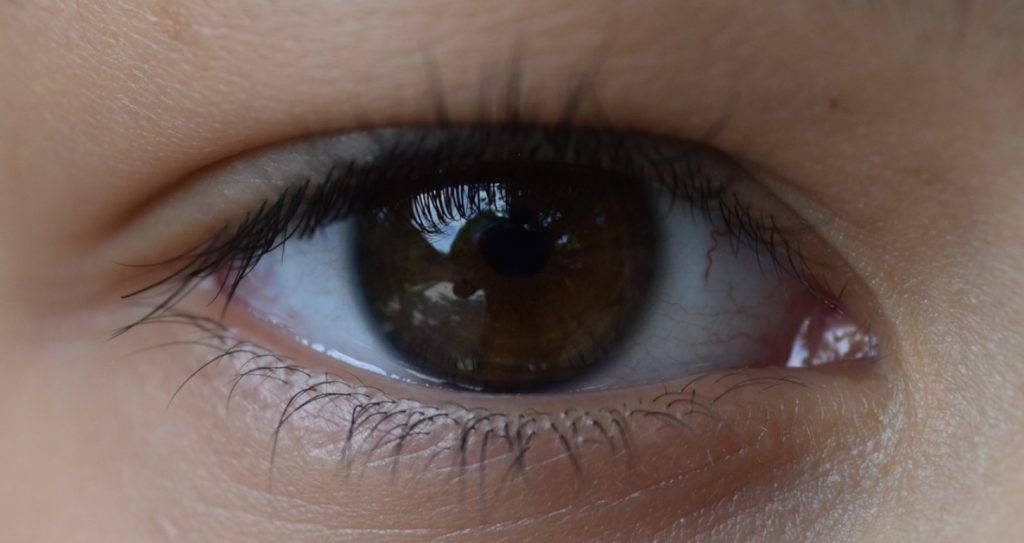
So, in our case, the extra gene eventually accumulated mutations that caused it to produce cone cells that were sensitive to a different range of wavelengths and this led to a lineage of mammals with a third kind of color receptors. Specifically, the new receptors allow primates to see red and orange hues.

It’s important to note that this new type of cone cell is not a reappearance of the one that was lost when mammals became nocturnal and burrowing. It’s an independently evolved photoreceptor that extends the color vision of primates and Old World monkeys.

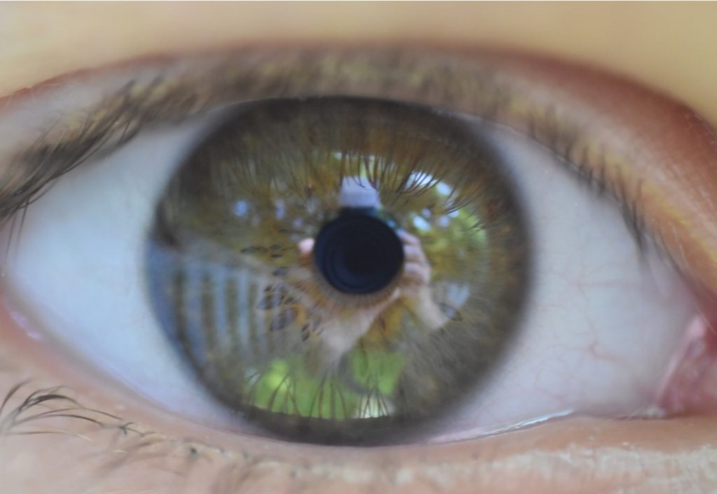
Homo sapiens
Homo sapiens

This third cone cell would not have been maintained in the lineage if it didn’t give its owners some advantage. What might that advantage have been?

Color and Climate 61

A howler monkey is a type of Old World monkey that eats mostly leaves. Young leaves tend to be the most nutritious. They also tend to be more reddish or orange, because they haven’t developed enough chlorophyll to mask the colors of the red and orange pigments. (These are the same pigments that make leaves change color in the Fall as they lose their chlorophyll.) In 1999, a team of scientists working in a national park in Uganda spent ten months studying what the various monkeys in the park ate. The researchers found that the howlers were using their extended color vision to selectively eat the most nutritious leaves.

So having trichromatic vision appears to have given howler monkeys an evolutionary advantage. But most monkeys today eat fruit, not leaves. Why would the ancestors of Old World monkeys and primates have evolved an adaptation to eating leaves in the first place?

Molecular studies of the photoreceptor proteins in primates have allowed scientists to estimate when the gene duplication that led to trichromatic vision occurred. It turns out that the lineage of mammals with three color genes goes back to between 40 and 30 million years ago.

It was around this time that the Earth was starting to get significantly cooler. Vast ice sheets pressed down from the poles, pushing back the tropical forests, full of palms and figs, and replacing them with grasslands and deciduous forests with more leafy trees. Gone were the large, abundant fruits, so now our ancestors had to rely much more on eating leaves.

The ice sheets have advanced and receded multiple times in the last 40 million years, but the Earth has never gone back to a climate where tropical forests covered most of the planet. It appears that we have that cooler climate to thank for our ability to experience such a colorful world.

The Story of Eyes: Part 1

A while back, I wrote a blog post about the parietal eye of lizards. (See Mysteries of the Third Eye.) In the course of doing research for that article, I read a bit about the evolution of eyes in general and I realized that this would make a fascinating topic for a future post — fascinating and challenging. The first eyes probably evolved around 600 million years ago and natural selection has been diversifying and refining them ever since. The variety of eyes and eye-like structures in modern organisms is truly mind-boggling. In this article and the one that follows, I’m going to cover a broad sampling of visual organs and try to give you some idea how they came to be.

Evolution of the Eye in a Nutshell 55 56

Let’s start with a quick overview. Most eyes consist of soft tissue, which rarely fossilizes, so their fossil record is very sparse. Nevertheless, scientists have a pretty good idea of the major stages in eye evolution. These are shown in the illustration below. An explanation of the various stages follows the figure.

Figure 1. Steps in the evolution of the eye.
Author: Matticus78 at English Wikipedia
Permission is granted to copy, distribute and/or modify this document under the terms of the GNU Free Documentation License, Version 1.2 or any later version published by the Free Software Foundation; with no Invariant Sections, no Front-Cover Texts, and no Back-Cover Texts.

Stage 1: Sensing the presence of light. (Figure 1-a) One of the earliest ‘proto-eyes’ to evolve was the eyespot. An eyespot consists of a patch of photoreceptive cells. This relatively simple organ is enough to let an organism know whether it’s in light or darkness. This may allow the organism to regulate it’s circadian rhythms. It may also allow it to move around at random until it finds the level of light or darkness that suits it.

Stage 2: Sensing the direction of light. (Figure 1-b) A very small organism, especially a microscopic one, is often transparent, so light can reach its eyespot either by shining directly on it or by first passing through the body. Eventually, some organisms evolved a layer of opaque pigment underneath the eyespot. Now, only light coming from outside the body could reach it. This enabled organisms to get some sense of the direction from which the light was coming.

Eventually, in some organisms, the eyespot became depressed, forming an eyecup. If light shone on the cup at a sharp angle, the rim of the cup would block part of it, so that only some of the photoreceptors in the eyecup were exposed. This allowed the animal to get a better sense of the light’s direction. Over many generations, the depression deepened into a pit and the organism’s ability to sense the direction of light steadily improved.

Stage 3: The pinhole camera eye. (Figure 1-c) Eventually, a limit was reached beyond which increasing the depth of the pit no longer helped. Now, however, evolution was in a position to take a very simple, but incredibly important step — the opening of the pit started to narrow, forming a pinhole camera.

Maybe you played around with one of these in science class back in the day. Take a shoe box and punch a small hole in one side. Light entering the box through this hole will form an image on the opposite wall of the box. (You can cut a larger hole in the top to peek at the image.)

So, as the opening got smaller, the pit started to act as a pinhole camera and a crude image began to form on the layer of photoreceptors lining the pit. The image got sharper as the opening got smaller, but, unfortunately, it also got dimmer. This is a serious limitation of pinhole cameras. Nevertheless, we now have our first true eye.

Step 4: Lenses and other refinements. (Figure 1 d-f) Covering the opening with a transparent layer of cells, displacing the water with a transparent ‘humor’, and adding a lens to collect more light (and thus improve sensitivity) led to the first ‘lens eyes’. Many other enhancements followed, some of which we’ll be covering later.

How Long Did All This Take?

So how long does it take to go from eyespots to eyes? Not as long as you might think, according to a paper by Dan-E. Nilsson and Susanne Pelger.59 These researchers estimated the time required to evolve an eye based on things like the rate of mutation and the relative advantage to the organism. They came up with a conservative estimate that the vertebrate eye could have evolved from a patch of photoreceptor cells in just 364,000 years — so, not even half a million years! This is a remarkably short period of time from an evolutionary standpoint and it helps to explain why there are so many varieties of eyes in modern animals.

Eyespot of the Planarian

Planeria are small, flat, worm-like animals, commonly called flatworms. What looks rather ridiculously like two crossed eyes, are actually eyespots. The dark areas are pigment cells that shade the photosensory neurons from one side, allowing the planarian to detect the direction of light.62 The eyespots are also slightly depressed, forming very shallow eyecups. This further aids the animal in sensing direction.63 I was able to find one site that shows the anatomy of a planarian eye. Click here if you want to check it out.

Phototaxis is the tendency of an organism to move either toward or away from light. Planaria exhibit negative phototaxis, meaning they tend to move away from it.64 65

Planaria
Kumeyaay Lake, San Diego County 7/17/17

A Quick Note

In the course of these two articles, I’ll be interspersing non-technical sections with slightly more technical ones. The latter will have a cyan background. Feel free to skip over the more technical parts, if you’re not feeling ambitious.

The First Photoreceptors and the Evolution of Multi-cellular Life 66

Bacteria are prokaryotes — single-celled organisms that lack a cell nucleus. Cyanobacteria, as the name suggests, are a type of bacteria. They’ve been around for between 3.75 and 2.6 billion years. Cyanobacteria use the photoreceptive compound chlorophyll for photosynthesis. Chlorophyll may have been the first photoreceptive molecule used by living things, although there are other possibilities.

Another important group of prokaryotes is the Archaea. Archaea are descended from bacteria and look very similar. In fact, they were originally thought to be bacteria. When scientists finally got around to doing molecular studies on them, however, it was found that they are very different genetically. In fact, Archaea are more closely related to eukaryotes — organisms that do have nuclei in their cells. Eukaryotes include single-cell Eukaryoptes called protists (amoebae, paramecia, etc.) and all multi-cellular organisms, including us.

One type of Archaea, halobacteria, uses a photoreceptive molecule called halorhodopsin both as an energy source and to detect light. This could have been the first photoreceptor used for sensory purposes. Halorhodopsin is a member of an important family of photoreceptors called rhodopsins. Various rhodopsins are used in the eyes of all animals.

Eukaryotic cells are much larger and more complex than prokaryotic cells. In addition to the nucleus, they contain various other membrane-enclosed structures called organelles that serve specialized functions within the cell. All eukaryotic cells, for instance, contain mitochondria. These are the ‘powerhouses’ of the cell. They produce the energy that facilitates high-energy activities such as locomotion. Another organelle, the chloroplast, is found only in plant cells. Chloroplasts, like cyanobacteria, contain chlorophyll, so they are used to harvest energy from sunlight.

Molecular studies of mitochondria and chloroplasts have found compelling evidence that both of these organelles originated as free-living bacteria. Chloroplasts, in particular, were originally cyanobacteria. At some point in evolutionary time, a larger single-cell organism must have either engulfed a cyanobacterium in an attempt to eat it or a cyanobacterium tried to parasitize the larger cell. Instead of the cyanobacterium being digested or the larger cell being killed, they somehow formed a ‘truce’. The bacterium reproduced inside the larger organism and, when that organism reproduced by dividing, the descendants of the captured bacterium got distributed to the new cells. Eventually, the cyanobacteria evolved a mutually beneficial relationship with the enclosing cells — they became chloroplasts. This process — of a captured cell being incorporated into a larger cell as an organelle — is called endosymbiosis.

Chloroplasts (and also mitochondria) have their own DNA, separate from the DNA in the nucleus of the parent cell. (This DNA, by the way, is very similar to bacterial DNA, which is what first gave researchers the clue that they originated as bacteria.) They reproduce independently and are passed down to the daughter cells in the cytoplasm. Over time, many of the genes that were originally in the bacterial DNA have been incorporated into the DNA of the parent cell, leaving the chloroplasts and mitochondria with much reduced, but still recognizably bacterial, genomes.

Recent research suggests that it was an Archaean that formed the first endosymbiosis with a bacterium, thus leading to the first eukaryotes and, eventually, to the first multi-cell organisms.

Volvox is a genus of single-cell organisms that forms large colonies. A Volvox colony behaves much like a single organism. It was probably such a colony that evolved to form the first truly multi-cell organism. Volvox contain a photosynthetic rhodopsin compound that is very similar to the rhodopsins in animals. It’s likely that all the rhodopsins used by animals have a common origin.

Figure 2. Euglena viridis, a type of Protist
Note the eyespot attached to the motor apparatus (the flagellum).
Author: Gevictor
This file is made available under the Creative Commons CC0 1.0 Universal Public Domain Dedication.

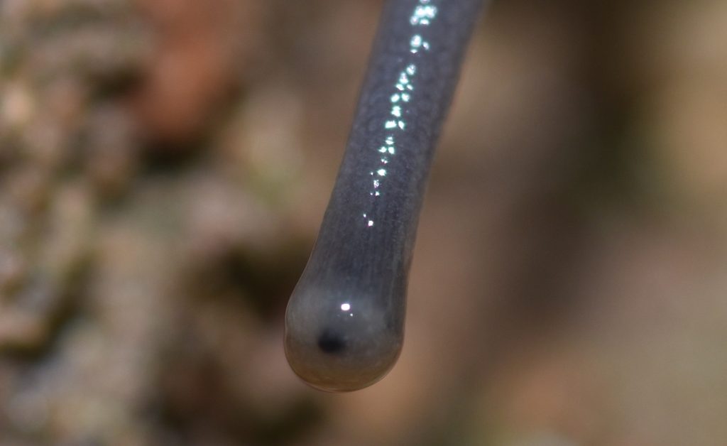
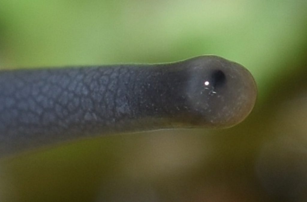
Eyespot of the Threadsnake 67

Not every animal that uses eyespots is ‘primitive’. Threadsnakes (a.k.a. blindsnakes) spend most of their time underground, so they don’t have much use for full-blown eyes. Their ancestors had eyes, of course, but living underground eliminated the selection pressures on them. What does this mean? Suppose a mutation occurs in one of the genes that’s responsible for the development of part of the eye, resulting in poorer vision. Well, it doesn’t matter, because the snake isn’t using its eyes, anyway. Over thousands of generations, such mutations will tend to accumulate, because natural selection is no longer weeding them out. Eventually, the threadsnake is left with nothing but eyespots.

Does this mean that they will eventually lose the eyespots, too? Threadsnakes do occasionally come to the surface — usually at night or late in the day — so it’s helpful if the snake can at least tell night from day. Eyespots are good for that. So it’s possible that future generations of threadsnakes will keep the eyespots.

Southwestern Threadsnake - Rena humilis humilis
Southwestern Threadsnake (Rena humilis humilis)
Southwestern Threadsnake - Rena humilis humilis
Southwestern Threadsnake (Rena humilis humilis)
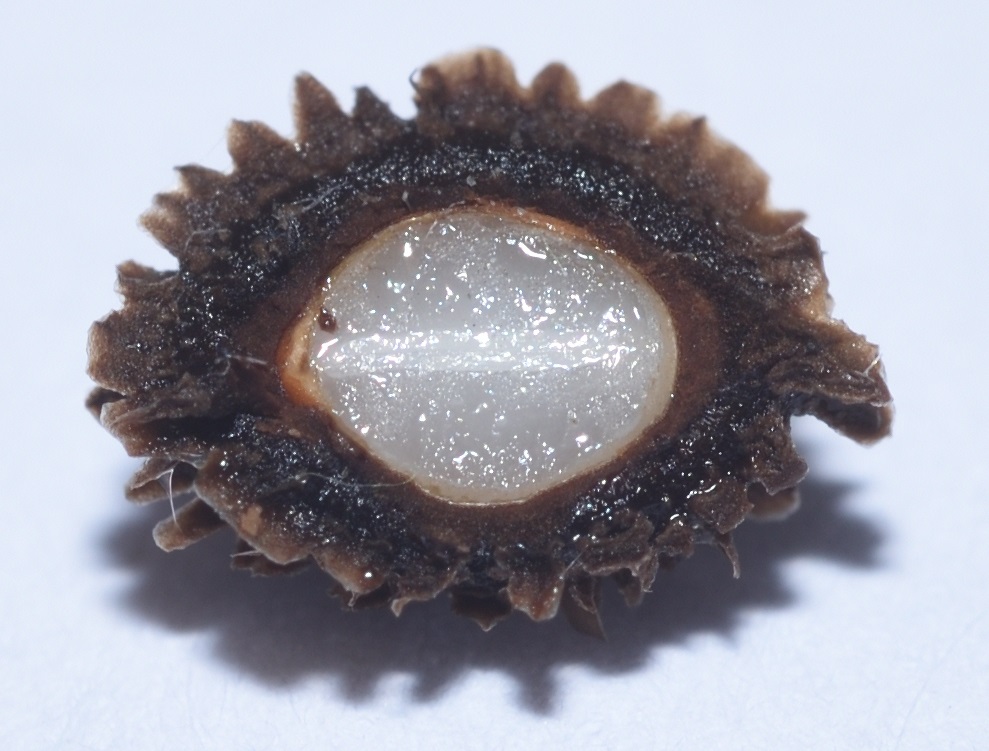
and some kind of seed

The Compound Eye 68

Eye evolution has taken two major directions — simple eyes (an eye with one lens) and compound eyes. All vertebrates have simple eyes (if they have any at all). Invertebrates may have either or both. We’ve already seen how simple eyes evolved. Compound eyes evolved by duplication. That is, a simple eye got duplicated multiple times to form a cluster and nerve impulses from all the eyes in the cluster were integrated in the brain to form a single image.

A simple lens eye doesn’t have to be very good. In many animals, the main purpose of a lens is to improve sensitivity by concentrating light. The ability to form an image is secondary. (The first eyes evolved in the depths of the ocean, so just detecting light would have been a priority.) Another way to increase sensitivity is to evolve multiple eyes, so that even more light is collected.

In this way, some animals evolved clusters of simple eyes. Now, suppose that such a cluster happens to form a bulge. Light hitting the bulge at a sharp angle would be blocked by the bulge itself, so that the light couldn’t reach all of the eyes. In this way, the bulge would give the organism a sense of the direction of the light. Increasing the bulge would improve that sense of direction.

So now we have a cluster of lens eyes, with each one oriented in a slightly different direction. In other words, we have the beginnings of a compound eye. In a true compound eye, the individual simple eyes — now just sub-units of the compound eye — are called ommatidia. (See figure 3 for a simplified illustration of an ommatidium.)

As the simple eyes evolved into ommatidia, they stretched out, becoming thin and deep, so that only the rays of light that hit the top of an ommatidium more or less directly were able to penetrate into its depths to reach the photoreceptors. In this way, each ommatidium received light from only a small section of the visual field. Input from all the ommatidia was integrated in the brain to form an image.

Eye of the Millipede 69

Millipedes are arthropods, like crustaceans, spiders, and insects. Some are eyeless; some have compound eyes; others, like the one pictured here, have patches of simple eyes that are only loosely integrated and maybe don’t quite qualify as fully compound.

Millipede (Tylobolus sp.)
Mission Trails Regional Park, San Diego County 12/03/19
Same millipede

Eye of the Crayfish 70

(Note: Part of this section is taken from my earlier post Crayfish Journeys.)

Crayfish are crustaceans, like crabs, lobsters, and shrimp. Like most crustaceans, they have compound eyes. Each eye consists of thousands of thin, columnar structures called ommatidia. The main parts of an ommatidium are a cornea, a lens, and photoreceptor cells. (Figure 3 shows a generic drawing of an ommatidium.) Each of the tiny, individual facets visible in the following photo is the cornea of an ommatidium. When excited by incoming light, the photoreceptor cells produce electrical impulses representing the intensity and color of the light. These impulses are integrated by the nervous system and brain of the crayfish to form an single image.

Red swamp crayfish (Procambarus clarkii)
Sweetwater River near Jamul, CA
Red swamp crayfish (Procambarus clarkii)
Sweetwater River near Jamul, CA
Figure 3 – A generic ommatidium
A and B represent the cornea and lens, respectively. E is the rhabdom, which contains the photosensitive proteins.
This image is in the public domain. It was downloaded from Wiki Commons at this url.

The Cambrian Explosion 71 72

The beginning of the Cambrian explosion is dated to about 541 million years ago and it lasted from around 13 to 25 million years. It was a remarkable period of diversification in which most of the major groups that we see in modern animals appeared. It marks the beginning of the Cambrian Period.

The Ediacaran Period, which immediately preceded the Cambrian, is sparcely represented in the fossil record, probably because most of the organisms at the time had soft bodies that rarely fossilized. In the Cambrian, by comparison, we see the emergence of animals with hard shells or exoskeletons. We also see the emergence of animals with fully developed eyes.

Trilobites, for instance, were a very large, diverse, and highly successful group of arthropods with a wide range of visual organs. Some were blind, but many others had compound eyes with rigid lenses made out of pure, transparent calcite. Each lens was an elongated prism. Some trilobites had only one such lens in each eye (in which case, I suppose it was really a simple eye, not a compound one). Others had thousands of lenses in each eye, rivaling some modern insects. Calcite is a mineral, by the way, so you could argue that trilobite eyes were made of rock (or, at least, the lenses were)!

Trilobites didn’t have corneas. The lenses were fixed to the exoskeleton of the animal. This means it was impossible to focus it, so trilobites were unable to accommodate for distance. Otherwise, though, some of them appear to have had surprisingly good vision. Studies have shown that some types of trilobite eyes had excellent depth of field and minimal spherical aberration.

Scientists are still debating what led to the proliferation of animal forms during the Cambrian. One likely explanation is the evolution of eyes. Having the ability to see other animals at a distance would have been a game changer. Suddenly, predators were able to see and stalk their prey. Their prey, in turn, evolved hard coverings to protect themselves. Of course, all of this took time. The changes that eventually led to the types of animals we see in the Cambrian explosion must have gotten their start in the Ediacaran. According to some estimates, true eyes may have evolved as early as 600 million years ago.

A Silurian scutelluid trilobite
Author: Kennethgass
This file is licensed under the Creative Commons Attribution-Share Alike 4.0 International license.
A fossil of a more elaborate trilobite

Eye of the Nautilus 73 74 75

Nautilus is a genus of cephalopods — the group that also includes squids and octopuses. They are one of the few surviving members of an ancient lineage that evolved in the late Cambrian. The best known species is the chambered nautilus (Nautilus pompilius), pictured below.

N. pompilius is most famous for its shell, which is usually displayed split down the middle to reveal the beautiful geometry of its interior. What doesn’t get as much attention is its large, pinhole camera eyes. These eyes have no cornea or lens. The pupil takes the form of a circular hole near the center of the eye, with a narrow slit trailing down vertically from it. This slit will close completely as the pupil contracts. In life, the eye is filled with a gelatinous substance that is probably similar to the vitreous body of our eyes, but the pupil has no covering, so seawater can freely pass in and out.

Nautilus pompilius
Source: http://opencage.info/pics/large_3628.asp
This file is licensed under the Creative Commons Attribution-Share Alike 2.5 Generic license.

The nautilus lives in the Pacific at depths ranging between 150 and 600 meters. It’s active mostly at night and in twilight, so it’s most important source of light is probably bioluminescence. A nautilus can only see a few meters and it’s vision must be rather blurry, but living as a scavenger and opportunistic predator, that’s apparently good enough. Also, its hard shell offers pretty good protection, so it doesn’t have much need to see predators coming a long way off.

Fish (and Their Eyes) Evolve 76

The earliest fish, the ostracoderms, were jawless fish related to the lampreys. The first fish with jaws were the Acanthodii. They showed up around 410 million years ago during the Silurian. Not much is known about their eyes.

We know a bit more about the eyes of another group of jawed fish, the placoderms, which also appeared during the Silurian. These fish were very heavily armored with bony plates on their heads and bodies. They had more bony plates in their jaws that served as cutting teeth. One of the largest placoderms, Dunkleosteus terrelli, grew up to 6 meters (almost 20 feet) in length. When it opened its jaws, the suction would be strong enough to suck in even large prey.

Skull of a Dunkleosteus sp. at Sam Noble Museum
This work has been released into the public domain by its author, Mitternacht90 at the Wikipedia project. This applies worldwide.
In case this is not legally possible:
Mitternacht90 grants anyone the right to use this work for any purpose, without any conditions, unless such conditions are required by law.

The eyes of some placoderms were encased in bony capsules. The molding of these capsules have allowed paleontologists to study their extraocular muscles (the muscles that attach to the outside of the eye and control its movement). It’s also allowed them to study the choroidal gland — a layer of connective tissue and blood vessels that supplies the outer layers of the retina with oxygen and nourishment. This layer allows an animal to develop a much thicker and more powerful retina than would be possible otherwise.

Although the eyes of modern ray-fin fish (Actinopterygii) are not encased in bone, they are enclosed in a ‘cartilaginous cup’. Many also have scleral ossicles. These are bones that are embedded in the sclera (the white of the eye) and form a ring around it, kind of like an iris of bone (but larger than the actual iris and unrelated in its function).

The scleral bones in fish likely serve as protection for the eye. In reptiles and birds, they serve the additional purpose of preventing distortion of the eyeball during focusing, which the animal does by changing the shape of the lens and cornea. (Fish focus by moving the lens back and forth and the cornea doesn’t change at all, so they don’t have this problem.)

Both the cartilaginous cup and the scleral ossicles are probably homologous to the bony casing of the placoderms. That is, they likely evolved from a similar structure that was present in the common ancestor of the two groups.77

Placoderms, by the way, provide the earliest evidence of a ‘third eye’ or parietal eye. (See Mysteries of the Third Eye.) So this organ must have evolved quite early in vertebrates. The lamprey, which probably evolved earlier than the placoderms (and is still around today), has a pineal gland, which is closely associated with the parietal eye. One source suggests that the parietal eye or something like it could even have evolved before the lateral eyes.78

Eye of the Moray 79

The moray eel, as its name implies, is a type of eel. Despite the unusually elongated and flexible body, eels are ray-finned fish — the most common type of fish. The moray’s eyes are rather small and it probably doesn’t have very good vision. It looks wicked scary, though, so I had to include it. (Weird nostrils, too!)

Moray eel

The Extraocular Muscles 80

Most vertebrates have six or seven ‘extraocular’ muscles that attach to the eye from the outside. Six of these muscles are used for the obvious purpose of changing the direction the eyes are looking. (The seventh, if present, is used to retract the eye. I’ll write more about this in the section on amphibians in part 2.) They serve another purpose that’s even more important, though. If a set of photoreceptors receives continuous stimulation, it becomes “fatiqued” — essentially, it runs out of the chemicals that are responsible for producing electrical impulses and stops working. To compensate for this, extraocular muscles move the eye in microsaccades — fine, fast movements that prevent the light from stimulating the same receptors over and over. This is true of humans, as well. Even when you stare fixedly at an object, your eyes are continually moving in microsaccades. Somehow your brain is wired to ignore these movements, so you’re totally unaware of them.

Eye of the Barreleye 81

Barreleyes are small, deep-sea fish in the family Opisthoproctidae. The name comes from their bizarre, barrel-shaped eyes. These eyes can be directed straight up when the fish is resting on the bottom or rotated to point forward when it’s hunting.

Various barreleyes
Source: https://www.flickr.com/photos/biodivlibrary/7413852664
Author: Chun, Carl
This image is in the public domain because it is a mere mechanical scan or photocopy of a public domain original, or – from the available evidence – is so similar to such a scan or photocopy that no copyright protection can be expected to arise.

At least one species of barreleye, Micropinna microstoma, has its eyes totally enclosed inside a transparent skull! Supposedly, even its brain is visible (although, in the video that I link to below, they say something about two large scales behind the eyes, so maybe they block the view of the brain). I so wish I could show you a photo of this insane creature. Unfortunately, I haven’t been able to find one that’s in the public domain, so all I can do is provide a link to a video. Click here to watch it. I strongly urge you to check it out. You’d be hard put to find a stranger animal.

The Lens and Cornea in Vertebrate Eyes 82

Most land vertebrates, such as us humans, use the cornea in conjunction with the lens to refract light and form an image on the retina. In fact, the cornea often does the bulk of the work. The lens serves mostly for accommodation — that is, changing the focus. The eye accomplishes this by using muscles attached to the lens to distort it — making it thicker or thinner as needed. The lens in land animals also tends to be less thick, since it doesn’t have to do all the imaging by itself.

In aquatic animals, this system doesn’t work, because the index of refraction of the cornea is almost the same as that of the surrounding water, so light is not bent as it passes through. The animal has to rely entirely on the lens to form the image. For this reason, the lens tends to be close to spherical in shape and the cornea is usually rather flat. The lens is also stiffer. Focusing is accomplished, not by distorting the lens, but by moving it forwards and backwards, like the lens in a telescope.

In cartilaginous fish (sharks, rays, etc.), the resting state of the lens is toward the front of the eye, which means that the fish are naturally focused on more distant objects. The fish focuses on near objects by pulling the lens backwards, closer to the retina. Bony fish focus in exactly the opposite way — the resting state of the lens is closer to the retina, meaning that the fish is naturally focused on nearby objects. To focus on distant objects, it has to move the lens away from the retina. This marked difference in focusing mechanisms suggests that the eyes of the common ancestor of cartilaginous and bony fish had little or no ability to accommodate. This ability must have evolved independently in the two lineages.

Eye of the Flatfish

Flatfish are members of the order Pleuronectiformes. They include such familiar fish as sole, halibut, and flounder. As far as I know, there’s nothing especially noteworthy about the anatomy of a flatfish’s eye or how it functions. What is noteworthy is the remarkable journey one of the eyes undergoes as the fish grows from a newborn fry to an adult.

In the adult, both eyes are on the same side of the head. The fish lies on the bottom, where it blends in almost perfectly with the seafloor. It may even cover itself with a thin layer of sediment, so that only its eyes are visible.

It doesn’t start out like this, however. A flatfish begins life as a very tiny and very ordinary looking fry (the standard term for a fish larva) with its eyes in the usual position, swimming around as part of the plankton. After a few weeks, though, a bizarre metamorphosis occurs. The bones of the fish’s skull distort as one of its eyes slowly migrates to the opposite side of the head. The fish’s sense of balance seems to undergo a transformation, as well, and it begins to tip over, swimming at an angle.

The side of the fish that ends up with both eyes develops a speckled coloration, matching the seafloor. The other side, now eyeless, loses pigmentation, turning pallid. Eventually, the fish settles to the bottom as the adult flounder we all know and love. Click here to check out Ikumi Kayama’s excellent illustration of the metamorphosis of a summer flounder (Aralichthys dentatus).

An adult flounder
Author: User:Moondigger
This file is licensed under the Creative Commons Attribution-Share Alike 2.5 Generic license.

This is all very interesting, but how on Earth did such a weird fish evolve? Evolution requires intermediate forms and it’s difficult to imagine how any such intermediate form could have been useful. What would be the evolutionary benefit of an eye that was only part-way migrated towards the opposite side of the body?

In a paper published in Nature in 2008, 83 Matt Friedman re-examined two types of fossil fish, Heteronectes and Amphistium, both about 45 million years old, found at a site in Italy. Prior to this, no one had thought that these fossils were related to flatfish, but Friedman used CT scanning to examine the fossils in great detail and found evidence that, not only were they members of this group, but that they represented intermediate steps in flatfish evolution. In both cases, one of the fish’s eyes had migrated towards the top of the head, but had not yet passed over it to the other side. (Various characteristics indicated that the fossils were of adult fish, so these were not fry in an intermediate stage of development.)

Friedman points out that modern flatfish often use their dorsal and anal fins to prop up their bodies above the substrate. Flatfish are ambush predators and such behavior would have allowed their ancestors to “peek” at their surroundings with the bottom eye, increasing their field of vision. A partially migrated eye would have further improved the fish’s view. (I do wonder why this “propping up” behavior persists in modern fish now that the migration has been completed, but maybe it’s still useful for tilting their field of vision towards something that attracts their attention.)

One last thing about flatfish — in most vertebrates, the inner ear contains structures that give the animal a sense of balance. Most fish use their inner ears in combination with their eyes to maintain an upright position. Even in the dark, they can tell which way is up, just as we can. Flatfish seem to be an exception. Flatfish larvae that have been blinded swim on their sides, as if their eyes had already migrated. One researcher used infrared imaging to film fry in complete darkness. Without light, the larvae were incapable of remaining upright. Some bobbed around with their heads pointing down. Older larvae leaned over and drifted to the bottom, landing on the side that would eventually be eyeless.

So in flatfish, the eyes seem to overrule the inner ear. This neatly explains why a fry swims leaning over as one of its eyes is in the process of migrating.

Rods and Cones and More 84

Photoreceptor cells are basically nerve cells that are used to detect light. In most animals, they are located in the retina. Humans and other mammals have three types of photoreceptor cells: rods, cones, and intrinsically photosensitive retinal ganglion cells. (I’m guessing you didn’t cover that last one in science class. In fact, they were only discovered in the 1990s.)

Cone cells are so called because they are roughly cone-shaped. The three types of cone cells in humans are referred to as S-cones, M-cones, and L-cones because the photoreceptor proteins they contain respond more readily to short, medium, and long wavelength light, respectively. Any of the cone types can respond to any of the visible wavelengths of light, but they are much more likely to respond to light in their particular category. In other words, it’s a statistical thing.

As you’ve probably guessed, Rod cells are roughly rod-shaped. While cone cells require a relatively high level of light exposure before they fire, a rod cell can be triggered by a single photon of light. When I was a kid, the numerals of alarm clocks were often painted with a substance that contained radium-226. The radioactive decay of radium produced a faint glow that made the numerals visible in the dark. (So I was probably getting a tiny amount of radiation poisoning, but I guess it wasn’t enough to have any long-term consequences. Knock on wood.) Lying in bed at night, if I stared at the numbers on the clock, I could see the tiny flash of a photon produced by a single decaying atom! 85
The human retina contains around 120 million rods and 6 million cones. Rods and cones are intermingled on the retina, but the cones are concentrated more towards the center — the part directly opposite the lens. At the direct center of the retina is a small pit called the fovea, which contains only cone cells. This is the area of highest visual acuity. When we look directly at an object, we’re positioning it in our visual field so that light from that object falls directly on the fovea. Because cones are much less sensitive than rods, a very dim star may disappear completely when you look at it directly, but reappear when you look off to the side.

A ganglion cell is usually defined as any nerve cell that’s located outside of the brain and spinal cord.86 The impulses produced by the rods and cones feed into bipolar cells (which I won’t get into) and then into ganglion cells, which do some preprocessing before the signals are sent on their way to the brain. Around 1% of the ganglion cells in the retina are intrinsically photosensitive retinal ganglion cells (ipRGCs). The first evidence that there was a third type of photoreceptor cell in the eye was discovered in 1923 when it was found that the pupils of mutant mice born without rods and cones still responded to light by contracting. Recent research suggests that ipRGCs support both the pupillary reflex and circadian rhythms.

As far as I can tell from my reading, most animals possess rods and cones of some sort. The ratio of rods to cones can vary greatly, however. A nocturnal animal will probably have very few cones compared to rods. The cone types can vary greatly, as well. Many birds and reptiles have four types of cones, while most mammals have only two. Some mammals, including humans, have independently evolved a third type. (I’ll cover this in more detail in part 2.)

Eye of the Four-eyed Fish 87

The four-eyed fish (genus Anableps) is a fish with… wait for it… two eyes. They only appear to have four because each eye contains two pupils, one above the other. Four-eyed fish live in Mexico and Central and South America. They inhabit mostly fresh and brackish water. The fish usually holds its eyes right at the surface of the water, so that one pupil is above the water line and the other is below it. Internally, different parts of the eye handle the two views separately. In this way, the fish can ‘keep an eye on’ things going on in both environments.

Among insects, whirligig beetles (family Gyrinidae) independently came up with the same trick. Their compound eyes are each divided into two parts — one for looking above the surface and one for looking below.88

Anableps anableps
Source: Reptiles, amphibia, fishes and lower chordata
Author: unknown
This work is in the public domain in the United States because it was published (or registered with the U.S. Copyright Office) before January 1, 1925.

End of Part 1

The Story of Eyes will be continued in the next post…