Category Archives: Fungi

A Gallery of Galls

If you’re a hiker, you might be familiar with oak apples. These aren’t really apples, of course. They’re plant galls, and they’re caused by a tiny wasp.

A large “oak apple” on a scrub oak. Caused by the oak gall wasp or California gall wasp (Andricus quercuscalifornicus). This type of gall forms a hard, dry “shell” when it’s fully formed.
Harvey Moore Trail near Descanso, CA. San Diego County 7/3/22

A gall is a plant growth that’s caused by, and serves as home to, some other type of organism. Most gall-formers are insects, but they can also be mites, nematodes1, fungi, bacteria, viruses, rotifers,2 or phytoplasmas.3 4 The gall occupant or its parent initiates the growth of the gall, usually by injecting a substance into the plant that mimics the plant’s hormones, hijacking its cellular machinery. Once started, a gall might grow into its final form on its own, or it might need additional stimulation from its occupant(s).5

A gall can be very simple or highly-structured, and galls of many different shapes, sizes, textures, and colors can grow on the same species of plant. Somehow, the gall-former forces the plant to grow the gall to its own specifications!

Many types of galls are found on oak trees, and most of them are caused by wasps. The gall in the previous photo was caused by a California gall wasp (Andricus quercuscalifornicus).6 Here’s a photo of the wasp. It’s only 5 mm long.

The oak gall wasp (Andricus quercuscalifornicus)
Author: USGS Bee Inventory and Monitoring Lab from Beltsville, Maryland, USA
Source:
Andricus quercuscalifornicus usda buffington

This image is in the public domain in the United States because it only contains materials that originally came from the United States Geological Survey, an agency of the United States Department of the Interior. For more information, see the official USGS copyright policy.

Here’s a photo of another oak gall of the same type. This one was still soft, so I cut it open.

An oak apple caused by the California gall wasp (Andricus quercuscalifornicus).
San Diego County 7/9/22

Inside, I found a wasp larva.

A cross-section of the gall in the previous photo, showing a tiny wasp larva.
A closeup of the larva

How an Insect Causes a Gall

There are two ways an insect can initiate a gall. It can injure the plant’s cells, causing an unorganized mass of protective tissue called plant callus to grow at the site of the wound, 7 8 or it can inject some substance into the plant that “instructs” it to form a gall. This substance is most likely a cocktail of chemicals that mimic the hormones the plant uses to control its growth.

Sometimes an adult insect uses its ovipositor9 to inject a substance that stimulates the growth of the gall. It then lays one or more eggs at or near the site. When the eggs hatch, the larvae find their way into the growing gall (or the gall simply grows around them). Sometimes the adult insect uses its ovipositor to inject both the larvae and the gall-stimulating substance at the same time.

In other cases, it’s the larva itself that initiates the growth of the gall. Often, its saliva contains a gall-stimulating substance. Even if the adult insect initiates the gall, the chewing of the larvae might be necessary to keep it growing. In more complex galls, the gall’s structure changes as it grows. Presumably, this means that the chemicals in the gall-former’s saliva are also changing as the larva develops.

For an amazing timelapse video of an aphid gall forming, check out this link.

Galls can take on some very strange shapes. Take, for example, the gall of the crystalline gall wasp in the following photos.

Gall of a crystalline gall wasp (Andricus crystallinus) on a California scrub oak (Quercus berberidifolia)
San Diego County 10/15/17
A closeup

There were some more galls on the same plant that were probably crystalline galls in earlier stages of development.

Some more galls on the same plant

Here are some more examples of crystalline galls.

Galls formed by the crystalline gall wasp (Andricus crystallinus) on a California scrub oak (Quercus berberidifolia)
San Diego County 10/6/22
A closer look at the galls from the last photo

In case you’re wondering, the two white, flower-shaped objects on the bottom leaf are not galls. They’re the pupal cases of a whitefly.

There is another type of gall in the photo, however. It’s in the fold of one of the leaves. I haven’t been able to identify it, but here are some closeups.

A closeup of the tiny, unidentified gall hidden among the crystalline galls in the last photo.
Closer still

What Counts as a Gall?10

I already defined galls earlier in the article, but to reiterate, galls are “growths on plants formed of plant tissue but caused by other organisms”.11 Of course, like most things in biology, it’s not quite that simple.

For starters, the host isn’t always a plant. It usually is, but it could also be a fungus, a lichen, or an alga. I won’t be covering any of those other, rarer, types of hosts, though. For the purposes of this blog post, gallers gall plants.

Also, the above definition isn’t complete. It’s not enough for an organism to cause a growth on a plant. It must also benefit in some way from that growth. It isn’t always easy to tell if this is the case. When an herbivore feeds on a plant, the damage alone is often enough to induce the plant to grow a different kind of tissue around the site of the injury ─ the plant callus I mentioned earlier. Usually, a galler or its parent injects substances into the plant to control the grow of new tissue, but not always. Sometimes the galler simply benefits from feeding on the callus, and that’s enough to classify the callus tissue as a gall.

On the opposite end of the spectrum, some galls can be quite complex, consisting of multiple layers, some nutritive, others protective. Often, the substances injected by the organism cause the plant to divert extra resources to the gall, making it (or a layer within it) more nutritious than other parts of the plant.

The fuzzy, little round gall in the next photo was formed by the aptly-named round-gall wasp, Burnettweldia washingtonensis.12

Burnettweldia washingtonensis gall on an oak
Oakoasis County Preserve, San Diego County 7/2/22
A closeup

Looking back at some of my pics on bugguide.net, I found a series of photos from 2018 of what looks like an older Burnettweldia washingtonensis gall. It was on a scrub oak, and it was definitely caused by some kind of gall wasp. I know this because when I cut it open, I found the wasp pupating inside! This was in February, so this species of wasp probably overwinters as a pupa inside the gall and exits as an adult in Spring.

A gall wasp gall on a scrub oak. This may be a Burnettweldia washingtonensis gall ─ the same kind shown in the earlier series of photos, but from later in the year.
Encinitas, CA 2/24/28

When I first cut the gall open, I was perplexed to find what appeared to be a loose nut inside. When I cut the “nut” open, I found the pupa.

The same gall cut open

The nut was actually a hardened inner layer of material ─ an “inner gall” ─ that formed around the pupa to protect it. This layer probably formed after the larva consumed all the tissue in the nutritive layer and decided it was time to pupate. The inner gall forms loose inside the outer gall because this makes it more difficult for a parasitoid13 to attack the pupa. The parasitoid uses its long, thin ovipositor to bore through the outer gall without too much problem, but when it tries to get a purchase on the inner gall, it’s like trying to spear a grape with a fork.

Here are several photos showing the wasp pupa from different angles.

The inner gall and the wasp pupa
Another view
And another

A spined turban gall wasp (Cynips douglasii) formed this next gall on the underside of an oak leaf.

Gall of a spined turban gall wasp (Cynips douglasii)
San Diego County 7/2/22
Another view

Types of Galls

Many kinds of plants are galled by many kinds of organisms. And, as we’ve seen, those galls can be very simple or very complex. There are several other ways that galls differ from each other.

First, different types of gall form on different parts of the plant. Some galls only grow on roots, for example. Others only grow on stems, leaves, buds, or flowers.14

Second, galls vary in size. They can be almost microscopic or bigger than your head.

Finally, and perhaps most obviously, they vary in form ─ from a simple swelling to a round ball to weird grows like the crystalline galls we saw earlier.

A particular organism doesn’t necessarily cause only one type of gall. Many insects, for example, have alternate generations ─ usually, a sexual one and an asexual one (where the insect reproduces without mating). Sometimes one of the generations forms galls and the other doesn’t. In other cases, both generations form galls, but on different types of plants, or on different parts of the same type of plant, or even on different parts of different types of plant.

Scientists who study galls (and who are called cecidologists) have come up with many descriptive terms for them. What follows is a list of some of the most common ones. I’m not going to show you examples of each one ─ that would make this post way too long. If you’re interested, check out “Plant Galls” by Margaret Redfern. Much of the info in this article comes from that book.

Fasciations, phyllathies, phyllodies, and virescences ─ Plants sometimes exhibit weird growth patterns called fasciations, which are caused by errors in their development. These can be genetic errors or they can result from environmental factors. If they aren’t caused by other organisms, then they don’t count as galls.15 A few organisms, however, cause fasciations and feed on them. These do count as galls.

A phyllody, to quote Wikipedia, is “the abnormal development of floral parts into leafy structures. It is generally caused by phytoplasma or virus infections, though it may also be because of environmental factors that result in an imbalance in plant hormones.”16

Phyllathy seems to be just another word for phyllody.

Virescences are “oddly shaped parts of plants, where shoots, roots, leaves, or flower parts are distorted, multiplied or bunched together.”17 When they occur on trees, virescenses are sometimes called witches’ brooms. Phyllodies are a type of virescence.

Solid swellings ─ These can be single enlarged cells in algae. In plants, they can be much larger ─ up to a meter or more on trees.18 Crown gall is a plant disease caused by the bacterium Agrobacterium tumefaciens. It results in tumor-like growths up to a foot in diameter on more than 600 different plant species, including both woody and herbacious plants.19 This is one way in which bacterial gallers differ from most other types of gall-formers. Most gallers gall only one or a few types of plants.

Erinea galls ─ Simple, hairy galls caused by eriophyid mites. The mites live among the hairs. Erinea galls often look like patches of velvet on the undersides of leaves. Sometimes they are reddish in color.20

Folds and rolls ─ These are simple leaf galls. The galler causes the edge of the leaf to fold over or roll up. It does this by stimulating cells on one side of the leaf to proliferate more than those on the other side.21 The galler benefits from the protection that the fold or roll provides, so, simple as they are, these do count as galls. Many types of mites and insects cause fold or roll galls.22

Pits, blisters, and pouches ─ These, like folds, rolls, and erinea galls, are open galls. And, like folds and rolls, they are fairly simple distortions of the leaf. Various mites and insects cause these types of galls.23

Big buds, cigars, pineapples, and rosettes24 ─ These types are lumped together because they all develop in buds, and they all form from young modified leaves that bunch together because their petioles (leaf stems) fail to develop. These are open galls. The mites or insects that cause them escape through openings between the leaves. Pineapple galls are sort of an exception. These are temporarily sealed, but changes in the gall tissue eventually provide openings for the insects to escape.

Closed chambered galls ─ These galls, as the name suggests, contain one or more closed chambers in which the gall-former(s) reside. These tend to be the most highly-structured types of galls.25

Burro-weed (Ambrosia dumosa), aka white bursage, is a type of ragweed that grows in the desert.26 The next two photos show an interesting gall that I found on a burro-weed. I think the gall-former is Procecidochares kristineae, a type of tephritid fly.27

A gall on burro-weed (Ambrosia dumosa), probably caused by Procecidochares kristineae.
Sonoran Desert, San Diego County 11/26/22
Another view

Cutting it open, I found that the occupant had already pupated and left, leaving behind its pupal skin.

Inside the gall. The dark object in the left section is the pupal skin. It was just a dry husk.

The Australian eucalyptus leafgall wasp (Ophelimus maskelli) galls the leaves of eucalytus trees. The galls are tiny red swellings.28

A eucalyptus leaf almost covered with galls of the Australian eucalyptus leafgall wasp (Ophelimus maskelli)
San Diego County, California, USA 11/5/22

Let’s zoom in on a few of those galls.

A closeup
Closer still

I used an x-acto knife to slice one of the galls open, and to my surpise, I managed to do it without injuring the baby wasp inside.

This isn’t the first time I’ve seen these particular galls. Back in May of 2020, I collected some eucalyptus leaves with galls, most of which had exit holes.

Empty O. maskelli galls on a eucalyptus leaf
San Diego County 5/4/20

A few still had the insect inside, though. I managed to capture this adult wasp after it exited one of the galls.

Australian eucalyptus leafgall wasp (Ophelimus maskelli)
San Diego County 5/4/20

Manzanita (genus Arctostaphylos) is a very common plant in the chaparral of Southern California. Occasionally, you’ll come across one that’s absolutely covered in bright red fold galls. These are caused by Manzanita leaf gall aphids (Tamalia coweni). They mostly form on the edges of leaves, but they can also form on midribs and on the inflorescenses (flower clusters).29

Fold galls formed by Manzanita leaf gall aphids (Tamalia coweni)
San Diego County 7/10/16
A closeup of a couple of galls on the same leaf
A small fly on one of the galls. I don’t know of any association between the fly and the gall, I just like the photo.

Here are some more aphid galls on a manzanita. I saw these in Tulare County in central California.

Aphid galls on manzanita
Tulare County, CA 7/10/16
A cross-section of one of the galls
A slightly closer look
One of the aphids
Another aphid

I found the next gall on a cottonwood tree. It was formed by poplar leaf-petiole gall aphids (Pemphigus populicaulis)30

Gall of poplar leaf-petiole gall aphid (Pemphigus populicaulis)
San Diego County 11/29/17

Aphids in various stages of development were inside. The winged one is an adult.

The same gall cut open

Here’s a gall I found on a California sagebrush (Artemisia californica). The gall-former is probably a gall midge31 in the genus Rhopalomyia.

Gall on California sagebrush (Artemisia californica), probably caused by Rhopalomyia sp., a gall midge.
San Diego County 2/24/19
Another view
A closeup

This next gall was on a Great Basin sagebrush (Artemisia tridentata). It was caused by a sponge gall midge (Rhopalomyia pomum). The gall was, indeed, soft and spongy.

Gall caused by a sponge gall midge (Rhopalomyia pomum) on Great Basin sagebrush (Artemisia tridentata)
San Diego County 4/28/18

One of the most common plants in the Sonoran Desert is the creosote bush (Larrea tridentata).32 It’s not uncommon to see these unusual-looking galls on them. They are caused by creosote gall midges in the genus Asphondylia.

Gall on a creosote bush (Larrea tridentata). Caused by a gall midge, Asphondylia sp.
Sonoran Desert, San Diego County 12/16/17
A closeup

This next gall was formed by a wasp on a wild rose.

Diplolepis sp. gall on a wild rose
Northern San Diego County 4/28/18

Nancy Asquith on bugguide.net identified the gall-former as “definitely Diplolepis“. She thinks it was probably a spiny leaf gall wasp (Diplolepis polita), which is very common in California. This type of gall would normally have many long spikes instead of one short one. Ms. Asquith thinks that it could have been a D. polita gall altered by inquilines.

And what’s an inquiline, you ask…

Gall Ecosystems

According to Wikipedia, an inquiline is “an animal that lives commensally in the nest, burrow, or dwelling place of an animal of another species.”33 So, in the context of galls, an inquiline is an animal that moves into the gall after the gall-former forms it. The “commensally” part of the definiton means that the inquiline neither benefits nor harms the gall-former.

In fact, entire ecosystems of organisms often form inside a gall. In addition to inquilines, there are parasites, parasitoids, and predators. A parasite feeds on the gall-former (or one of the other occupants) without killing it; a parasitoid feeds on the gall-former (or one of the other occupants) and eventually kills it; a predator feeds on the gall-former (or one of the other occupants), kills it, and then moves on to prey on another occupant (or an occupant of another gall, or something else entirely). Some predators eat one or more of the occupants and then feed on the gall itself.

Of course, gall occupants are not just fed on by other occupants. Birds and mice will break open galls to get to the insects inside. Herbivores like caterpillars will feed on a gall and unintentionally eat the occupants in the process.

Even if the gall-formers make it to adulthood and exit the gall, other organisms might move in later. If nothing else, empty galls eventually serve as food for bacteria and fungi.

Some species of sawfly are gallers of willows. The next series of photos show galls of the willow apple gall sawfly (Pontania californica).

Galls of willow apple gall sawflies (Pontania californica)
San Diego County 8/20/22
The same galled leaf showing the top (dorsal) surface.
The bottom (ventral) surface.
A section through one of the galls. You can see that it has cavities for multiple larvae. One of the larvae is visible.

And here’s a live oak apple gallfly gall on a live oak.

Gall of live oak apple gallfly (Callirhytis quercuspomiformis)
San Diego County 9/24/17

The beaked twig gall wasp (Disholcaspis plumbella) makes brightly-colored galls. This one is a bit out-of-focus, sadly. It has three exit holes, so it looks like at least some of the occupants have already left.

Gall of a beaked twig gall wasp (Disholcaspis plumbella) on a scrub oak
San Diego County 9/24/17

This gall on coyote brush (Baccharis pilularis) was caused by a gall midge ─ probably a coyote brush bud gall midge (Rhopalomyia californica).

Gall midge gall on coyote brush
San Diego County 7/16/22
Another view. Lots of exit holes, so it must have had multiple occupants.

I think that what looks like long, whitish husks protruding from a few of the holes are the exuviae (discarded exoskeletons) left behind by adult midges emerging from the gall.34

This is a bud gall, meaning that it formed from tissue that would have formed into a bud if the galler hadn’t gotten there first. You can see in the cross-section below that it has a structure similar to a bud. I didn’t find any larvae or pupae inside, but a small larval cavity is visible in the right half.

A very common plant in my area is another species of baccharis ─ broom baccharis (Baccharis sarothroides).35 It’s also frequently galled by midges. I see them very often on my local hikes.

Unidentified gall midge galls on broom baccharis (Baccharis sarothroides).
San Diego County 7/6/22

I don’t know what caused the galls on this leaf. Possibly mites.

Unidentified galls. I think the plant is called basket bush.
San Diego County 10/7/17

The vast majority of gallers are parasites. In other words, the galler benefits from the relationship, but the host plant is harmed by it. Sometimes the damage is severe. A galled plant might be stunted or produce fewer seeds. If the infestation is bad enough, it might even die. In other cases, the plant hardly seems to notice.36

It’s very rare for gallers to form mutualistic relationships with a host, but it does happen. One well-known example is the fig wasp.

Fig Wasps37 38

Fig wasps are wasps that spend their larval stage in figs. There are many different kinds of fig wasps, but the ones we’re interested in are in the family Agaonidae. These are the ones that act as pollinators for fig trees. The trees cannot reproduce without the wasps and the wasps cannot reproduce without the trees, so this is a clear case of mutualism.

A fig is not actually a fruit. It’s a syconium. An article on the Britannica website describes it as an “urn-shaped inflorescence”. The article goes on to explain that the syconium probably originated from the “invagination of a flat flower-bearing capitulum”.39 Maybe you’re having trouble making sense of this explanation, so let’s dissect it a bit.

An inflorescense is a cluster of flowers on a stem.

A capitulum is “An inflorescence that consists of closely packed flowers or florets which have no stalks and arise on a flattened axis, all at the same level.”40

An invagination is a surface that has folded into itself to form a cavity, pouch, or tube.41

So, Britannica seems to be saying figs evolved from an inflorescense of small, tightly-packed flowers that folded in on itself to form a pouch with the flowers inside. This makes sense because unripe figs actually do have flowers inside them! So, where do the wasps come in?

A female wasp enters a fig through a small opening. She has to force her way through this opening, and, in the process, she usually loses her wings and most of her antennae. In other words, she won’t be leaving. A female fig wasp only gets to pollinate one fig.

Once inside, the wasp lays eggs on some of the flowers. A flower that has an egg deposited on it develops a gall instead of a seed. The wasp deposits pollen that she picked up from her “home” fig (the one she hatched out in) on some of the other flowers.

When a male fig wasp hatches out of a gall, it searches in the same fig for galls containing females. When he finds one, he chews a hole in it and mates with the unhatched female. That’s right… the unhatched female. The female emerges from her gall already pregnant. The male dies inside the fig, but before he does (in many species, at least) he chews an escape hole for the female. When females emerge from their galls, they crawl over ungalled flowers on their way to the exit holes, picking up pollen as they go. Once outside the fig, they waste no time looking for a new, unripe fig in which to lay their eggs and die. What a life!

There are many kinds of fig wasps in the family Agaonidae. The description that I just gave of the fig wasp life cycle is somewhat generalised. Details vary from species to species. If you want to do some further reading, a good place to start is the Britannica article that I referenced earlier. It’s short, but it has a nice illustration of the life cycle. Click here to read it.

The Evolution of Gall-forming

Unsurprisingly, we don’t know a lot about how gall-forming evolved. One thing that’s certain is that it arose independently many times.42 Despite this, gall-forming is not all that common an adaptation. Apparently, it’s a successful enough strategy to have evolved many times, but not successful enough to be the “go-to” strategy for any large group like mites or insects.43

Although we don’t know many details, it’s not hard to imagine how an organism might evolve to form galls. At least, this is how I imagine it…

When an insect larva feeds on plant tissue, the plant typically responds by growing callus tissue44 to cover the wound. This might be a better source of nutrients than the regular tissue, so the insect proceeds to feed on it. Maybe some chemical in the larva’s saliva happens to stimulate the plant to change something about the way it grows this tissue. If the change is beneficial, the insect’s descendants are more likely to survive and more likely to keep secreting that substance in their saliva. Then, maybe a mutation occurs that causes a new chemical to be included in their saliva. If it also proves beneficial, the new mutation might be retained. And so on. Insects use many chemicals that are simular to plant hormones (they probably have similar evolutionary origins), so it’s not as unlikely as it might sound.

Similarly, if an insect lays an egg by injecting it into plant tissue, various substances get injected along with it. Some of these substances are probably meant to protect the egg from the plant’s defences, for example. A side effect of some of the substances might be to alter the way the plant grows tissue in the vicinity of the egg. Maybe this altered tissue benefits the larva in some way. And so on.

This just my take on how gall-forming might have evolved.

Evolution of gall morphology is affected by the pressure put on the gall-formers by their enemies. For example, an insect deep inside a large gall is often safer than an insect in a small gall or one farther from the center, simply because it’s harder for parasites, parasitoids, and small predaters to get to them. On the other hand, a large gall is more conspicuous, so there’s more chance that a large predator, such as a bird, will notice it.45 So, it’s a give and take. The gall-former will evolve in whichever direction gives the best results.46

Addendum

In the short time that’s passed since I published this article, I’ve already found a number of new galls. Also, my friend, Meridith Erickson, sent me a great photo that she took of a large, old oak apple. So, I figured I’d add a section at the end of the post for new photos.

I’ll start with Meridith’s photo.

Meridith photographed this gall while hiking in Daley Ranch ─ a preserve in Escondido, California (San Diego County). 12/3/22
Lots of exit holes in this one.
Photograph © Meridith Erickson. Used with permission.
Here, I’ve zoomed in on the top of the gall, where it looks like something tried to chew its way in.
Photograph © Meridith Erickson. Used with permission.

This next gall was on the stem of a grass called big galleta (Hilaria rigida). I haven’t been able to identify it, yet. It was a large gall ─ around an inch long.

Unidentified gall on Hilaria rigida.
Colorado Desert, San Diego County 1/29/23
Another view
The same gall cut open

I found this gall on a California sagebrush (Artemisia californica). It was about a quarter of an inch long.

Unidentified gall on California sagebrush (Artemisia californica)
San Diego County 2/7/23
Another view

When I cut it open, I found the larva still inside. I think it’s probably some kind of gall midge larva, but I’m not sure.

Cut open
A closeup of the larva
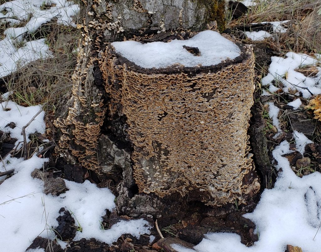
Hairy Curtain Crust (Stereum hirsutum)

A Deeper Dive into Fungi

I’ve covered fungi before. See Zooming in on Fungi and In Forests of Lichen. Those articles only scratched the surface, however. In this post, I’m going to dive a bit deeper, examining what we know (or suspect) about the evolution of fungi and their relationships to other life forms.

Just to refresh your memory, here’s a short excerpt from my first post on the topic:

…fungi are a separate kingdom of life — like the animal kingdom or the plant kingdom. They include yeasts, molds, and mushrooms. Actually, a mushroom is just the fruiting body (reproductive part) of the associated fungus. The main body consists of very thin filaments called hyphae (singular hypha). These hyphae are hidden in the substrate — that is, in the soil or inside the wood or whatever — that the mushroom is growing on. Collectively, the hyphae form an extensive network called a mycelium (plural mycelia).39

I mentioned in that excerpt that fungi are a kingdom. A kingdom, of course, is a way of grouping living things. But how, exactly, is a kingdom defined? To answer that, we’ll have to delve into a bit of taxonomy.

The Domains of Life and the Origin of Eukaryotes44 47

Biological taxonomy – the practice and science of categorizing living things – is constantly in flux. This isn’t because there’s anything inherently wrong with the science. Our classification systems are tools – ways of describing and keeping track of the organisms we’re talking about. As we learn more, we refine our tools.48

In modern taxonomy, the highest taxonomic level is the domain. All life is divided into three domains: Bacteria, Archaea, and Eukarya. Bacteria, as you probably know, are single-celled organisms. Archaea are also single-celled organisms. They look a lot like bacteria, but are quite different genetically. Both bacteria and archaea are prokaryotes. That is, their cells lack a nucleus.

Organisms whose cells do contain a nucleus make up the third domain, Eukaryotes. The purpose of a nucleus is to enclose the organism’s DNA, isolating it from the rest of the cell. This protects all that precious genetic material from being damaged by the complex chemical reactions going on outside the nucleus.

The nucleus is not the only organelle (specialized, membrane-bound subunit) to be found in a eukaryotic cell. There are many others. For example, there are the all-important mitochondria — the famous “powerhouses” of the cell. Mitochondria got their start when some single-celled organism — possibly an archaea — absorbed a bacterium. Perhaps it was trying to eat the bacterium. Or perhaps the bacterium was parasitic and it was trying to do the eating. Either way, something went wrong (or right, depending on your point of view). Neither cell was able to eat the other. Instead, they both survived, with the smaller cell continuing to live inside the larger one. The smaller cell even reproduced inside the larger cell. And when the larger cell itself eventually reproduced by splitting in two (fission), some of the bacterium’s progeny ended up in both of the new cells.

Over millions of years, the smaller cells evolved to specialize in producing energy, while the larger cells evolved to handle all the other functions — moving around, finding food, etc. In other words, the smaller cells became mitochondria and the larger cells became eukaryotes.

Probably the next most-famous organelle is the chloroplast. These are mostly found in plants. Chloroplasts have a similar origin to mitochondria, except that they’re all descended from a captured cyanobacterium. Cyanobacteria are photosynthetic, so they ended up specializing in capturing energy from sunlight, which they pass on to the host cell.

So, in summary, there are three domains, two of prokaryotes and one of eukaryotes. The following phylogenetic tree49 illustrates this.

A phylogenetic tree showing the three domains of life
Source: NASA Astrobiology Institute, found in an article
Author: This vector version: Eric Gaba (Sting, fr:Sting), Cherkash
Permission: Public domain

The Six Kingdoms of Life (or five or seven or four, depending on who you ask)50 51

The second highest taxonomic level is the kingdom. While the number of domains is pretty much agreed upon, the number of kingdoms is a bit more up in the air. Carl Linnaeus (1707–1778), who invented the first modern system of taxonomy, recognized only two kingdoms — plants and animals. (Actually, he included a third kingdom — minerals — but nobody much used that one.)

Later, when microscopic organisms were discovered, they were put into a third kingdom. Still later, as biologists learned more about how living organisms differ from one another, other systems were introduced with four, five, six, and even seven kingdoms. Today, the number of kingdoms depends on who you talk to, with some scientists wanting to do away with the term altogether. So, we should probably regard ‘kingdom’ as an informal term, used more for convenience than for any rigorous scientific reason. For our purposes, it doesn’t much matter which of the more modern definitions we go with, so I’ll pick one that’s popular here in the United States.52

The six kingdoms are (for our purposes):

  • Bacteria
  • Archaea
  • Protozoa
  • Chromista
  • Plantae
  • Fungi
  • Animalia

As we’ve already seen, the first two of these kingdoms — Bacteria and Archaea — consist of single-celled prokaryotic organisms; Protozoa and Chromista are single-celled eukaryotes; Plantae and Animalia (plants and animals) are multi-cellular eukaryotes; and Fungi are eukaryotes that are mostly multi-cellular, but include some single-celled types.

Why Fungi Get Their Own Kingdom53 54

For a long time, fungi were included in the plant kingdom. This is probably because, like plants, they are mostly sessile organisms. That is, they don’t move around. However, when genetic tools started coming into their own in the latter half of the 20th century, scientists analyzed the genetic code of fungi and discovered that, not only are they very different from plants, they are actually more closely related to animals!

Let’s examine some of the crucial differences between plants, animals, and fungi.

The most obvious difference is the way they make their living. Most plants photosynthesize. They convert light energy from the sun into chemical energy, which they store and use as fuel. Animals can’t make their own food. They have to get it from other organisms. They do this by engulfing organisms or parts of organisms or their remains. In other words, they eat stuff. Inside their bodies, animals use digestive juices to break down this organic matter into usable nutrients.

Like animals, fungi don’t make their own food. Unlike animals, they don’t break down their food inside their bodies. Instead, they secrete digestive enzymes into their surroundings. These enzymes break down organic matter in the environment and the fungi absorb the externally digested nutrients.

Beyond this fundamental difference in behavior, there are structural differences. Animal cell membranes contain cholesterol, whereas fungal cell membranes contain ergosterol and some other polymers. Animals don’t have cell walls (stiff, thick layers that cover the membranes). Plants and fungi do have cell walls, but plants use cellulose to stiffen them, while Fungi use chitin.55

Scientists have found that plants lack the gene sequences that are necessary for animal development; animals lack the gene sequences for plant development; and fungi have none of the sequences for plant or animal development. These findings underline the fundamental differences between the three kingdoms. Of course, these differences didn’t come about overnight. At some point, the three branches of life must have diverged from one another. More on that in a bit.

Anatomy of a Fungus56

Let’s take a break from taxonomy to discuss some anatomy. Since most fungi grow as hyphae, we’ll concentrate on those types. The following image shows a species of Penicillium with the various parts labeled.

Penicillium sp.
Author: Penicillium_labeled.jpg: Y_tambe
Derivative work: Adrian J. Hunter (talk)
This file has been extracted from another file
: Penicillium labeled.jpg
This file is licensed under the Creative Commons Attribution-Share Alike 2.5 Generic, 2.0 Generic and 1.0 Generic license.

Here’s the legend for the image:

  1. hypha – A thin filament. A web of these hyphae is a mycelium.
  2. conidiophore – “a specialized hyphal branch of some fungi that produces conidia”57
  3. phialide – A flask-shaped projection from the dilated part of the top of the conidiophore.
  4. conidia – Asexual spores (singular conidium).
  5. septa – Walls that divide the hyphae into compartments.

There are two kinds of hyphae –  septate and coenocytic. (A particular fungus will have one or the other type, but not both.) Septate hyphae are divided into compartments by their septa. Each compartment has one or more nuclei. Septa have pores that allow some of the contents to pass between compartments. Sometimes this even includes nuclei. Coenocytic hyphae have no compartments, so there’s no division into cells. For this reason, they are sometimes described as multinucleate supercells.

The following diagram shows a blow up of the tip of a septate hypha.

Cells at the tip of a hypha
Author: AHiggins12
This file is licensed under the Creative Commons Attribution-Share Alike 3.0 Unported license.

I won’t bother including a legend for the various parts. They are mostly the usual things found in a typical cell. If you want the details, though, you can click here for the Wikipedia article.

The most important part in the diagram is the septum, which is labeled 2.

Reproduction58

Reproduction in fungi gets complicated, and there’s plenty of variation between species. Basically, a fungus may reproduce either sexually or asexually. Asexual reproduction can occur either through vegetative spores (conidia) or mycelial fragmentation. Mycelial fragmentation just means that a part of the mycelium breaks off and grows into a separate fungus. A spore is a special cell that can develop into a clone of the fungus. The nice thing about spores is that they are easy to disperse. Some fungi produce mushrooms, which are basically specialized structures for dispersing spores.59

Sexual reproduction in fungi is complicated, confusing, and fascinating. I won’t go into a lot of detail, since it’s not all that relevant to what I want to cover here, but, basically, when hyphae from two different mycelial networks meet, they fuse, exchanging genetic material. The fused hyphae go on to branch and recombine, forming more mycelium and eventually producing spores containing crossed genetic material. Unlike most animals, fungi do not have sexes per se. Instead, they have mating types. According to Wikipedia, mating types are the “the microorganism equivalent to sex in higher organisms”. An organism might have two mating types, and only organisms of different types are able to successfully mate, but it will probably not be obvious which of the types should be the “female” and which should be the “male”. So the different types are typically designated by letters, numbers, or just “+” and “−”. Different groups of fungi have different sets of mating types and, in some groups, there are no types at all – any member of the group can mate with any other member.60 61

Fossil and Molecular Clues about Fungus Evolution62 63

We don’t have a lot of fossils of fungi, but there are some. What we know about fungal evolution is based on the few fossils that we do have, combined with genetic studies.

The earliest fossils that show some of the typical features of fungi are very old – they date back to around 2.4 billion years ago. These were bottom-dwelling ocean organisms that had filamentous structures suggestive of hyphae. These structures, like hyphae, were capable of recombining, forming a network.

In 2020, two interesting papers were published about possible fungus fossils. The first study, published in Nature, examines an approximately one billion year old fossil from the Canadian Arctic that may be that of a microscopic fungus. Tests done on this fossil showed evidence of chitin, while the same tests showed no evidence of cellulose. The morphology (form) of the fossil was suggestive of hyphae with septa and terminal spores. While not conclusive, this certainly sounds like a fungus.64

The second study, published in Science Advances, used a more rigorous set of chemical tests to examine a fossil from the Democratic Republic of the Congo that is 810 to 715 million years old. The fossil exhibits a woven mesh of filaments, and the study used three different techniques to show that the filaments contain chitin on their exteriors. Not many organisms produce chitin, and, of those that do, only fungi form the kind of mesh exhibited by this fossil.65

Studies from 2009 used comparisons of the rate of evolution in closely related groups to estimate the origin of fungi at 760 to 1060 million years ago (mya). These first fungi were probably aquatic and remained aquatic throughout the Paleozoic Era, 542–251 mya. They may have been similar to modern-day chytrids. Chytrids (division Chytridiomycota) are single-celled fungi. They are unusual among fungi in that they have mobile cells, called zoospores, that move using a posterior whip-like structure (flagellum).

Note that this seems to contradict the fact that the fossil organisms mentioned earlier already exhibited networks of filaments. This isn’t really a contradiction, though. Fungi might have originated as single-celled, chyrid-like organisms which evolved during this time period into multicellular mesh-like forms. The single-celled varieties would have continued to exist alongside them. In fact, phylogenetic studies indicate that most fungi lost the flagellum early in their evolutionary history, and most modern species don’t have them.

Loss of the flagellum may have been an adaptation to terrestrial or semi-terrestrial life. That is, the early, aquatic fungi were all single cells with flagella, and only evolved into flagellum-less, multicellular organisms while making the transition onto land. From what I can tell, it’s not clear exactly what kind of environment the organisms in the two studies from 2020 lived in. They may have lived on the margins of a body of water or in very shallow water.

The Last Common Fungal Ancestor

What I would really like to know, and the reason I started researching this post, is how it all started. What type of organism did fungi evolve from? In other words, what was the last common fungal ancestor (LCFA)?

According to Wikipedia, a sister group or sister taxon “comprises the closest relative(s) of another given unit in an evolutionary tree.66 This is an important concept, because, if two closely related groups share a particular characteristic, it’s likely that they inherited it from the last common ancestor of the two groups. So if we can find the sister group to fungi, it will give us important clues as to what the LCFA must have been like.

One paper I found, published in 2019,18 offered some fascinating insights into early fungal evolution. According to this paper, the sister group to Fungi is the Nucleariida. This is a group of amoebae that are commonly found in marine environments. As mentioned earlier, fungi are estimated to have originated 760 to 1060 mya. This is long before life began to colonize the land. These two facts together point to an aquatic origin for fungi. Most modern lineages of fungi are terrestrial; however, genetic studies have shown that all terrestrial fungi form a monophyletic group, meaning they all evolved from a single ancestor. Studies also suggest that the flagellum was lost only once. In other words, one lineage of fungi invaded the land, lost their flagella in the process, and evolved into mycelia.

Summarizing the Origin of Fungi

I found a number of scientific papers that discussed the origin of fungi. Trying to sort through all the details in these papers and present the results in a clear, concise manner proved quite difficult. But after a number of aborted attempts, I stumbled upon a website (which you can view by clicking here) that gives a nice summary.

According to this site, animals, many single-celled eukaryotes, and fungi make up a supergroup called Opisthokonta within Eukarya. All the single-celled organisms in this group have a single flagellum (whip-like filament) on the rear end of the cell. This flagellum is used for propulsion. While most fungi lost the flagella at some point in their evolutionary history, and most animal cells don’t have them, there are some holdovers. Animal sperm cells still have flagella, and the mobile gametes and spores of some types of fungi do, as well.

Opisthokonta is subdivided into two groups: Holozoa, which includes animals and their relatives, and Holomycota, which is made up of fungi and their relatives. Both groups include some examples of single-celled species, so it’s likely that multicellularity evolved independently in the two groups.

The closest living relatives to animals are choanoflagellates, a group of free-living single-celled and colonial eukaryotes that possess posterior flagella. Animals are believed to have evolved from something similar to this. In fact, sponges, which are some of the most primitive animals around, have cells that strongly resemble choanoflagellates.

Choanoflagellates and choanocytes
Choanoflagellate cells closely resemble sponge choanocyte cells. The beating of the flagella of these cells in the sponge draws water through it.
Author: Clark MA, Choi J and Douglas M
This file is licensed under the Creative Commons Attribution 4.0 International license.

Fungi, on the other hand, are most closely related to an order of single-celled eukaryotes called Nucleariida. (Maybe. See the next section) These are amoeba-like organisms that extend thread-like pseudopods,67 which they use to move themselves around and also to detect and consume food. According to this website, the main difference between fungi and nucleariids is that, whereas all fungi have chitin in their cell walls, nucleariids don’t. This suggests that the last common ancestor of Holozoa and the Holomycota probably had chiton in its cell walls, but this characteristic was lost in the nucleariid lineage.

Nuclearia thermophila (order Nucleariida)
Author: ja:User:NEON / User:NEON_ja
This file is licensed under the Creative Commons Attribution-Share Alike 2.5 Generic license.

Another Possibility

The idea that the nucleariids are the closest relatives of fungi doesn’t sit well with me. I don’t see how you get from thin pseudopods to flagella. They seem like very different things. Another paper,68 published in 2018, argues for a different sister group – the phylum Aphelida. Aphelids are parasites of algae. An aphelid starts out as a free-swimming single cell with a posterior flagellum. When this cell comes into contact with an algal cell, it attaches to it and forms a cyst. The cyst grows a tube that penetrates the cell wall of the host and injects its contents. The aphelid feeds on the host cell’s cytoplasm, growing and producing extra nuclei until it has completely filled the host cell. At this point, the aphelid divides into mobile spores which rupture the host cell and disperse to find new hosts.69 The paper reports on a genetic study suggesting that fungi evolved from an ancestor similar to an aphelid, possibly also parasitic on algae. Aphelids are phagocytes, meaning they engulf their food. Somehow, in the course of their evolution into fungi, this aphelid-like ancestor must have transitioned from phagocytosis to osmocytosis – secreting digestive enzymes into the environment and absorbing the already digested nutrients.

How this transition took place is unknown, but the 2019 paper70 mentioned earlier seems to suggest that the tube this parasite used to penetrate algal cell walls could have been the origin of the hyphae that fungi use for penetration and anchorage. Fungi are very good at penetrating solid surfaces and digesting cellulose and other biopolymers. This paper summarizes its conclusions by saying “This suggests that the ancestor of all fungi was probably a parasitoid of microalgae, with phagotrophic capabilities, showing both amoeboid and flagellar motility, and possessing chitin cell walls, at least in some life stages.”

This is interesting, but it sounds like we still don’t have a good idea how the ancestors of fungi transitioned from phagocytosis to osmocytosis.

Fungi and Plants71 72

While it’s true that fungi are genetically more closely related to animals than plants, that’s not to say that fungi have not been a major factor in plant evolution. Far from it. Fungi and plants formed close partnerships early on and it may have even been fungi that allowed plants to colonize the land.

Many fungi have formed symbiotic relationships with plants. A fungus’s hyphae will grow around a plant’s roots. In some cases, they will even penetrate the roots. The fungus and plant are then able to exchange water, nutrients, and sugars. A fungus’s mycelium is able to cover a much larger area that the root system of an associated plant, and the hyphae that form the mycelium are able to absorb nutrients, especially phosphorus, more efficiently than the roots. So the fungus passes on water and nutrients to the plant, greatly increasing its ability to absorb these things from its surroundings. In return, the plant manufactures sugars, which it passes on to the fungus. As I mentioned in one of my earlier posts, it has even been suggested that vascular plants (plants that have specialized conducting tissues to transport minerals and water) evolved from non-vascular plants when the hyphae of a fungus grew into the root system of a plant and never left.

In recent years, scientists have learned some remarkable things about mycorrhizal fungi (fungi that form a mutualistic relationship with plants). For one thing, it’s been shown that if a mycelial network is associated with a number of trees, and some of those trees are struggling to survive – maybe they aren’t getting enough sunlight to manufacture the sugars they need, for example – the fungus will take sugar from the trees that are doing well and transfer it to the trees that are struggling. Some authors have made it sound like this is an example of nature acting in an altruistic way – trees helping other trees. More likely, the fungus is just helping itself. Keeping its associated trees happy and healthy will be good for the fungus in the long run. Still, it’s a remarkable finding.

A Few More Landmarks in Fungal History

Most of us tend to ignore fungi. They just don’t seem all that important compared to plants and animals. This is certainly not true. Although they live mostly in the soil or inside other substrates and, as such, are not very visible, fungi are everywhere. They are quietly performing vitally important functions in the ecosystem.

That said, there have been times in Earth’s history when fungi would have been a lot more noticeable, had there been anybody there to notice. As I mentioned in an earlier post, around 470 to 360 mya, there existed a fungus called Prototaxites whose fruiting bodies grew to a height of 8 meters or 26 feet.73 Since the earliest trees evolved only around 370 mya,74 for most of its time on Earth, Prototaxites would have easily been the tallest organism around. Imagine actual forests of giant, tree-like fungi. I couldn’t find a recent artist rendering of Prototaxites that was in the public domain, but here’s a link to a reddit image. I don’t know how close it is to what these organisms actually looked like – probably not that all that close – but it should at least give you an idea how bizarre a land dominated by such ancient fungi must have been.

There has been at least one other time in prehistory when fungi were the most noticeable organisms around. In the aftermath of the asteroid impact that wiped out the non-avian dinosaurs, the land would have been full of rotting corpses of plants and animals – a virtual treasure trove for fungi that mostly feed on dead organic matter. Unsurprisingly, paleontologists have found evidence of a great increase in the prevalence of fungi at this time. Basically, all that death led to a huge fungal bloom, turning the land into “a massive compost heap”75, as one author put it.

Zooming in on Fungi

I absolutely refuse to make any jokes about fungi being “fun guys”. People who do that should be shot. Really. Fungi is the plural of fungus. The fungi are a separate kingdom of life — like the animal kingdom or the plant kingdom. They include yeasts, molds, and mushrooms.

Hairy Curtain Crust (Stereum hirsutum)
Near Lake Morena, Campo, CA 2/24/2019

Actually, a mushroom is just the fruiting body (reproductive part) of the associated fungus. The main body consists of very thin filaments called hyphae (singular hypha). These hyphae are hidden in the substrate — that is, in the soil or inside the wood or whatever — that the mushroom is growing on. Collectively, the hyphae form an extensive network called a mycelium (plural mycelia).

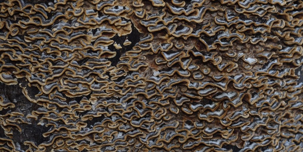
Hairy Curtain Crust (Stereum hirsutum)
Gills of the hairy curtain crust mushroom (Stereum hirsutum)

Because fungi don’t move around like animals, most people assume they’re closely related to plants. In fact, geneticists have found that fungi are more closely related to animals! This makes sense when you consider that fungi don’t use photosynthesis to make their own food like most plants do. Like animals, they feed off detritus (rotting organic matter) or off other organisms. The difference is, instead of ingesting their food and then digesting it inside their bodies, fungi secrete digestive juices into the surrounding environment. Their food gets broken down by these secretions and the fungi absorb the resulting nutrients.

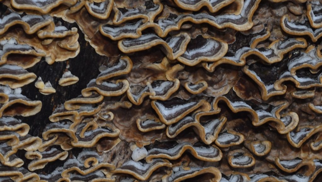
Hairy Curtain Crust (Stereum hirsutum)
Closeup

It’s long been known that fungi serve an important role in ecosystems as decomposers and recyclers, but, in recent years, scientists have uncovered other roles that are nothing short of mind-blowing. It turns out that most plants form mutualistic relationships with certain types of fungi. These fungi, called mycorrhyzal fungi, grow around and often into the roots of their host plant. The fungal network (mycelium) then acts as an extension of the plant’s root system, greatly increasing the plant’s ability to absorb water and minerals from the soil. In return, the plant supplies the fungus with carbohydrates, such as glucose and sucrose. If a plant is forced to grow without its usual fungal symbionts, it will be much smaller and less healthy, if it survives at all. Similarly, some mycorrhizal fungi will not survive unless a host plant is present.

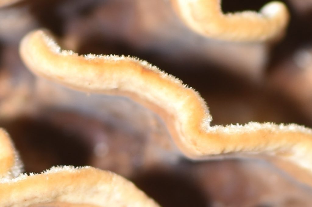
Hairy Curtain Crust (Stereum hirsutum)
Extreme closeup

That’s pretty interesting, but it gets better. It turns out that mycelia form a kind of communication and transportation network between plants. If a tree is not getting enough sunlight because it’s being shaded by larger trees, the fungal network may actually transfer food from the larger trees to the smaller one to supplement its diet. Remarkably, the fungi sometimes even transport food between different species of tree! All this is not meant to suggest that plants or fungi are actually intelligent; although, some people have suggested that there may be a kind of intelligence or even consciousness involved. That seems unlikely to me, but, either way, the relationship between plants and fungi is certainly much more complicated than anyone expected and we’re still learning more.

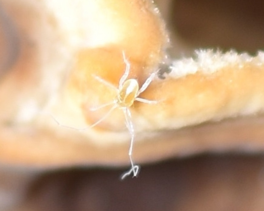
Mite
Mite

One final note… As I just mentioned, many fungi send their hyphae into the roots of their host plants. There is some evidence that the vascular systems of plants evolved from such a relationship. In this scenario, fungal hyphae would have formed a relationship with some non-vascular plant, living inside the plant just as fungal hyphae often do today. Eventually, these hyphae became a part of the plant, evolving into the vessels of the plant’s vascular system!

This isn’t quite as far-fetched as it might sound. It’s been shown that many of the organelles in plants and animals, such as chloroplasts, protoplasts, and mitochondria started out as free-living microbes that were ingested by a larger microbe. Instead of being digested, they somehow managed to survive inside the larger cell, eventually forming a symbiotic relationship with it. This “dual” cell eventually evolved into a multi-cell organism. This would be a nice subject for a future post, but, for now, suffice it to say that such relationships lend credence to the idea that the vascular system in plants could have started off as a network of invading fungal hyphae.

I’ve been talking mostly about the fungi that form mushrooms. I shouldn’t end this post without a brief mention of the other two types of fungus. Molds are similar to the mushroom fungi, but they have much smaller and, presumably, simpler reproductive structures. Yeasts are single-cell fungi. Some yeasts do form chains of cells, but they don’t form mycelial networks.

This post has been about an entire kingdom of living things, so, obviously, we’ve barely scratched the surface. I’m sure there’ll be many more posts to come about this fascinating group of organisms!

.