In one of my earlier posts, I covered woodlice — a group of crustaceans with a remarkable number of delightfully whacky common names. (See Cheesy Bobs.) In this article, we’ll take a look at another group of crustaceans with some very odd names — the amphipods.
Amphipods are closely related to woodlice. And like woodlice, they’re numerous, widespread, and largely ignored. If you spend a lot of time at the beach, you might know them as sand fleas or sand hoppers; if you’re an angler, you might use them as bait and know them as scuds, sideswimmers, or freshwater shrimp (they’re not shrimp); or, if you have an aquarium, you might know them as scuds and use them as fish food.1 Otherwise, there’s a good chance you don’t know them at all.
While woodlice are isopods, amphipods are… well, amphipods — members of the order Amphipoda. Like the isopods, they’re a diverse group, having more than 9,950 described species as of 2016. Many of them are marine organisms, but a large number live in freshwater, and a few have even adapted to life on land.2
My First Encounter with an Amphipod
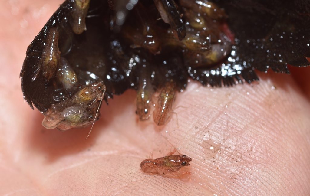
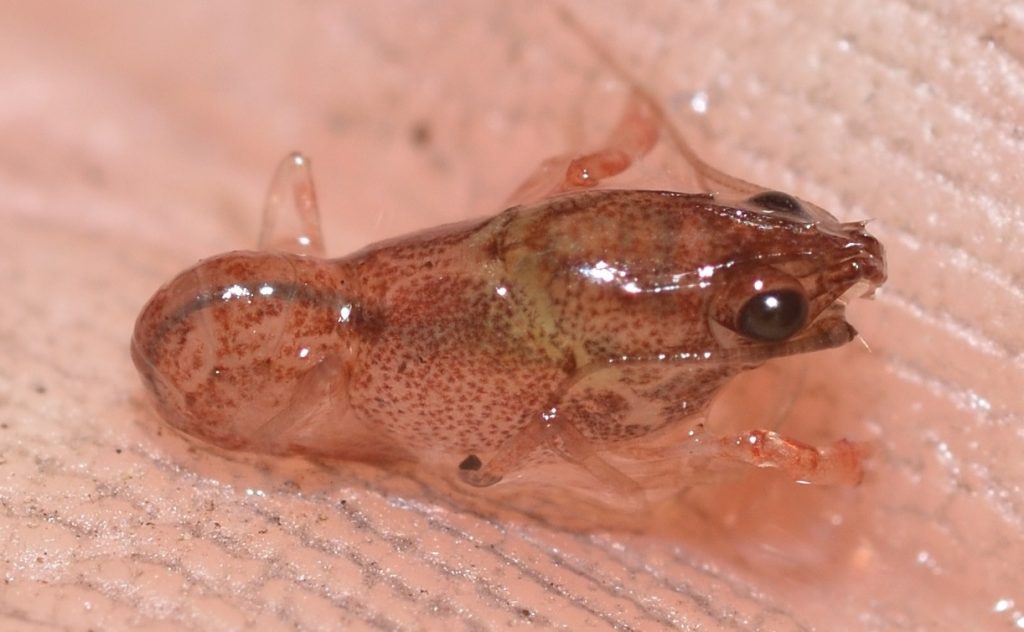
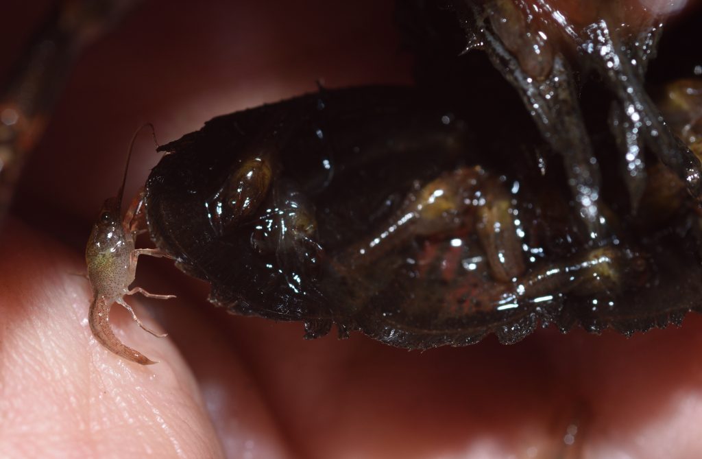
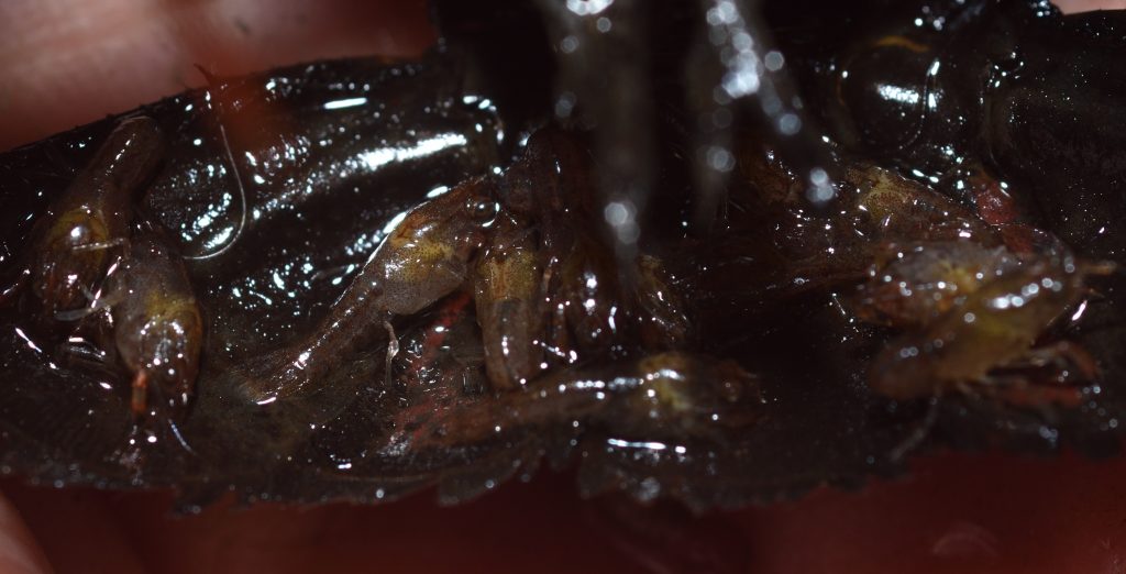
The first time I saw an amphipod, I had no clue what it was. I was examining some mud at the edge of a stream and saw this ‘thing’ that looked a like a really tiny shrimp. I was pretty sure it wasn’t a shrimp, though. For one thing, it wasn’t in the water.3 It was also hopping around a lot (which shrimp don’t typically do). I took some photos and posted them on bugguide.net, hoping that someone would identify it. I wasn’t disappointed.4 It was, I was given to know, an amphipod — something I’d never heard of before.

San Diego County. 8/19/17


Anatomy and Classification5
Figure 1 shows the body plan of an amphipod. If you read my previous posts about crayfish and woodlice, some of this anatomy might look familiar. That’s not surprising, given that they’re all crustaceans. They’re also all members of the largest class of crustaceans, the Malacostraca. Members of Malacostraca have bodies divided into head, thorax, and abdomen.6 Usually, the body is further divided into about 20 segments, but in amphipods, there are only 13. (Woodlice may have as few as 11.) 7 8 Note that amphipods lack a carapace — that hard covering or shell that protects the thorax of most crustaceans.9

This file is licensed under the Creative Commons Attribution-Share Alike 4.0 International license.
Attribution: © Hans Hillewaert
Within Malacostraca, amphipods and woodlice (but not crayfish) are included in the superorder Peracarida. The main characteristic of this group is the brood pouch or marsupium in females. The marsupium is an egg chamber formed by appendages called oostegites, which are attached to the first segments of the front pair of legs. The eggs are laid directly into the brood pouch. After hatching, the young remain in the pouch for several molts. When they finally emerge, they look like miniature adults and are called mancae.10 11
The brood pouches of woodlice, by the way, are probably one of the things that enabled them to adapt to a life out of the water. The marsupium is able to hold enough water to keep the eggs and newborns alive until they develop the book lungs that will enable them to extract oxygen from just a film of water. As long as they can keep their book lungs wet, woodlice can adapt to just about any terrestrial environment.12 13 Some amphipods seem to be following in the footsteps of the woodlice and using their marsupia to help them adapt to a terrestrial life.
In both amphipods and crayfish, the legs attached to the thorax are called pereiopods (or periopods). ‘Swimming legs’ called pleopods are attached to their abdomens, and flattened appendages called uropods are located at the tail end. In crayfish and shrimp, the uropods (one pair in crayfish and two in shrimp) fan out into wide tail flippers, which the animals can use to rapidly propel themselves backward. Amphipods have three pairs of uropods. They don’t fan out into a wide tail like they do on crayfish and shrimp, and they don’t use them to propel themselves backwards. However, many amphipods do use their tails to jump, so I suppose the uropods probably help with that.14 Notice that in a couple of the photos the amphipod has its tail curled under its abdomen, ready to flip down, propelling the animal up and away.
The pereiopods of amphipods are uniramous — that is, they aren’t branched. Compare this with crayfish, whose legs are all biramous (two branches). You can read my blog post on crayfish to learn more about biramous appendages.
One characteristic of amphipods that make them easy to recognize is that their bodies are flattened laterally. That is, they’re deeper from top to bottom than they are wide. This may have originally been an adaptation for swimming between dense vertical fronds of aquatic vegetation, such as kelp. (At least, I remember reading that somewhere. I really need to take better notes.) Most species usually swim with their bodies oriented on their sides — hence the name sideswimmers. I’m guessing they do this because of that odd lateral compression of their bodies. It seems like that would just naturally tend to make them tip over while swimming.
Some amphipods are blind, but most have large, flat compound eyes. Unlike crayfish and shrimp, the eyes are not on stalks.
Some Phylogenetic Trees15
For those of you who would like to get a bit more into the weeds, here’s a quick overview of the phylogeny of the class Malacostraca. Phylogeny, simply put, is “the evolutionary history of a kind of organism”.16 A phylogenetic tree is a branching diagram that shows that history.17
The one thing that scientists are pretty sure about regarding the group Malacostraca is that it’s monophyletic. This means that the group is composed of the common ancestor of the group and all its descendants. It’s a natural grouping, in other words.18 Beyond that, the relationships between the various members are not well-established. Before molecular genetics, phylogenetic trees were constructed using morphology or sometimes the biochemical characteristics of the different organisms, with the assumption that the more similar the organisms, the more closely related they must be. This, however, doesn’t take into account convergent evolution, which can lead to similarities even between organisms that are not at all closely related. Today, we can use genetic data, together with bioinformatics19, to determine genetic relationships. Even these techniques are not foolproof, though. Plus, they’re expensive, so not as many such studies have been done as we would like. Long story short, we still don’t know exactly how amphipods, crayfish, woodlice, and other malacostracans are related.
Figure 2 shows a number of phylogenetic trees that have been proposed for the group over the years. The first three were constructed using morphological data. The last three are based on molecular data, so I would tend to have more confidence in those, but, obviously, more studies need to be done.

Author: Squidonius (talk)
From the Wiki Commons page: “I, the copyright holder of this work, hereby release it into the public domain. This applies worldwide.
If this is not legally possible:
I grant any entity the right to use this work for any purpose, without any conditions, unless such conditions are required by law.“
A Bit of Microscopy
Recently, I bought a cheap USB microscope that attaches to my smart phone. (I’ve had several of these. They tend to break easily, but they’re good while they last.) I mainly bought it so that I could show my grandnephew some of the things you can see with a microscope.
The first thing I did was to collect some water, along with some bottom sediment, from the local river. The water contained a number of different organisms that were visible to the naked eye, including flat worms or planaria (which are awesome!), snails, and some kind of long thin worm. The snails laid eggs on the sides of the jar, which were pretty cool to look at, too. But the most numerous organisms, by far, were the amphipods. The water was full of them. We managed to catch a few with an eyedropper, so we were able to get a closer look. Here’s a video of one trapped in a drop of water. (The green oval is the petal of a duckweed plant.)
Here’s another video taken through the glass of the jar. A snail was gliding along the side, scraping it with it’s mouth, probably for algae. An amphipod decided to hitch a ride.
If you’d like to buy a microscope for a kid…
A lot of toy and hobby stores sell cheap, plastic microscopes for children. Don’t waste your money. These types of microscopes are hard to use, and you can’t see much with them. They won’t hold your child’s interest (or yours) for very long. A USB microscope is a much better investment. A lot of companies sell them. Make sure you get one that goes up to a magnification of 1000x. If the magnification is less than that, it can still be okay, but why skimp? The difference in price isn’t that much.
Click here to see the microscope that I bought on Amazon. You’ll probably also need a USB-C to USB adapter cable. Click here for a link for one of those. (I don’t have any affiliation with Amazon or with whatever company sells these products — they’re just the ones I purchased most recently. I’ve owned a couple of USB microscopes from another company and they worked just as well.)
You’ll need to download a free app on your phone. I would suggest CameraFi2. (I tried one other app, but it did something funky with the colors.)
My grandnephew is currently about four and a half years old. That’s probably around the minimum age for this kind of thing — his attention span is short, but not too short. By the way, don’t try to push your kid to be more interested. You can gently encourage their interest — “Hey, look at this! This is cool! — but young kids have short attention spans, and they want to explore things in their own way. You may be convinced that they would have more fun if they listened to you and followed your directions, but they don’t see it that way.
Here’s a photo of my USB microscope attached to my smartphone.

So what can you look at with your new microscope? Bugs are always a good choice. Of course, they tend to move around a lot if they’re alive, and maybe you just don’t have any handy. Another good choice is fabric. Focus the microscope on the sleeve of a shirt or on your pants leg (blue jeans are especially interesting). You’ll get a nice closeup view of the weave. Your kid should be impressed.
You can look at feathers. You can look at the hairs on your head or the fur on your cat. My nephew’s beard hair contained a lot of really thick black hairs that were very cool to see. You can look at the ridges on the tips of your fingers that form your fingerprints. You can look at leaves and flowers. You can… well, you get the idea.
The app allows you to take photos and videos. If you upload a good video to YouTube, be sure to post a link in the comments.
And now, back to scuds…
The Vast Variety of Amphipods
Amphipods, as we’ve seen, inhabit a wide variety of environments. Unsurprisingly, they have an equally wide variety of lifestyles. When it comes to feeding, there are herbivores, carnivores, omnivores, detritivores, and parasites. The family with the most species is Grammaridae, the members of which are called gammarids.20 The gammarids appear to be the most primitive of the amphipods, with other families showing more specialization in form, behavior, and ecological relationships.21 I think all the amphipods I’ve seen in my area have been gammarids.
While I haven’t encountered a lot of different kinds of amphipods in my own journeys, Wiki Commons has been a good source of public-domain photos of some of the more interesting species out there.
Hunchback Amphipods22
These beautiful amphipods go by the scientific name Iphimedia gibba. They are marine amphipods found only off the coast of South Africa. Hunchback amphipods are very small — at most 5 mm (0.20 in) long. As the name suggests, the body is hunched, which causes the head to face down.

Cape Peninsula, South Africa. Taken in 20 meters of water
Author: seascapeza
This file is licensed under the Creative Commons Attribution 3.0 Unported license.
Epimeriids23
The amphipod pictured below, Epimeria pandora, is in the family Epimeriidae. Members of this family are found in cold oceans all over the world. In fact, this particular species was found close to Antarctica. Epimeriids tend to be large amphipods. Their head-and-body length ranges from 0.8 to 8 cm (0.3–3.1 in). (Over three inches! That’s a big amphipod. Even 0.3 inches is pretty huge compared to the ones I see in my area.)

Bransfield Strait (near Antarctica)
Author:
d’Udekem d’Acoz, C., Verheye, M.L. 2017. Epimeria of the Southern Ocean with notes on their relatives (Crustacea, Amphipoda, Eusiroidea). European Journal of Taxonomy. 359: 1-553. DOI: 10.5852/ejt.2017.359
This file is licensed under the Creative Commons Attribution 3.0 Unported license.
They come in a variety of colors and some have striking patterns. Some, like the one pictured, have spines along their back and sides. These may help protect them against being eaten by fish, or they could serve as a form of camouflage, breaking up their outline.
Most epimeriids are predators or scavengers. They feed on various small, bottom-dwelling invertebrates. One species, Epimeria parasitica, may be parasitic on sea cucumbers.24
Lawn Shrimp and Beachhoppers25
Lawn shrimp (a.k.a, landhoppers) are not really shrimp, of course. They are amphipods in the family Talitridae. Unlike other amphipods, which don’t do well out of water, lawn shrimp are able to survive for long periods on land, and some species in the Southern Hemisphere are entirely terrestrial. That, by the way, is not to say that they’re only found there. Some of those south-of-the-equator species have been introduced here in California, where they can be found in urban and agricultural environments. In Golden Gate Park and other parks in San Francisco, for example, an Australian lawn shrimp, Arcitalitrus sylvaticus, is found in large numbers in the leaf litter of Eucalyptus and other trees.26
Most members of Talitridae are only semi-terrestrial, though. They inhabit damp sand beaches, where they feed on seaweed that has been tossed onto the beach by the tide. For this reason, they are often called beachhoppers.

Wakulla County, Florida 3/5/2017
Author: Andrew Cannizzaro
This file is licensed under the Creative Commons Attribution 2.0 Generic license.

I think this might be a fully terrestrial amphipod because 1) it doesn’t appear to be in water and 2) it was in New Zealand, which is in the Southern Hemisphere. The Wiki Commons post doesn’t list the species name, though, so I’m just guessing.
North Dunedin, Otago, New Zealand 8/30/2018
Author: Donald Hobern from Copenhagen, Denmark
This file is licensed under the Creative Commons Attribution 2.0 Generic license.
Skeleton Shrimp27
Also known as ghost shrimp, skeleton shrimp are (yet again) not shrimp. They’re amphipods in the family Caprellidae.

Collected from the Belgian continental shelf in 1999
Photographer: Hans Hillewaert
Attribution: © Hans Hillewaert
This file is licensed under the Creative Commons Attribution-Share Alike 4.0 International license.
In case you’re having a hard time believing these bizarre-looking creatures are really amphipods, figure 2 shows a generalized sketch of their anatomy, which you can compare to figure 1, which shows the anatomy of a more typical amphipod. As you can see, they have the same basic parts, arranged in the same basic order; although their abdomens are greatly reduced, while other parts have been stretched, distorted, or lost altogether.

Source: This file was derived from: Caprella mutica 3.jpg
Author: Obsidian Soul
This file is licensed under the Creative Commons Attribution-Share Alike 3.0 Unported license.
Most of the species in Caprellidae are predators, ambushing their prey in much the same way as a praying mantis. The amphipod will attach itself to a frond, adapting a pose that, together with its protective coloration, makes it resemble another frond. There it will remain, motionless, waiting for something small and delectable to wander by. Often, this will be a small worm or a protozoan, which it snatches up in its gnathopods.
Other caprellids have more varied diets, feeding on detritus, protozoa, diatoms, the larvae of crustacea, or smaller amphipods.

Author: Caprella_mutica_3.jpg: Hans Hillewaert
derivative work: Obsidi♠nSoul
This is a retouched picture, which means that it has been digitally altered from its original version. Modifications: adapt. The original can be viewed here: Caprella mutica 3.jpg: . Modifications made by Obsidian Soul.
This file is licensed under the Creative Commons Attribution-Share Alike 3.0 Unported license.
The weirdness of skeleton shrimp is not limited to their appearance — it extends to their mating habits. First, the female can only mate just after she has molted and before her new exoskeleton has hardened. I have no idea why this is or why in the world it would have evolved to be like this. Maybe there are other arthropods that have this trait, but I’ve never heard of any.
Second, in some species, the female will kill the male after mating. She does this by injecting it with venom from a claw on her gnathopod. I assume she does this because he represents an easy meal, helping to ensure her survival and the survival of any offspring that result from their mating. (From an evolutionary point of view, that’s a win-win.)
Whale lice, as you have probably surmised, are not actually lice. They are amphipods in the family Cyamidae, and they’re closely related to skeleton shrimp, which we covered in the last section. Whale lice live their entire lives as external parasites on whales, dolphins, or porpoises.

This amphipod was collected on 9/3/2006 from Humpback whale (Megaptera novaeangliae) stranded on the Belgian coast.
Photographer: Hans Hillewaert
Attribution: © Hans Hillewaert
This file is licensed under the Creative Commons Attribution-Share Alike 4.0 International license.
They attach themselves to places on the host body where they’ll be protected from water currents, including natural body openings and wounds. The lice range in size from 5 to 24 mm (0.2 to 1 inches). Most species parasitize a single species of whale, and almost every species of whale has a whale louse that’s specific to it! Remarkably, when it comes to sperm whales, the relationship is sex-specific. Cyamus catodontis lives only on male sperm whales, and Neocyamus physeteris lives only on females and calves.
In one sense, whale lice live up to their namesake (lice) — on average, a single whale is host to around 7,500 of them. Surprisingly, though, they seem to be commensals. That is, although they are parasites, they don’t do any significant harm to their hosts. They mostly eat the algae that settles on the host’s body. They also usually eat the dead skin as it flakes off. Whale lice often settle in open wounds, and they can cause some minor skin damage, but they don’t usually cause significant illness.
As already mentioned, whale lice spend their entire lives on their host — they have no free-swimming life stage. Given their large numbers on any single whale, you’d think they would be a constant source of irritation. I don’t know for sure that they’re not. But whales and whale lice have been evolving together for a very long time, so I suspect that whales have evolved to ignore them.
Hyperiids and Their Bizarre Eyes30
The amazing image below was created by Hans Hillewaert by focus stacking 35 images.31

Photographer: Hans Hillewaert
This file is licensed under the Creative Commons Attribution-Share Alike 4.0 International license.
Attribution: © Hans Hillewaert
The photo is of an amphipod named Hyperia galba in the family Hyperiidae. These amphipods live in the bells of various types of jellyfish. H. galba may or may not be an ectoparasite (a parasite that lives on the outside of the body of its host). Some scientists believe it’s a food parasite, but there’s no direct proof of this. It may seem like common sense that it must be parasitizing the jellyfish in some way, but it’s possible that the jellyfish just provides a good home for it. If nothing else, hitching a ride on a larger organism saves it the trouble of having to swim around on its own. In fact, some scientists maintain that the only truly parasitic amphipods are the whale lice (family Cyamidae) which I covered in the last section.32
But I’m wandering far afield of what I wanted to go in this section. In the section teaser, I mentioned something about bizarre eyes, and, as you can see in the photo, the eyes of H. galba don’t look much like the eyes of the other amphipods we’ve seen up to now. In fact, members of the family Hyperiidae sport a remarkable variety of innovative eyes. To quote Karen Osborn of the Smithsonian’s National Museum of Natural History, “With about 340 known species, hyperiid amphipods have every variation on eye shape that you can imagine and several you would never think of, from no eyes, to eyes that take up nearly half their body, eyes out on long needles, mirrored eyes, to eyes that can see in 360 degrees.”33 The reason this particular family of amphipods have evolved such weird eyes is that they live in the dark depths of the ocean — a place where what little light is available filters down from the surface or is provided by bioluminescence — light produced chemically in the bodies of various organisms. With such little light to work with, they’ve had to get creative, and creative they’ve certainly got! Click here to view the article that I based this section on. You don’t have to read it (although it is very accessible) — just look at some of the wonderful photos of the different hyperiid species.
The article focuses on one particular species, Paraphronima gracilis. This amphipod has 16 retinas in each eye! Its two large compound eyes envelop its head “like a space helmet”, to quote the article. Each eye is divided up into ommatidia (see The Story of Eyes: Part 1). Each ommatidium is topped by a facet that focuses the light. The light travels down a long, thin, fiber optic-like piece of guide tissue that ends at a segment of orange retina.
This isn’t too different from the compound eyes of most crustaceans, except that in all other cases, the retina forms a continuous sheet. Each ommatidium just focuses light one particular spot on the retina. So normally, an organism with two compound eyes would have two retinas — not 32! Scientists know of no other animal with compound eyes that splits up the retina in this way.
So why has P. gracilis evolved such an unusual eye design? It’s not completely understood, but this design seems to help them see better in dim light. Apparently, splitting up the retina in this way allows the eye to collect lots of light without sacrificing resolution — that is, the ability to see details — which is a problem that other light-adapted organisms have. I don’t know for sure how this works, but I would guess that splitting up the retina in this way prevents light falling on one segment of retina from stimulating neighboring sections. That is, it it better isolates the light collected by the individual ommatidia.
P. gracilis feeds on siphonophores, strange relatives of jellyfish that I would love to go in to detail about.34 Alas, this is an article about amphipods, so I’ll leave it to you to google them. Anyway, as it swims, P. gracilis points its eyes upwards, so that any siphonophores swimming above are silhouetted against the down-welling light. A small portion of each eye also points to the side. Scientists believe this allows them to detect the bioluminescence of animals swimming nearby.
Oh, and one other thing. Again, according to Osborne, “…we think they may be able to switch between grouping light signals at depth or at night when there is less light and not grouping signals when in shallower water where there is more light.” Amphipods are so cool!
A number of aquatic amphipods live in silken tubes. According to one paper, “The tube-building corophioid amphipod Crassicorophium bonellii produces from its legs fibrous, adhesive underwater threads that combine barnacle cement biology with aspects of spider silk thread extrusion spinning.”38 I won’t go into any detail here on silk production, since I already discussed it (at great length, of course) in another blog post. (See Silk…) I was surprised to learn about these silk-producing amphipods, though, as I hadn’t come across them when researching that previous article.
Flesh-Eating Amphipods39
Amphipods don’t often attack humans. In fact, it’s almost unheard of — almost. I did come across one bizarre instance of amphipods with a taste for human flesh.
The incident happened in Australia, because… of course it did. As reported in an article in The Guardian in 2017 (you can read the original here), a teenager was soaking his legs in the water at Brighton beach when the creatures “attacked”. The amphipods, a species in the family Lysianassidae, started feeding on the flesh around his ankles, causing a significant amount of bleeding, which the teen didn’t notice until he was walking away from the water. The article I linked to has a rather graphic photo of his bleeding legs. There’s also an interesting video. The kid actually ended up being taken to the hospital, because the bleeding didn’t stop when they wiped away the blood. (I don’t think he was in any real danger. His parents were just being cautious. Or maybe they were a little freaked out.)
Apparently, these amphipods will routinely nip at people in the water, but they don’t normally do any real damage. In this case, the boy kept his legs still for a long time, giving the wee animals a chance to dig in. It also helped that the cold water probably numbed his legs enough to prevent him from feeling their bites.
Plastic in the Depths40
You may have found the last section on “flesh-eating” amphipods rather disturbing. This section, I’m afraid, is also disturbing, but in a much more serious way.
I wasn’t kidding when I said amphipods are widespread — they’ve been found even in the Mariana Trench — the deepest ocean trench in the world. The maximum confirmed depth of the trench is about 10,984 meters or 6.8 miles. Unconfirmed measurements put it even deeper. I don’t know if any amphipods have been found at those depths, but the species we’re going to discuss here was found at 6,500 meters (over 3.7 miles), which is plenty deep. Its name is Eurythenes plasticus. A female specimen is pictured below.

Author: WESTON, J.N.J. et al. 2020. New species of Eurythenes from hadal depths of the Mariana Trench, Pacific Ocean (Crustacea: Amphipoda). Zootaxa 4851(1): 151–162.
Source: https://doi.org/10.11646/zootaxa.4748.1.9
This file is licensed under the Creative Commons Attribution 4.0 International license.
When this species was discovered, one of the specimens collected had plastic particles in its body. Plastic pollution is a very serious problem in our oceans. The plastic that ends up as trash in our landfills never decomposes, but it does break up into finer and finer particles, and many of those particles end up in the ocean, where they are ingested by marine life. Finding such particles in this new species of amphipod serves to highlight the seriousness of the problem. The plastic that our species has produced in such abundance and discarded so carelessly has found its way into the deepest depths of the ocean. There, some of it was ingested by an animal that we didn’t yet even know existed. The species name, plasticus, was chosen to draw attention to this disturbing fact.
Amphipods and the Origin of Wings41 42
“Insects got there first.” That’s a common refrain in articles about the evolution of flight, because insects were the first group of animals to evolve wings. So what does that have to do with amphipods, which are crustaceans — not insects — and definitely do not have wings?
Scientists used to think that insects evolved from myriapods (millipedes and centipedes). This presented a problem because those animals don’t have any structures that could have served as good starting points for the development of wings. So scientists figured that insects must have evolved wings from scratch. Then, in 2010, genetic studies revealed that insects were actually most closely related to crustaceans.
That’s where our amphipods come in. A paper published in 202043 describes research that compared the genetic instructions that control the development of legs in the amphipod Parhyale hawaiensis to the genetic instructions that control the development of legs in insects, such as the fruit fly Drosophila and the beetle Tribolium. All these organisms share five leg-patterning genes that control the development of the six leg segments farthest from the body wall.44 The researchers discovered this by disabling the genes and noting that the last six segments failed to develop.
Insects only have six segments in their legs, but crustaceans, such as Parhyale, have seven. One of the researchers, Heather Bruce, wondered where this seventh segment went when crustaceans evolved into insects. Bruce started digging into the literature, and she eventually unearthed an old idea from 1893 that the ancestors of insects had re-enforced their body wall by incorporating the part of the leg region closest to it.
It turns out that Parhyale shows evidence that one of its former leg segments was incorporated into its body wall. Insects, however, incorporated two leg segments. If you click here to open an article from “UChicago News” and then scroll down a bit, you’ll see some nice illustrations which compare the leg segments of crustaceans and insects and shows how some of those segments ended up as part of the body wall.
So how does this relate to wings? Bruce kept reading and managed to find another theory developed in the 1980s which suggested that little lobes on a pair of these incorporated segments eventually migrated onto the back where they evolved into the wings. The genes for the musculature for those segments were already there — they just had to be reactivated and adapted to produce flight muscles instead of leg muscles.
The issue probably isn’t settled yet — I’m sure many more studies will follow — but the evidence is mounting that a segment in the leg of crustaceans was first absorbed into the body wall and later extended out of the wall again in another direction and developed into a wing! There are more details from other papers that I could get into, such as how “proto-wings” might have served as thermo-regulators before being adapted for gliding and eventually flight, but I’ll leave it to you to explore the topic further, if you so desire. Happy googling!
Why Amphipods?
It’s always fun to introduce people to an animal they’ve never heard of. The crazy thing about amphipods, though, is that they’re not uncommon. Yes, most of them are pretty small, but they’re not usually microscopic. When I collected that first jar of river water to explore with my grandnephew, it was full of them, and they were very noticeable — as long as you bothered to look.
And that’s the crux of it — most of us just don’t look. We dream of travelling to distant, exotic lands, while never noticing the worlds in our own backyards. That’s one of the reasons I started this blog. I hope that by giving you a close look at various organisms — both common and uncommon — you’ll come to realize just how much there is to explore — right in your own backyard.