Tag Archives: crustacean

Here in the Age of Barnacles

I recently made a trip to Florida to visit family. Walking on a beach there, I came across a small piece of driftwood that was covered in marine life. There was a sponge, some globs of algae, and a bunch of barnacles. It was the barnacles that caught my attention.

I don’t spend a lot of time at the beach, so I don’t have much experience with barnacles. Prior to researching this article, I knew about as much about them as most people do, which is to say, next to nothing. I did know they were sedentary animals, so, zooming in for a close shot with my camera, I was surprised to see them moving. They were extending some kind of feelers and swaying back and forth on their stalks.

Here’s a video I recorded. I later identified them as Lepas anserifera, a species of goose barnacle.

I guess I shouldn’t have been so surprised. Sedentary just means rooted to one spot – it doesn’t mean the animal can’t move at all. A barnacle has to get its food somehow, and that means some kind of movement.

A weird, boring animal that most people know nothing about? Obviously, a blog post was in order. Speaking of not knowing much about them, it’s possible that some of you might not have even realized that the animals in the video were barnacles, if I hadn’t told you. You probably think of barnacles as looking like tiny volcanos. These are the acorn barnacles, and there are certainly plenty of them around. Here’s a photo of some of these more ‘traditional’ barnacles that I saw on a beach in California.

Little brown barnacles (Chthamalus dalli)
Swami’s Beach, Encinitas, CA. 5/8/2022

So what is a barnacle?1

A barnacle is an arthropod, like insects, spiders, and scorpions. More specifically, it’s a crustacean, which means it’s related to things like crabs, crayfish, and shrimp. That might come as a surprise. At first glance, a barnacle certainly doesn’t look very shrimp-like. However, if you were to pry apart the hard plates, you would see that the animal inside actually does look a bit like a shrimp lying on its back with its legs sticking up in the air. The legs are what it uses to sweep the water for small animals and other edible particles. The great Victorian zoologist Louis Agassiz described it beautifully – the barnacle, according to Agassiz, is “nothing more than a little shrimp-like animal, standing on its head in a limestone house and kicking food into its mouth.”2

Those legs, modified in the adult for filter feeding, are called cirri (singular cirrus). Here’s a diagram of a cross section of a typical acorn barnacle.

If you were to see a barnacle larva, its crustacean nature might be more apparent. A barnacle starts out life as an egg, which hatches into a nauplius (plural nauplii). Nauplius is the name used for the first larval stage of many types of crustaceans. In this stage, the larva has three pairs of legs and one eye. It has no thorax or abdomen – just a head and a telson. The telson is the last segment of the body – the one that would come after the abdomen, if it had one. The legs are attached directly to the head.3 4

The nauplius drifts around as part of the zooplankton,5 feeding and growing and occasionally molting. A barnacle’s last larval stage is called a cyprid or cypris. In this form, it doesn’t eat. It’s sole purpose is to find a nice place to settle down.

Source/photographer: English: Calman, W. T. (1911
) Life of Crustacea, Category:New York: the MacMillan Company
This is a photograph from the Freshwater and Marine Image Bank at the en:University of Washington. Materials in the Freshwater and Marine Image Bank are in the public domain. No copyright permissions are needed. Acknowledgement of the Freshwater and Marine Image Bank as a source for borrowed images is requested.
The nauplius of a bay barnacle (Balanus improvisus)
Author: Minami Himemiya
This file is licensed under the Creative Commons Attribution-Share Alike 3.0 Unported license.
The cyprid of a bay barnacle (Balanus improvisus)
This file is licensed under the Creative Commons Attribution-Share Alike 3.0 Unported license.

Cyprids have to be picky about where they settle. Since they are sedentary in their adult forms, it’s important to find a place where there’s plenty of food, hopefully not too much predation, and lots of other barnacles of the same species around for mating purposes. It searches its environment by probing with its antennules (the first pair of antennae in crustaceans6) and by sensing a chemical released by others of its kind.7 A cyprid can live from days to weeks, depending on the species. As it begins to exhaust its energy supply, it becomes less fussy and may settle for a site that’s less than optimal.

Once it picks a location, the cyprid cements itself headfirst to the spot, using a cement excreted from its antennae. Then it metamorphoses into its adult form. In the case of acorn barnacles, it grows four, six, or eight hard, calcareous8 plates to surround and protect it.

Anatomy of a Barnacle9

This is the best image I could find on Wiki Commons for barnacle anatomy. It would be perfect if the labels weren’t in French. Still, you can probably figure out what most of the terms mean.

Anatomy of an acorn barnacle
Author: Auguste Le Roux
Permission is granted to copy, distribute and/or modify this document under the terms of the GNU Free Documentation License, Version 1.2 or any later version published by the Free Software Foundation; with no Invariant Sections, no Front-Cover Texts, and no Back-Cover Texts. A copy of the license is included in the section entitled GNU Free Documentation License.
This file is licensed under the Creative Commons Attribution-Share Alike 3.0 Unported license.

In an acorn barnacle, two of the plates form an operculum, which may be recessed into the ring formed by the other plates. The operculum opens to let the barnacle extends its cirri for feeding. In species of barnacles that inhabit tidal areas, the operculum closes up when the tide goes out to keep the barnacle from drying out.

Click here to see an article that has a nice diagram of the arrangement of plates for a typical acorn barnacle. It also has a nice diagram of the internal anatomy. For an article that shows both acorn and goose barnacle anatomy, click here. The latter article is written for kids, but it’s worth a read. It sums up a lot of the stuff I cover here in a very simple and approachable way.

Classifying Barnacles10

As with many other groups of organisms, barnacle taxonomy has changed quite a bit over the years and is still changing. To avoid going down a taxonomic rabbit hole, I’m going to stick to the larger, hopefully more stable, groupings. I’m also going to stick mostly to the classification scheme presented by Wikipedia.11

As of 2021, barnacles are classified as follows:
Phylum: Arthropoda
Subphylum: Crustacea
Class: Thecostraca
Subclass: Cirripedia12

The most important grouping here is the subclass Cirripedia. Barnacles make up this subclass, so scientists often refer to them as cirripedes. Cirripedia is further broken down into three infraclasses: Thoracica, Rhizocephala, and Acrothoracica. Since these are super important, I’ll give each of them their own section.

Thoracica13

Infraclass Thoracica includes both the acorn barnacles and the goose barnacles. Goose barnacles are also called gooseneck barnacles or stalked barnacles. They attach to the substrate via a thick, muscular stalk called a peduncle. For this reason, they are also sometimes called pedunculate barnacles. Goose barnacles used to make up order Pedunculata, but researchers have now reclassified them into multiple orders (which I won’t get into).

I included a video of some goose barnacles that I saw in Florida at the beginning of this post. Here are a few still photos.

Goose Barnacles (Lepas anserifera) on a piece of driftwood
Lantano Municipal Beach, Lantano, FL 5/3/2022
Notice the tiny barnacle of the same species next to the two larger ones. When the cyprid first metamorphoses into its adult form, that form is very small. Probably much smaller even than this one.
This species of barnacle is wide, thin, and delicate-looking. Unlike the more robust species of goose barnacle pictured later in this section.
Extending the cirri
A close look at those cirri
If the barnacle was underwater, the cirri would be spread out, instead of clumped together.

Soon after I got back from Florida, I drove to Swami’s Beach in Encinatas, California, just 30 miles from where I live. I spent some time taking pictures of the gooseneck barnacles (Pollicipes polymerus) in the tide pools there. I included a few of photos below. As you can see, they are quite a bit more robust-looking than the goose barnacles I saw in Florida. This is because they’re a tidepool species and have to withstand the pummeling of the waves. Life attached to a piece of driftwood is usually a lot more laid back.

Gooseneck Barnacle (Pollicipes polymerus)
(There are a few little brown barnacles visible on the face of the rock on the left-hand side.)
Swami’s Beach, Encinitas, CA 5/8/2022
A closeup with my fingers for scale

Up close, the many small plates give these barnacles a rough, kind of “prehistoric” look, like they’d be right at home with a few dinosaurs splashing around in the surf.

In the right half of the photo, you can see a bunch of dark, almost black, mussels. The barnacles have to compete with the mussels for space on the rocks.
An even closer look. Notice the tiny scales on the stalk. This is a very well-armored barnacle.
Here, you can see some of the barnacles extending their cirri. The waves were occasionally crashing over them, so I guess it was worth their time to try to filter out plankton from the spray.

Barnacle Geese14 15 16

You might be wondering why pedunculate barnacles are called goose barnacles. It’s an interesting story. The word ‘barnacle’ was originally the name of a type of goose. The barnacle goose, Branta leucopsis, breeds mainly on the Arctic islands of the North Atlantic. From there, they migrate to Europe. Since they were never seen to nest or lay eggs in Europe (and it had not yet become generally known that birds migrate), people in the Middle Ages concluded that they must develop from barnacles through spontaneous generation.17

The idea was apparently inspired by the similarity in shape and color between these two otherwise wildly dissimilar organisms. Here’s a picture of some barnacle geese.

Barnacle geese (Branta leucopsis) in Naantali, Finland
Author: Diana Ringo
This file is licensed under the Creative Commons Attribution 4.0 International license.

And here’s one of some goose barnacles.

Goose barnacle (Lepas anatifera), taken in Playa Ponzos, Ferrol, Spain
Author: Franciscosp2
This file is licensed under the Creative Commons Attribution-Share Alike 3.0 Unported license.

You can see the similarity. As if more evidence were needed, the tuft of brown cirri that protrudes from a goose barnacle’s capitulum (shell) even resembles the down on an unhatched gosling. (I guess even people in the Middle Ages knew that other types of geese hatch from eggs.)

Since goose barnacles are often found on driftwood, people figured they must have been attached to branches that later fell into the water. One has to wonder how they explained the fact that they never found these branches while they were still attached to the tree, but, in any case, this was the explanation given by the archdeacon of Brecon, Gerald of Wales, in his literary work, Topographia Hiverniae,18 written around 1188.19

All this led to the English names “goose barnacle” and “barnacle goose”. Even the scientific name reflects this strange belief. The “anserifera” part of Lepas anserifera comes from the Latin word for goose: anser. As late as 1678, one writer wrote of “multitudes of little Shells; having within them little Birds perfectly shap’d, supposed to be Barnacles. (By “Barnacles”, he meant the birds.)

Lest we conclude that our ancestors were not very scientific in their thinking, here’s an old illustration that shows how they imagined the stages of development from barnacle to goose.


The process of development of the barnacle goose from the shell-like bud to the fully developed goose.
This file comes from Wellcome Images, a website operated by Wellcome Trust, a global charitable foundation based in the United Kingdom. Refer to Wellcome blog post (archive).
Source/photographer: https://wellcomeimages.org/indexplus/obf_images/8a/84/026cec1244a6a48b1c9bc2407f22.jpg
Gallery: https://wellcomeimages.org/indexplus/image/M0005646.html
Wellcome Collection gallery (2018-04-03): https://wellcomecollection.org/works/ndfpyunw CC-BY-4.0
This file is licensed under the Creative Commons Attribution 4.0 International license.

Some Catholics reasoned that since barnacle geese were “neither flesh, nor born of flesh”, they could get away with eating them on days when eating meat was forbidden by the church. Not every Catholic agreed. The Holy Roman Emperor Frederick II, for one, examined some barnacles, and, noting that there was no evidence of any embryonic birds in them, decided the whole thing was basically horse puckey. (Not his words.)20

While we’re on the subject, I just couldn’t resist including the following photo. It’s a taxidermy exhibit in the Zoological Museum in Copenhagen illustrating the myth of the barnacle goose.

Description: Rogue taxidermy illustrating the barnacle goose myth, Zoological Museum, Copenhagen.
Author: FunkMonk
This file is licensed under the Creative Commons Attribution-Share Alike 4.0 International license.

Moving on, acorn barnacles form the order Balanomorpha.21 They are a type of sessile barnacle. Sessile barnacles make up the group Sessilia. According to Wikipedia, “Sessilia is an unranked clade of barnacles, comprising the barnacles without stalks, or acorn barnacles. They form a monophyletic group and are probably derived from stalked or goose barnacles..” Don’t worry… I’m not going to try to break down that quote. I only include it because it mentions that acorn barnacles probably evolved from goose barnacles.

I showed you a photo of some acorn barnacles earlier in the post. Here’s another one, which shows a few more of the same species.

Little brown barnacles (Chthamalus dalli)
These are much smaller than either of the goose barnacles in the earlier photos.
Swami’s Beach, Encinitas, CA 5/8/2022
Notice how the barnacles are clumped together. A cyprid always does its best to settle next to other barnacles of the same species. Otherwise, its chances of reproducing are pretty slim.
A closeup of one of the barnacles. Notice how the outer shell is divided into plates. Because this species of barnacle lives in the tidal zone, the waves, along with their load of sand and other particles, are constantly eroding the barnacle’s shell, making it irregular and dirty-looking. Acorn barnacles that live below the tide line, tend to have much more pristine shells.
As I was placing these images on the page, I noticed that there are some individuals of what might be a different species of acorn barnacle in the lower right of this photo. It’s possible that they are just a more immature form of the same species, but the shells look very different in both shape and color, so idk.

Stalked and acorn barnacles are not the only members of infraclass Thoracica. Wart barnacles make up the group Verrucomorpha, which is either an order or a family (different articles give it different rankings). These are the asymmetrical barnacles. They differ from the other sessile barnacles in having the plates asymmetrically arranged, often giving them a lop-sided appearance.22 Then there’s the family Neoverrucidae. I’ve found very little info online about this group. Apparently, it contains the genus Neoverruca, whose members are found around hydrothermal vents.23

Rhizocephala24

The barnacles in infraclass Rhizocephala are all parasitic on other crustaceans. Some of them even parasitize thoracican barnacles. Rhizocephalans are fascinating, if somewhat gruesome. As nauplii, they don’t feed at all, although they do go through several molts. The cyprid also does not feed. In the (now deprecated) subgroup Kentrogonida, the female cyprid settles on a host and metamorphoses into a form called a kentrogon. The kentrogon has no appendages other than the antenna that it uses to attach to the host, and its body has no visible segmentation. Its only purpose is to inject a mass of cells called a vermigon into the host’s body. It does this through a stylet on its head. In the (also now deprecated) subgroup Akentrogonida, the kentrogon stage seems to have been lost. The cyprid injects the vermigon through one of its antennules.

Inside the body of the host, the vermigon grows into a network of root-like threads, called an interna, mostly centering on the host’s digestive system. It feeds by absorbing nutrients directly from the hemolymph (what passes for blood in crustaceans). The barnacle then grows an externa, a sac-like structure that extrudes from the host’s abdomen. This, as you might have guessed, is the reproductive part of the female. The following photos show the exerna of barnacles protruding from a couple of crabs.

A parasitic barnacle (Sacculina carcini) on a female swimming crab, from the Belgian coastal waters.
Attribution: © Hans Hillewaert
This file is licensed under the Creative Commons Attribution-Share Alike 4.0 International license.
A double infection of a parasitic barnacle (Sacculina carcini) on a male swimming crab, from the Belgian coastal waters
Attribution: © Hans Hillewaert
This file is licensed under the Creative Commons Attribution-Share Alike 4.0 International license.

The externa is located where the host’s egg sac would normally be. The barnacle castrates the host and chemically alters its behavior, so that it cares for the externa as if it were its own egg sac (even if the host happens to be male!)

So where is the male barnacle while all this is going on? The male cyprid is attracted by pheromones released by the female. What happens next depends on the particular the type of rhizocephalan barnacle. In the order Kentrogonida, the male enters the externa through a special oriface called the mantle departure, which leads to two receptacle passageways. Once inside, a very short-lived male stage called a trichogen emerges from the body of the cyprid through the antennule opening. The trichogen is the homologue of the female kentrogon, which is to say, they both evolved from the same structure. But in the male, this structure has been “reduced to an amoeboid unsegmented cuticle-covered mass of cells consisting of three to four cell-types”.25 The trichogen sheds its cuticle before reaching the end of the passageway. Having two receptacles, the externa can accept two males. This has the advantage of increasing the genetic variability of the offspring.

Other subgroups of Rhizocephala do variations on the same theme. For example, in the order Akentrogonida, there is no trichogen stage. The male cyprid injects its cell mass through an antennule directly into the externa.

In injecting this mass of cells into the externa, the male barnacle is basically injecting itself. And, since the injected stage consists of just sperm-forming cells, it follows that, in this stage of its development, the male rhizocephalan is the simplest known form of male in the animal kingdom.26

Acrothoracica27 28

Members of Acrothoracica are called burrowing barnacles because, in their cyprid forms, they bore into various kinds of calcareous29 materials, including mollusk shells, coral, crinoids, gastropod shells (both of live gastropods and those inhabited by hermit crabs), thoracican barnacles, bryozoans, limestone, and chalk. The cyprids have chitinous teeth that they use for boring. Once ensconced in their self-excavated burrows, they metamorphose into adult barnacles and live out the rest of their lives as filter feeders, much like acorn and goose barnacles. Burrowing barnacles, however, have no need of thick, calcareous protective plates – they have their burrows to protect them.

Individual acrothoracican barnacles are either male or female. The males are dwarfs and are sometimes found attached to the female or to the wall of her burrow.

Burrowing barnacles are smaller than most other barnacles, tending to be only a few millimeters long. Since many types live on the shells of other creatures, they can be thought of as having a symbiotic relationship with a host. In most cases, the relationship is probably a mutualism, in that the burrower benefits, but the host isn’t significantly harmed.30

Barnacle Sex

I’ve already briefly discussed sex in rhizocephalan and acrothoracican barnacles. In the thoracicans – the acorn and gooseneck barnacles – things work a little differently. For starters, most species are hermaphrodites. That is, every barnacle has both male and female sex organs. So when two thoracican barnacles meet, they can fertilize each other.

As I mentioned earlier in the article, when a cyprid is looking for a place to settle, it favors locations where there are already other barnacles of the same species. Both acorn and goose barnacles are found in large, crowded masses. Even so, you might think that internal sex would be out of the question for such sessile organisms, especially in the case of acorn barnacles. Not so. The thoracicans have come up with a very simple solution to the problem – the longest penises in the animal kingdom! – relative to body size, of course. A barnacle’s penis can be up to eight times its body length!31

I was so hoping to find a nice, copyright-free image of a barnacle penis, but I wasn’t able to find good one. Failing that, I found an article that contains an excellent photo of one. It’s in “Deep Sea News” and it’s by Dr. J. Matt Hoch. The article is short, very interesting, and has an awesome title. Click here to check it out. (Dr. Hoch, by the way, claims that a barnacle’s penis can stretch as much as ten times the length of its body. Since he has personally researched the subject, I suppose he should know what he’s talking about.)

But why settle for a photo when you can watch a video? Click here for a very strange music video about barnacle sex.

Whale Barnacles32

Barnacles don’t just attach themselves to nonliving substrates. They also attach to many types of marine animals. A few species of acorn barnacles have evolved to attach themselves to whales. Some species can live on a variety of whales. Others attach mostly to one species. Altogether, barnacles have managed to colonize around a dozen different whales species.

Some kinds of whale barnacle can grow quite large (for barnacles) – around the size of a small coffee cup. They also tend to be more spherical than cone-shaped. The following photo shows some barnacles attached to the underside of a dead humpback whale calf.

Barnacles attached to the ventral pleats of a humpback whale calf (photo taken during necropsy). Alaska, Peril Strait, Baranof Island. 2005 October 18
Author: Aleria Jensen, NOAA/NMFS/AKFSC.
Source: NOAA Photo Library: anim1032
This file is licensed under the Creative Commons Attribution 2.0 Generic license.
This image is in the public domain because it contains materials that originally came from the U.S. National Oceanic and Atmospheric Administration, taken or made as part of an employee’s official duties.

Here’s a close-up. You can see the cirri inside the opercula. (Notice the whale lice hanging out among the barnacles. See my earlier post: Scuds, Skeleton Shrimp, and Whale Lice.) Since whale barnacles don’t normally need to worry about drying out, at least during their host’s lifetime, their opercula can’t close up.

A close-up
Author: Aleria Jensen, NOAA/NMFS/AKFSC.
Source: NOAA Photo Library: anim1032
This file is licensed under the Creative Commons Attribution 2.0 Generic license.
This image is in the public domain because it contains materials that originally came from the U.S. National Oceanic and Atmospheric Administration, taken or made as part of an employee’s official duties.
This image was produced by cropping the original.

When it comes to whale barnacles, one unanswered question is how the tiny larvae – each one about the size of a grain of salt – find a whale to attach to. Scientists do have a hypothesis. Barnacle larvae go through several developmental stages. It can take up to two weeks before they’re ready to find a whale and settle down, so they need to have whales in close proximity during that time. It seems likely that the adult barnacles wait until the whales gather in coastal areas for breeding to release their larvae. When the larvae are finally ready to settle, a chemical signal probably leads them to a nearby whale. This signal could be a pheromone emitted by adult barnacles, or it could be some chemical that sloughs off the skin of the whale itself.

Once it finds a whale, the larva doesn’t attach just anywhere on the whale. A whale barnacle eats the same plankton as its host, so it needs to settle someplace where it will have full access to the clouds of plankton that the whale swims through. This generally means the whale’s forehead, its tail, or the leading edges of its flippers.

How does such a tiny larva find the correct spot? While we don’t know for certain, the process is believed to go something like this: The larva attaches itself temporarily to the whale’s body by squeezing a drop of sticky polymer out of one of its antennules. It then squeezes a drop out of the other antennule, sticking that one down. It releases the first antennule and swings itself to a new spot. Then it does it again. And again. In this way, the larva can slowly grope its way all over the whale until it finds where it wants to be. The article I got this from describes the larva as “leaving little gluey footprints” as it makes its way around. (I don’t know if researchers have actually found such traces or if the scientist quoted just liked the idea of a larva leaving little ‘footprints’.)

Whale barnacles are not easy to study. For starters, there’s not much grant money available. What little funding there is usually goes either to trying to figure out how to keep barnacles from attaching to the hulls of boats or trying to develop new adhesives based on the glues that barnacles use to attach themselves to substrates. The life history of whale barnacles just doesn’t seem to be much of a priority in the world at large.

Even if you manage to get funding for a project, you still have to find barnacles to study. You can’t just pry a barnacle off a living whale – they’re too firmly embedded in the whale’s flesh for that. About the only way to obtain them is to find a dead whale washed up on the shore and try to get to it before all the barnacles die. And since, as I already mentioned, whale barnacles can’t close up their opercula to keep from drying out, you have to get to the carcass pretty quick.

On top of all that, no one has figured out how to keep whale barnacles alive in the lab. Researchers in Japan were able to keep some larvae alive for about two weeks, but that’s as long as anyone has been able to do it.

So, unsurprisingly, there aren’t a whole lot of scientists studying whale barnacles. And this means there’s a lot that we still don’t know about them. Still, there are a few hardy researchers working to correct that. One such researcher is Larry Taylor. For his PhD thesis, Taylor somehow managed to convince his advisor to let him work on whale barnacles.

As an acorn barnacle grows, it adds concentric rings of material to the bottom of its shell wall. It gets this material by pulling dissolved oxygen and minerals from the water around it. These rings act much like tree rings, storing a record of the changing conditions the animal experiences. But unlike trees, which only add one ring of new wood per year, a barnacle can crank out a new ring in a much shorter amount of time – in as little as a few weeks.

Taylor’s idea for his thesis was to analyze the chemical composition of the rings in fossilized whale barnacle shells to try to determine the migration patterns of the whales that they had been attached to in life. This information could be important for a number of reasons. For one thing, scientists would like to know when and why baleen whales started migrating. One idea is that they started doing it around three million years ago when the ice age was making it too cold for whales in some of their northern habitats. It could also have changed the distribution of the plankton that the whales fed on. Taylor’s results could help to confirm or refute these ideas.

The changing climate would also have affected the distribution of plankton from season to season. This would be reflected in the migration patterns of the whales. So, having a record of these migration patterns could provide clues to how changes in climate affect the oceans.

Taylor was able to discern a pattern of changes in the rings that provided clear evidence for migration. This at least confirmed that baleen whales have been migrating for a very long time. It’s just a start, though. Taylor and his colleagues hope to analyze fossil barnacles from many other time periods, helping scientists build a better picture of how Earth’s climate has changed over millions of years.

In the interest of brevity, I’ve glossed over many fascinating details of Taylor’s work. For a more complete treatment, check out this article: What Whale Barnacles Know.

Move Over Finches33 34 35

Of all the many animals that Charles Darwin studied during his voyage on the Beagle, the Galápagos finches are probably the ones that get the most media attention. However, the influence of those finches on the development of Darwin’s ideas is probably exaggerated. (They didn’t even get a specific mention in “The Origin of Species”.) Surprisingly, the animal that played the biggest part in the development of his theory might very well have been – you guessed it – the barnacle.

On a beach in Chile, three years into his voyage on the Beagle, Darwin found a large shell that was peppered with hundreds of minute holes. Intrigued, he took it back to the ship and examined it with his microscope. He probed the holes with a needle and pulled out tiny, shell-less barnacles. These were the burrowing barnacles, infraclass Acrothoracica, that I described earlier in this post. Only, they didn’t have that name back then. In fact, Darwin was the first scientist to ever document them. He nicknamed the new type of barnacle Mr. Arthrobalanus. The name means “jointed barnacle”.

When Darwin returned to England in 1836, he was still a creationist. He had not yet formulated his famous theory. Then, Darwin read “Essay on the Principle of Population” by Thomas Malthus. The gist of the essay is that the human population always outgrows the available food supply, leading to a struggle for survival. Darwin realized that the same principle would apply to other organisms. It was this principle that led him to the idea of the survival of the fittest. If an organism varies from the other members of its species in a way that increases its chances of successfully reproducing, its lineage is more likely to continue. Further advantageous changes in some of its descendants could eventually lead to the emergence of a new species.

His theory was still far from fully developed, however. Within any given species, individuals seemed to be pretty uniform, at least to Darwin’s eye. This was a problem, because without variation, natural selection would have nothing to work with. Survival would be a matter of pure chance, not fitness. Not knowing how to solve this seeming flaw in his thinking, he wrote his ideas down, but didn’t publish them.

Years later, Darwin wrote a letter to his good friend, the botanist Joseph Hooker, sharing his theory on the origin of species. In response, Hooker pointed out that Darwin had never studied even one species in detail. This would make it difficult for him to get such a radical idea taken seriously by other scientists.

Hooker had a point. It may come as a surprise to some readers, but Darwin was an amateur scientist – a very serious, well-traveled, and accomplished amateur, to be sure – but he had never earned a degree in the natural sciences. And, as Hooker pointed out, he had never concentrated on one species. Darwin set out to remedy that. He decided to focus his efforts on his old friend, Mr. Arthrobalanus. He would spend a month or so describing the species, just to prove himself to the scientific establishment, and then he could get back to his theory.

To describe this new species, Darwin needed to know how it differed from other, previously described barnacle species. To that end, he began writing to museums, requesting specimens. He soon found out that existing barnacle studies left a lot to be desired. There were so many mistakes, gaps, and redundancies that Darwin realized he was going to have to clean up the mess himself, reclassifying everything, before he could even think about describing “his” barnacle.

He spent the next eight years doing just that. He requested more specimens, filling his study with piles of smelly boxes received from all over the world. He carefully dissected the specimens, studying them under his microscope, and making notes whenever he found something interesting. He worked so hard that his doctors warned him that we was ruining his health. He ignored them. He finally published his results in a series of exhaustive monographs in 1851 and 1854.36

Needless to say, eight years of tireless study made Darwin an expert on barnacles. (To this day, his monographs are still referenced.) And one result of becoming an expert was that he could now see all the subtle variations that his formerly untrained eye had missed. The variations were small, but they were enough for natural selection to work with. Training his eye to see variations in barnacles also made it easier for him to spot similar variations in other organisms. Moreover, by studying barnacles in such detail, Darwin was able to pick up on many of the tricks that evolution uses, such as repurposing an existing organ to serve a new function. There’s no doubt that the lowly barnacle played a great part in the development of Darwin’s ideas.

Just as with the last section, I’ve had to gloss over many fascinating details. If you want to read more about Darwin and his barnacles, click here. If you want to read much more, there’s an entire book, “Darwin and the Barnacle: The Story of One Tiny Creature and History’s Most Spectacular Scientific Breakthrough” by Rebecca Stott. (I actually haven’t gotten around to reading it myself, but it’s definitely on my list.)

The Age of Barnacles

Well, in the words of the great Britney Spears, “Oops, I did it again”. In doing research for this post, I read somewhere that Darwin had been so impressed with how widespread and successful barnacles were that he suggested our present time should be called the “Age of Barnacles”. I thought, wow, I could work that into a title for my blog post. I eventually settled on “Here in the Age of Barnacles”. Unfortunately, I neglected to make a note of where I read about Darwin’s remark, and, naturally, now I can’t find the source. So, I guess you’ll have to take my word for it.

A Little Something Extra

I’m going to leave you with a link to one of the freakiest videos I’ve ever watched. What happened to this guy was like something out of science fiction. Trigger warning: some of you might find this video disturbing. It does have some beautiful cinematography, though. Click here if your curiosity outweighs your squeamishness.

Scuds, Skeleton Shrimp, and Whale Lice

In one of my earlier posts, I covered woodlice — a group of crustaceans with a remarkable number of delightfully whacky common names. (See Cheesy Bobs.) In this article, we’ll take a look at another group of crustaceans with some very odd names — the amphipods.

Amphipods are closely related to woodlice. And like woodlice, they’re numerous, widespread, and largely ignored. If you spend a lot of time at the beach, you might know them as sand fleas or sand hoppers; if you’re an angler, you might use them as bait and know them as scuds, sideswimmers, or freshwater shrimp (they’re not shrimp); or, if you have an aquarium, you might know them as scuds and use them as fish food.19 Otherwise, there’s a good chance you don’t know them at all.

While woodlice are isopods, amphipods are… well, amphipods — members of the order Amphipoda. Like the isopods, they’re a diverse group, having more than 9,950 described species as of 2016. Many of them are marine organisms, but a large number live in freshwater, and a few have even adapted to life on land.20

My First Encounter with an Amphipod

The first time I saw an amphipod, I had no clue what it was. I was examining some mud at the edge of a stream and saw this ‘thing’ that looked a like a really tiny shrimp. I was pretty sure it wasn’t a shrimp, though. For one thing, it wasn’t in the water.26 It was also hopping around a lot (which shrimp don’t typically do). I took some photos and posted them on bugguide.net, hoping that someone would identify it. I wasn’t disappointed.37 It was, I was given to know, an amphipod — something I’d never heard of before.

A freshwater amphipod (possibly Hyalella sp.)
San Diego County. 8/19/17
Another view
A closeup showing one of the relatively simple compound eyes

Anatomy and Classification38

Figure 1 shows the body plan of an amphipod. If you read my previous posts about crayfish and woodlice, some of this anatomy might look familiar. That’s not surprising, given that they’re all crustaceans. They’re also all members of the largest class of crustaceans, the Malacostraca. Members of Malacostraca have bodies divided into head, thorax, and abdomen.39 Usually, the body is further divided into about 20 segments, but in amphipods, there are only 13. (Woodlice may have as few as 11.) 40 41 Note that amphipods lack a carapace — that hard covering or shell that protects the thorax of most crustaceans.42

Figure 1. Body plan of an amphipod
This file is licensed under the Creative Commons Attribution-Share Alike 4.0 International license.
Attribution: © Hans Hillewaert

Within Malacostraca, amphipods and woodlice (but not crayfish) are included in the superorder Peracarida. The main characteristic of this group is the brood pouch or marsupium in females. The marsupium is an egg chamber formed by appendages called oostegites, which are attached to the first segments of the front pair of legs. The eggs are laid directly into the brood pouch. After hatching, the young remain in the pouch for several molts. When they finally emerge, they look like miniature adults and are called mancae.43 44

The brood pouches of woodlice, by the way, are probably one of the things that enabled them to adapt to a life out of the water. The marsupium is able to hold enough water to keep the eggs and newborns alive until they develop the book lungs that will enable them to extract oxygen from just a film of water. As long as they can keep their book lungs wet, woodlice can adapt to just about any terrestrial environment.45 46 Some amphipods seem to be following in the footsteps of the woodlice and using their marsupia to help them adapt to a terrestrial life.

In both amphipods and crayfish, the legs attached to the thorax are called pereiopods (or periopods). ‘Swimming legs’ called pleopods are attached to their abdomens, and flattened appendages called uropods are located at the tail end. In crayfish and shrimp, the uropods (one pair in crayfish and two in shrimp) fan out into wide tail flippers, which the animals can use to rapidly propel themselves backward. Amphipods have three pairs of uropods. They don’t fan out into a wide tail like they do on crayfish and shrimp, and they don’t use them to propel themselves backwards. However, many amphipods do use their tails to jump, so I suppose the uropods probably help with that.47 Notice that in a couple of the photos the amphipod has its tail curled under its abdomen, ready to flip down, propelling the animal up and away.

The pereiopods of amphipods are uniramous — that is, they aren’t branched. Compare this with crayfish, whose legs are all biramous (two branches). You can read my blog post on crayfish to learn more about biramous appendages.

One characteristic of amphipods that make them easy to recognize is that their bodies are flattened laterally. That is, they’re deeper from top to bottom than they are wide. This may have originally been an adaptation for swimming between dense vertical fronds of aquatic vegetation, such as kelp. (At least, I remember reading that somewhere. I really need to take better notes.) Most species usually swim with their bodies oriented on their sides — hence the name sideswimmers. I’m guessing they do this because of that odd lateral compression of their bodies. It seems like that would just naturally tend to make them tip over while swimming.

Some amphipods are blind, but most have large, flat compound eyes. Unlike crayfish and shrimp, the eyes are not on stalks.

Some Phylogenetic Trees48

For those of you who would like to get a bit more into the weeds, here’s a quick overview of the phylogeny of the class Malacostraca. Phylogeny, simply put, is “the evolutionary history of a kind of organism”.49 A phylogenetic tree is a branching diagram that shows that history.50

The one thing that scientists are pretty sure about regarding the group Malacostraca is that it’s monophyletic. This means that the group is composed of the common ancestor of the group and all its descendants. It’s a natural grouping, in other words.51 Beyond that, the relationships between the various members are not well-established. Before molecular genetics, phylogenetic trees were constructed using morphology or sometimes the biochemical characteristics of the different organisms, with the assumption that the more similar the organisms, the more closely related they must be. This, however, doesn’t take into account convergent evolution, which can lead to similarities even between organisms that are not at all closely related. Today, we can use genetic data, together with bioinformatics52, to determine genetic relationships. Even these techniques are not foolproof, though. Plus, they’re expensive, so not as many such studies have been done as we would like. Long story short, we still don’t know exactly how amphipods, crayfish, woodlice, and other malacostracans are related.

Figure 2 shows a number of phylogenetic trees that have been proposed for the group over the years. The first three were constructed using morphological data. The last three are based on molecular data, so I would tend to have more confidence in those, but, obviously, more studies need to be done.

Figure 2. Some possible phylogenetic trees for Malacostraca
Author: Squidonius (talk)
From the Wiki Commons page: “I, the copyright holder of this work, hereby release it into the public domain. This applies worldwide.
If this is not legally possible:
I grant any entity the right to use this work for any purpose, without any conditions, unless such conditions are required by law.“

A Bit of Microscopy

Recently, I bought a cheap USB microscope that attaches to my smart phone. (I’ve had several of these. They tend to break easily, but they’re good while they last.) I mainly bought it so that I could show my grandnephew some of the things you can see with a microscope.

The first thing I did was to collect some water, along with some bottom sediment, from the local river. The water contained a number of different organisms that were visible to the naked eye, including flat worms or planaria (which are awesome!), snails, and some kind of long thin worm. The snails laid eggs on the sides of the jar, which were pretty cool to look at, too. But the most numerous organisms, by far, were the amphipods. The water was full of them. We managed to catch a few with an eyedropper, so we were able to get a closer look. Here’s a video of one trapped in a drop of water. (The green oval is the petal of a duckweed plant.)

Here’s another video taken through the glass of the jar. A snail was gliding along the side, scraping it with it’s mouth, probably for algae. An amphipod decided to hitch a ride.

If you’d like to buy a microscope for a kid…

A lot of toy and hobby stores sell cheap, plastic microscopes for children. Don’t waste your money. These types of microscopes are hard to use, and you can’t see much with them. They won’t hold your child’s interest (or yours) for very long. A USB microscope is a much better investment. A lot of companies sell them. Make sure you get one that goes up to a magnification of 1000x. If the magnification is less than that, it can still be okay, but why skimp? The difference in price isn’t that much.

Click here to see the microscope that I bought on Amazon. You’ll probably also need a USB-C to USB adapter cable. Click here for a link for one of those. (I don’t have any affiliation with Amazon or with whatever company sells these products — they’re just the ones I purchased most recently. I’ve owned a couple of USB microscopes from another company and they worked just as well.)

You’ll need to download a free app on your phone. I would suggest CameraFi2. (I tried one other app, but it did something funky with the colors.)

My grandnephew is currently about four and a half years old. That’s probably around the minimum age for this kind of thing — his attention span is short, but not too short. By the way, don’t try to push your kid to be more interested. You can gently encourage their interest — “Hey, look at this! This is cool! — but young kids have short attention spans, and they want to explore things in their own way. You may be convinced that they would have more fun if they listened to you and followed your directions, but they don’t see it that way.

Here’s a photo of my USB microscope attached to my smartphone.

USB microscope attached to a smartphone

So what can you look at with your new microscope? Bugs are always a good choice. Of course, they tend to move around a lot if they’re alive, and maybe you just don’t have any handy. Another good choice is fabric. Focus the microscope on the sleeve of a shirt or on your pants leg (blue jeans are especially interesting). You’ll get a nice closeup view of the weave. Your kid should be impressed.

You can look at feathers. You can look at the hairs on your head or the fur on your cat. My nephew’s beard hair contained a lot of really thick black hairs that were very cool to see. You can look at the ridges on the tips of your fingers that form your fingerprints. You can look at leaves and flowers. You can… well, you get the idea.

The app allows you to take photos and videos. If you upload a good video to YouTube, be sure to post a link in the comments.

And now, back to scuds…

The Vast Variety of Amphipods

Amphipods, as we’ve seen, inhabit a wide variety of environments. Unsurprisingly, they have an equally wide variety of lifestyles. When it comes to feeding, there are herbivores, carnivores, omnivores, detritivores, and parasites. The family with the most species is Grammaridae, the members of which are called gammarids.53 The gammarids appear to be the most primitive of the amphipods, with other families showing more specialization in form, behavior, and ecological relationships.54 I think all the amphipods I’ve seen in my area have been gammarids.

While I haven’t encountered a lot of different kinds of amphipods in my own journeys, Wiki Commons has been a good source of public-domain photos of some of the more interesting species out there.

Hunchback Amphipods55

These beautiful amphipods go by the scientific name Iphimedia gibba. They are marine amphipods found only off the coast of South Africa. Hunchback amphipods are very small — at most 5 mm (0.20 in) long. As the name suggests, the body is hunched, which causes the head to face down.

Hunchback amphipods (Iphimedia gibba) surrounded by ornate amphipods (Cyproidea ornata) on a bright red sponge.
Cape Peninsula, South Africa. Taken in 20 meters of water
Author: seascapeza
This file is licensed under the Creative Commons Attribution 3.0 Unported license.

Epimeriids56

The amphipod pictured below, Epimeria pandora, is in the family Epimeriidae. Members of this family are found in cold oceans all over the world. In fact, this particular species was found close to Antarctica. Epimeriids tend to be large amphipods. Their head-and-body length ranges from 0.8 to 8 cm (0.3–3.1 in). (Over three inches! That’s a big amphipod. Even 0.3 inches is pretty huge compared to the ones I see in my area.)

Epimeria pandora
Bransfield Strait (near Antarctica)
Author:
d’Udekem d’Acoz, C., Verheye, M.L. 2017. Epimeria of the Southern Ocean with notes on their relatives (Crustacea, Amphipoda, Eusiroidea). European Journal of Taxonomy. 359: 1-553. DOI: 10.5852/ejt.2017.359
This file is licensed under the Creative Commons Attribution 3.0 Unported license.

They come in a variety of colors and some have striking patterns. Some, like the one pictured, have spines along their back and sides. These may help protect them against being eaten by fish, or they could serve as a form of camouflage, breaking up their outline.

Most epimeriids are predators or scavengers. They feed on various small, bottom-dwelling invertebrates. One species, Epimeria parasitica, may be parasitic on sea cucumbers.57

Lawn Shrimp and Beachhoppers58

Lawn shrimp (a.k.a, landhoppers) are not really shrimp, of course. They are amphipods in the family Talitridae. Unlike other amphipods, which don’t do well out of water, lawn shrimp are able to survive for long periods on land, and some species in the Southern Hemisphere are entirely terrestrial. That, by the way, is not to say that they’re only found there. Some of those south-of-the-equator species have been introduced here in California, where they can be found in urban and agricultural environments. In Golden Gate Park and other parks in San Francisco, for example, an Australian lawn shrimp, Arcitalitrus sylvaticus, is found in large numbers in the leaf litter of Eucalyptus and other trees.59

Most members of Talitridae are only semi-terrestrial, though. They inhabit damp sand beaches, where they feed on seaweed that has been tossed onto the beach by the tide. For this reason, they are often called beachhoppers.

An unidentified member of Talitridae
Wakulla County, Florida 3/5/2017
Author: Andrew Cannizzaro
This file is licensed under the Creative Commons Attribution 2.0 Generic license.
Talitridae sp.
I think this might be a fully terrestrial amphipod because 1) it doesn’t appear to be in water and 2) it was in New Zealand, which is in the Southern Hemisphere. The Wiki Commons post doesn’t list the species name, though, so I’m just guessing.
North Dunedin, Otago, New Zealand 8/30/2018
Author: Donald Hobern from Copenhagen, Denmark
This file is licensed under the Creative Commons Attribution 2.0 Generic license.

Skeleton Shrimp60

Also known as ghost shrimp, skeleton shrimp are (yet again) not shrimp. They’re amphipods in the family Caprellidae.

A skeleton shrimp (Pariambus typicus)
Collected from the Belgian continental shelf in 1999
Photographer: Hans Hillewaert
Attribution: © Hans Hillewaert
This file is licensed under the Creative Commons Attribution-Share Alike 4.0 International license.

In case you’re having a hard time believing these bizarre-looking creatures are really amphipods, figure 2 shows a generalized sketch of their anatomy, which you can compare to figure 1, which shows the anatomy of a more typical amphipod. As you can see, they have the same basic parts, arranged in the same basic order; although their abdomens are greatly reduced, while other parts have been stretched, distorted, or lost altogether.

Figure 2. Generalized body plan of a skeleton shrimp
Source: This file was derived from: Caprella mutica 3.jpg
Author: Obsidian Soul
This file is licensed under the Creative Commons Attribution-Share Alike 3.0 Unported license.

Most of the species in Caprellidae are predators, ambushing their prey in much the same way as a praying mantis. The amphipod will attach itself to a frond, adapting a pose that, together with its protective coloration, makes it resemble another frond. There it will remain, motionless, waiting for something small and delectable to wander by. Often, this will be a small worm or a protozoan, which it snatches up in its gnathopods.

Other caprellids have more varied diets, feeding on detritus, protozoa, diatoms, the larvae of crustacea, or smaller amphipods.

A male Japanese skeleton shrimp (from Belgium)
Author: Caprella_mutica_3.jpg: Hans Hillewaert
derivative work: Obsidi♠nSoul
This is a retouched picture, which means that it has been digitally altered from its original version. Modifications: adapt. The original can be viewed here: Caprella mutica 3.jpg: . Modifications made by Obsidian Soul.
This file is licensed under the Creative Commons Attribution-Share Alike 3.0 Unported license.

The weirdness of skeleton shrimp is not limited to their appearance — it extends to their mating habits. First, the female can only mate just after she has molted and before her new exoskeleton has hardened. I have no idea why this is or why in the world it would have evolved to be like this. Maybe there are other arthropods that have this trait, but I’ve never heard of any.

Second, in some species, the female will kill the male after mating. She does this by injecting it with venom from a claw on her gnathopod. I assume she does this because he represents an easy meal, helping to ensure her survival and the survival of any offspring that result from their mating. (From an evolutionary point of view, that’s a win-win.)

Whale Lice61 62

Whale lice, as you have probably surmised, are not actually lice. They are amphipods in the family Cyamidae, and they’re closely related to skeleton shrimp, which we covered in the last section. Whale lice live their entire lives as external parasites on whales, dolphins, or porpoises.

A dorsal (top) view of a female Humpback louse (Cyamus boopis Lütken, 1870)
This amphipod was collected on 9/3/2006 from Humpback whale (Megaptera novaeangliae) stranded on the Belgian coast.
Photographer: Hans Hillewaert
Attribution: © Hans Hillewaert
This file is licensed under the Creative Commons Attribution-Share Alike 4.0 International license.

They attach themselves to places on the host body where they’ll be protected from water currents, including natural body openings and wounds. The lice range in size from 5 to 24 mm (0.2 to 1 inches). Most species parasitize a single species of whale, and almost every species of whale has a whale louse that’s specific to it! Remarkably, when it comes to sperm whales, the relationship is sex-specific. Cyamus catodontis lives only on male sperm whales, and Neocyamus physeteris lives only on females and calves.

In one sense, whale lice live up to their namesake (lice) — on average, a single whale is host to around 7,500 of them. Surprisingly, though, they seem to be commensals. That is, although they are parasites, they don’t do any significant harm to their hosts. They mostly eat the algae that settles on the host’s body. They also usually eat the dead skin as it flakes off. Whale lice often settle in open wounds, and they can cause some minor skin damage, but they don’t usually cause significant illness.
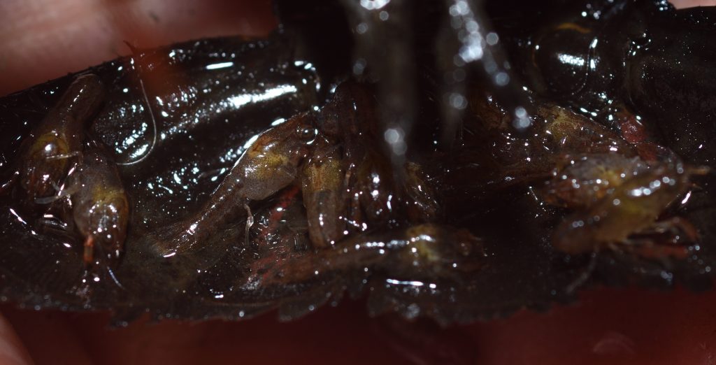
As already mentioned, whale lice spend their entire lives on their host — they have no free-swimming life stage. Given their large numbers on any single whale, you’d think they would be a constant source of irritation. I don’t know for sure that they’re not. But whales and whale lice have been evolving together for a very long time, so I suspect that whales have evolved to ignore them.

Hyperiids and Their Bizarre Eyes63

The amazing image below was created by Hans Hillewaert by focus stacking 35 images.64

Hyperia galba
Photographer: Hans Hillewaert
This file is licensed under the Creative Commons Attribution-Share Alike 4.0 International license.
Attribution: © Hans Hillewaert

The photo is of an amphipod named Hyperia galba in the family Hyperiidae. These amphipods live in the bells of various types of jellyfish. H. galba may or may not be an ectoparasite (a parasite that lives on the outside of the body of its host). Some scientists believe it’s a food parasite, but there’s no direct proof of this. It may seem like common sense that it must be parasitizing the jellyfish in some way, but it’s possible that the jellyfish just provides a good home for it. If nothing else, hitching a ride on a larger organism saves it the trouble of having to swim around on its own. In fact, some scientists maintain that the only truly parasitic amphipods are the whale lice (family Cyamidae) which I covered in the last section.65
But I’m wandering far afield of what I wanted to go in this section. In the section teaser, I mentioned something about bizarre eyes, and, as you can see in the photo, the eyes of H. galba don’t look much like the eyes of the other amphipods we’ve seen up to now. In fact, members of the family Hyperiidae sport a remarkable variety of innovative eyes. To quote Karen Osborn of the Smithsonian’s National Museum of Natural History, “With about 340 known species, hyperiid amphipods have every variation on eye shape that you can imagine and several you would never think of, from no eyes, to eyes that take up nearly half their body, eyes out on long needles, mirrored eyes, to eyes that can see in 360 degrees.”66 The reason this particular family of amphipods have evolved such weird eyes is that they live in the dark depths of the ocean — a place where what little light is available filters down from the surface or is provided by bioluminescence — light produced chemically in the bodies of various organisms. With such little light to work with, they’ve had to get creative, and creative they’ve certainly got! Click here to view the article that I based this section on. You don’t have to read it (although it is very accessible) — just look at some of the wonderful photos of the different hyperiid species.

The article focuses on one particular species, Paraphronima gracilis. This amphipod has 16 retinas in each eye! Its two large compound eyes envelop its head “like a space helmet”, to quote the article. Each eye is divided up into ommatidia (see The Story of Eyes: Part 1). Each ommatidium is topped by a facet that focuses the light. The light travels down a long, thin, fiber optic-like piece of guide tissue that ends at a segment of orange retina.

This isn’t too different from the compound eyes of most crustaceans, except that in all other cases, the retina forms a continuous sheet. Each ommatidium just focuses light one particular spot on the retina. So normally, an organism with two compound eyes would have two retinas — not 32! Scientists know of no other animal with compound eyes that splits up the retina in this way.

So why has P. gracilis evolved such an unusual eye design? It’s not completely understood, but this design seems to help them see better in dim light. Apparently, splitting up the retina in this way allows the eye to collect lots of light without sacrificing resolution — that is, the ability to see details — which is a problem that other light-adapted organisms have. I don’t know for sure how this works, but I would guess that splitting up the retina in this way prevents light falling on one segment of retina from stimulating neighboring sections. That is, it it better isolates the light collected by the individual ommatidia.

P. gracilis feeds on siphonophores, strange relatives of jellyfish that I would love to go in to detail about.67 Alas, this is an article about amphipods, so I’ll leave it to you to google them. Anyway, as it swims, P. gracilis points its eyes upwards, so that any siphonophores swimming above are silhouetted against the down-welling light. A small portion of each eye also points to the side. Scientists believe this allows them to detect the bioluminescence of animals swimming nearby.

Oh, and one other thing. Again, according to Osborne, “…we think they may be able to switch between grouping light signals at depth or at night when there is less light and not grouping signals when in shallower water where there is more light.” Amphipods are so cool!

Amphipod Silk68 69 70

A number of aquatic amphipods live in silken tubes. According to one paper, “The tube-building corophioid amphipod Crassicorophium bonellii produces from its legs fibrous, adhesive underwater threads that combine barnacle cement biology with aspects of spider silk thread extrusion spinning.”71 I won’t go into any detail here on silk production, since I already discussed it (at great length, of course) in another blog post. (See Silk…) I was surprised to learn about these silk-producing amphipods, though, as I hadn’t come across them when researching that previous article.

Flesh-Eating Amphipods72

Amphipods don’t often attack humans. In fact, it’s almost unheard of — almost. I did come across one bizarre instance of amphipods with a taste for human flesh.

The incident happened in Australia, because… of course it did. As reported in an article in The Guardian in 2017 (you can read the original here), a teenager was soaking his legs in the water at Brighton beach when the creatures “attacked”. The amphipods, a species in the family Lysianassidae, started feeding on the flesh around his ankles, causing a significant amount of bleeding, which the teen didn’t notice until he was walking away from the water. The article I linked to has a rather graphic photo of his bleeding legs. There’s also an interesting video. The kid actually ended up being taken to the hospital, because the bleeding didn’t stop when they wiped away the blood. (I don’t think he was in any real danger. His parents were just being cautious. Or maybe they were a little freaked out.)

Apparently, these amphipods will routinely nip at people in the water, but they don’t normally do any real damage. In this case, the boy kept his legs still for a long time, giving the wee animals a chance to dig in. It also helped that the cold water probably numbed his legs enough to prevent him from feeling their bites.

Plastic in the Depths73

You may have found the last section on “flesh-eating” amphipods rather disturbing. This section, I’m afraid, is also disturbing, but in a much more serious way.

I wasn’t kidding when I said amphipods are widespread — they’ve been found even in the Mariana Trench — the deepest ocean trench in the world. The maximum confirmed depth of the trench is about 10,984 meters or 6.8 miles. Unconfirmed measurements put it even deeper. I don’t know if any amphipods have been found at those depths, but the species we’re going to discuss here was found at 6,500 meters (over 3.7 miles), which is plenty deep. Its name is Eurythenes plasticus. A female specimen is pictured below.

Eurythenes plasticus — an amphipod found in the Mariana Trench
Author: WESTON, J.N.J. et al. 2020. New species of Eurythenes from hadal depths of the Mariana Trench, Pacific Ocean (Crustacea: Amphipoda). Zootaxa 4851(1): 151–162.
Source: https://doi.org/10.11646/zootaxa.4748.1.9
This file is licensed under the Creative Commons Attribution 4.0 International license.

When this species was discovered, one of the specimens collected had plastic particles in its body. Plastic pollution is a very serious problem in our oceans. The plastic that ends up as trash in our landfills never decomposes, but it does break up into finer and finer particles, and many of those particles end up in the ocean, where they are ingested by marine life. Finding such particles in this new species of amphipod serves to highlight the seriousness of the problem. The plastic that our species has produced in such abundance and discarded so carelessly has found its way into the deepest depths of the ocean. There, some of it was ingested by an animal that we didn’t yet even know existed. The species name, plasticus, was chosen to draw attention to this disturbing fact.

Amphipods and the Origin of Wings74 75

“Insects got there first.” That’s a common refrain in articles about the evolution of flight, because insects were the first group of animals to evolve wings. So what does that have to do with amphipods, which are crustaceans — not insects — and definitely do not have wings?

Scientists used to think that insects evolved from myriapods (millipedes and centipedes). This presented a problem because those animals don’t have any structures that could have served as good starting points for the development of wings. So scientists figured that insects must have evolved wings from scratch. Then, in 2010, genetic studies revealed that insects were actually most closely related to crustaceans.

That’s where our amphipods come in. A paper published in 202076 describes research that compared the genetic instructions that control the development of legs in the amphipod Parhyale hawaiensis to the genetic instructions that control the development of legs in insects, such as the fruit fly Drosophila and the beetle Tribolium. All these organisms share five leg-patterning genes that control the development of the six leg segments farthest from the body wall.77 The researchers discovered this by disabling the genes and noting that the last six segments failed to develop.

Insects only have six segments in their legs, but crustaceans, such as Parhyale, have seven. One of the researchers, Heather Bruce, wondered where this seventh segment went when crustaceans evolved into insects. Bruce started digging into the literature, and she eventually unearthed an old idea from 1893 that the ancestors of insects had re-enforced their body wall by incorporating the part of the leg region closest to it.

It turns out that Parhyale shows evidence that one of its former leg segments was incorporated into its body wall. Insects, however, incorporated two leg segments. If you click here to open an article from “UChicago News” and then scroll down a bit, you’ll see some nice illustrations which compare the leg segments of crustaceans and insects and shows how some of those segments ended up as part of the body wall.

So how does this relate to wings? Bruce kept reading and managed to find another theory developed in the 1980s which suggested that little lobes on a pair of these incorporated segments eventually migrated onto the back where they evolved into the wings. The genes for the musculature for those segments were already there — they just had to be reactivated and adapted to produce flight muscles instead of leg muscles.

The issue probably isn’t settled yet — I’m sure many more studies will follow — but the evidence is mounting that a segment in the leg of crustaceans was first absorbed into the body wall and later extended out of the wall again in another direction and developed into a wing! There are more details from other papers that I could get into, such as how “proto-wings” might have served as thermo-regulators before being adapted for gliding and eventually flight, but I’ll leave it to you to explore the topic further, if you so desire. Happy googling!

Why Amphipods?

It’s always fun to introduce people to an animal they’ve never heard of. The crazy thing about amphipods, though, is that they’re not uncommon. Yes, most of them are pretty small, but they’re not usually microscopic. When I collected that first jar of river water to explore with my grandnephew, it was full of them, and they were very noticeable — as long as you bothered to look.

And that’s the crux of it — most of us just don’t look. We dream of travelling to distant, exotic lands, while never noticing the worlds in our own backyards. That’s one of the reasons I started this blog. I hope that by giving you a close look at various organisms — both common and uncommon — you’ll come to realize just how much there is to explore — right in your own backyard.

Crayfish Journeys

When Charles Darwin published On the Origin of Species in 1859, he argued the case for evolution by presenting what he hoped would be an overwhelming amount of evidence. I think most scientists would agree that he succeeded admirably. Nevertheless, Darwin had to contend with the fact that much of the scientific knowledge we take for granted today didn’t exist at the time. Obviously, he knew nothing about the genetic code. Even the basics of heredity were almost unknown. (Gregor Mendel had made a great start, but his work was pretty much ignored until decades later.) So the mechanism underlying natural selection was a black box to him. This was a problem, but not an insurmountable one. Darwin simply had to present evidence that there must be some such mechanism; he didn’t actually need to find it.

A more serious problem was continental drift, a.k.a plate tectonics. There were many cases of identical fossils found on widely separated bodies of land with no obvious way that the organisms could have gotten from one body to the other. For instance, there were fossils of animals found on the west coast of Africa that were identical to fossils found on the east coast of South America. Today the explanation is obvious. We know that all the continents used to be joined together into one vast super-continent called Pangaea. Pangaea eventually broke up and the fragments drifted apart to form separate land masses. The animals found in both South America and Africa evolved there when the two continents were connected.

Darwin knew nothing about continental drift. He didn’t even know about radioactive elements (whose decay heats up the Earth’s mantle and produces the massive convection currents that move the continents.) No one at the time seriously believed that the continents could be moving. So Darwin was forced to come up with alternative explanations for such fossils. When I read On the Origin of Species as a young man, I remember thinking that the suggestions he put forth were not very convincing.66 71

So what does all this have to do with crayfish? Crayfish are basically small, freshwater lobsters, but, unlike their marine relatives, they can’t survive in the open ocean. Nevertheless, there are native crayfish found on all the continents except Africa and Antarctica. So crayfish are a textbook example of a population of organisms that evolved when all the continents were joined together and were broken up into separate populations when Pangaea broke up. Those populations continued to evolve after the breakup. I’ll be discussing this in more detail later, but, first, let’s get to know our subject a little better.

Red swamp crayfish (Procambarus clarkii)
Sweetwater River near Jamul, CA 8/19/2017
Claw (chela) of red swamp crayfish
Sweetwater River near Jamul, CA 8/19/2017

Crayfish, Crawfish, or Crawdad?

The word crayfish probably comes from the Frankish word krebitja, which is a diminutive of the word for crab.78 This evolved into the Old French word for crayfish — “escreviss” — and eventually into our version of the word. Crayfish are also called crawfish or crawdads in some localities. “Crawfish” is obviously just a variation on the pronunciation. The origin of “Crawdad” is a bit more mysterious. It could be related to the use of “dad” in “doodad”, but no one seems to know for sure.

Crayfish are crustaceans, which means that they are related to such animals as crabs, shrimps, prawns, and woodlice. Crustaceans are, in turn, a subgroup of Arthropods. Like other arthropods, such as insects and spiders, crustaceans have exoskeletons. Unlike other arthropods, they have biramous limbs. A biramous limb is an apendage that branches into two parts. The bottom part is often (but not always) used for locomotion, while the upper part is used for other purposes — gills, for instance.

Crayfish evolved from lobsters. Some authors treat them as a type of lobster; some don’t. It’s not important from a scientific point of view, since “crayfish” and “lobster” are both common names. Historically, crayfish have generally been classified as belonging to three families — Astacidae, Cambaridae, and Parastacidae. Together, these three families can be referred to as Astacida. So Astacida is the scientific name for crayfish as a group.79 The study of crayfish is called Astacology. 80

Crayfish in California

There is only one extant (not extinct) species of crayfish that’s native to California. That’s the Shasta crayfish (Pacifastacus fortis). It lives only in Shasta County in northeast California. It’s threatened by habitat loss and also by competition from an invasive species, the signal crayfish (Pacifastacus leniusculus).81 82

Another invasive species that’s found in California, including here in San Diego County, is the red swamp crayfish (Procambarus clarkii), which is in the Cambaridae family. P. clarkii is native to the southern and southeastern United States and northern Mexico. Its native range doesn’t extend much farther west than Texas, but it’s been introduced to many other places in the U.S. and around the world. In some places, it’s become a serious pest.83

The red swamp crayfish is the most widely farmed species of crayfish in the world. It’s used as food, as bait for fishermen, in the aquarium trade, and as research animals in schools and laboratories. All this accounts for its wide dispersal. In some cases, P. clarkii has been deliberately introduced into an area in hopes of an economic return. In other cases, schools or individuals have released them into the wild just because they didn’t know any better.

P. clarkii is the only crayfish that I’ve encountered in southern California. All the photos that I’m including with this post, unless otherwise noted, are photos of this species and all were taken of individuals that I found in the Sweetwater River near Jamul, CA.

Crayfish Anatomy84

Crayfish are a popular subject for dissection in high school and college biology classes. As a result, there are some excellent resource videos online. The easiest way to learn crayfish anatomy is to watch some of these videos. Click here for a good anatomy video. And here’s a couple of links to videos that actually show the dissection process: Crayfish Dissection 1, Crayfish Dissection 2

Just in case you don’t feel like watching a bunch of videos, I’ll give you a quick lesson. Figure 1 shows the basics of crayfish external anatomy.

Figure 1 – This image is in the public domain. It was downloaded from Wiki Commons at this url.

For the internal anatomy, refer to figure 2 below:

Figure 2 – This image is in the public domain. It was downloaded from Wiki Commons at this url.

Figure 1 shows a female crayfish in the genus Cambarus. Other crayfish species should have similar anatomies. I don’t know what species was used for Figure 2.

Crayfish are decapods, a word which means “ten-footed”.85 In the case of crayfish, this refers to the five pairs of walking legs or pereiopods. The first pair — the pair with the big claws — are also called chelipeds.

On the sides of the thorax, hidden by the carapace, are the gills. Each gill looks like a feathery appendage, but it’s actually the upper branch of one of the walking legs. Unfortunately, the gills are not shown in the illustrations, but if you watch the dissection videos, you’ll get a good look at them.

The head and thorax are joined and together make up the cephalothorax. This is covered on the top and sides by a shell called the carapace. The carapace includes the rostrum — the pointed “nose” or beak at the front of the head.

Crayfish have two pairs of antennae — one long pair and another much shorter pair. The shorter pair are called antennules. Each antennule is branched. That is, they are biramous appendages like the walking legs. Arthropods are believed to have evolved from annelids — segmented worms with non-branched (uniramous) appendages on each segment. As new species evolved from this ancestor, segments were added, lost, or fused, and became specialized in various ways. The appendages went on to differentiate into new forms that served different purposes. Some, for instance, evolved into mouth-parts and others became antennae.

In the crustaceans (or maybe in an earlier ancestor), the appendages evolved to be biramous. It’s likely that two segments were fused into one segment with two pairs of appendages and, later, the two pairs of appendages fused to become one biramous pair.86

Note the “pleopods” on the “tail” (really the abdomen) of the crayfish. These are also called swimmerets. These are thin, flimsy-looking, biramous appendages that are used by the female to hold her eggs to the bottom of her abdomen after she lays them. Despite the name, they’re not actually used for swimming, although they may help to keep water flowing over the gills. They may also help in steering.

In the male crayfish, the first pair of pleopods are enlarged and are used for transferring sperm during copulation. These more specialized pleopods are called gonopods.

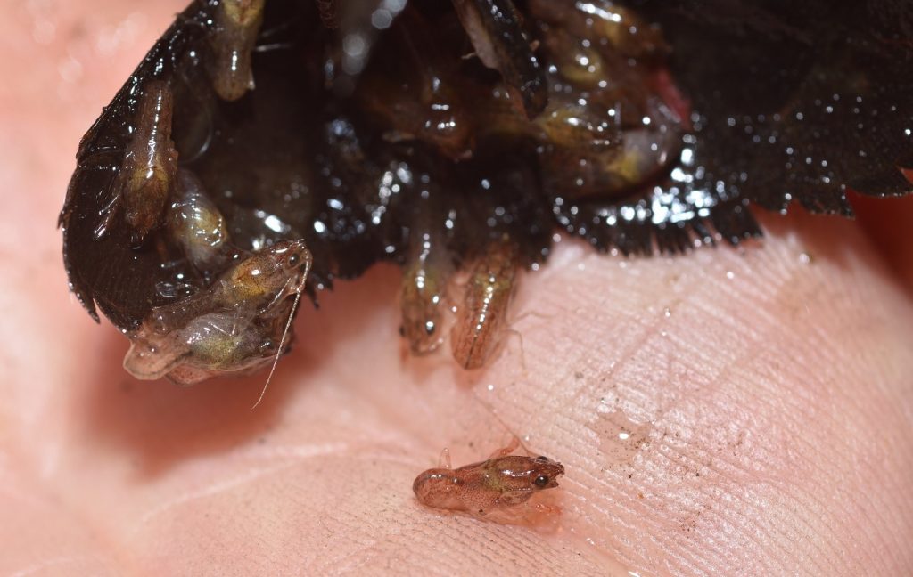
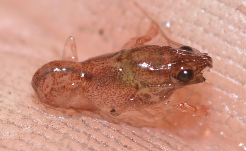
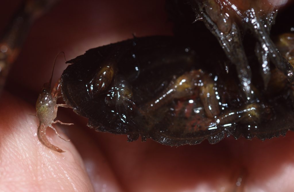
The tail is made up of the telson (which is actually the last segment of the body) and four paddle-like structures called uropods, two on each side of the telson. When frightened, a crayfish will often propel itself rapidly backwards by flipping its tail in an arc underneath its body.

On the underside of the head, just a little farther back than the eyes, are the openings to the nephridia. The nephridia (there are two of them) are glands that are present in many invertebrates that function like kidneys — they eliminate toxic substances and waste products from the urinary and circulatory systems.87 In crustaceans, the nephridia are also called green glands, antennal glands, or maxillary glands. The following two photos clearly show the openings of the green glands. The actual glands are not visible in the above illustrations, but one of the openings is labeled in each figure.

Red swamp crayfish (Procambarus clarkii)
The pink oriface is the opening of the green gland.
Sweetwater River near Jamul, CA 10/4/2019
Red swamp crayfish (Procambarus clarkii)
Showing the openings of the green glands.

In the above photos, you can see the largest of the three pairs of maxillipeds. These are appendages used to manipulate food. They’re also labeled in figures 1 and 2. The photo below gives you a more complete view.

Red swamp crayfish (Procambarus clarkii)

Also visible in the above photo are the mandibles. Those are the small, light tan structures between the maxillipeds. The mandibles are used for grinding up food.88 Here’s a better view of them:

In the following shot, you can see two of the pairs of maxillipeds from the side. (The third pair is hidden underneath the body.)

Red swamp crayfish, showing two of the three pairs of maxillipeds

The following video shows a crayfish using its maxillipeds to search the sediment for food.

In the next photo, you can see the rostrum, the antennae, the antennules, and the compound eyes. The eyes are mounted on short stalks called pedicels.

Red swamp crayfish (Procambarus clarkii)

Like the compound eyes of many insects (which probably evolved from crustaceans), a crayfish eye consists of thousands of ommatidia — thin, columnar structures that run from the surface of the eye to the retina. The ommatidium of a crayfish consists of a cornea, a lens, and photoreceptor cells. (Figure 3 shows a generic drawing of an ommatidium.) Each of the tiny, individual facets visible in the following photos is the cornea of an ommatidium. When excited by incoming light, the photoreceptor cells produce electrical impulses representing the intensity and color of the light. These impulses are processed by the nervous system and brain of the crayfish to form an image.

Crayfish eye
Figure 3 – A generic ommatidium
A and B represent the cornea and lens, respectively.
This image is in the public domain. It was downloaded from Wiki Commons at this url.

I mentioned earlier that arthropods are believed to have descended from a segmented worm with a pair of appendages on each segment. Originally, all the segments were probably identical externally, except for the head and tail, where the orifices for the mouth and anus would have been located. The body of the crayfish is still divided into segments, although these segments have been greatly modified, and each of the segments still has a pair of appendages.

To summarize, here’s a chart that lists the segments of the crayfish body and their associated appendages:

Head: 5 segments
Segment 1: Antennas
Segment 2: Antennules
Segments 3-5: Mouth-parts, consisting of 3 pairs of modified appendages (Only the mandibles are visible in the photos.)

Thorax: 8 segments
Segments 1-3: Three pairs of maxillipeds used for manipulating food
Segment 4: Chelipeds (the 1st pair of walking legs with the large claws)
Segments 5-8: The other 3 pairs of walking legs

Abdomen: 5 segments
Segments 1-5: Pleopods or swimmerets

Tail: 1 segment
Segment 1: Telson and uropods

Daily Life

Most crayfish live in bodies of freshwater, such as lakes, rivers, streams and ponds, although a few species can live in brackish water. They are mostly nocturnal, hiding under rocks or logs or in burrows during the day and coming out to forage at night. There are cave-dwelling species that have lost eyes and pigmentation. Most crayfish are omnivores, eating a variety of plants and small animals, both living and dead.

Many, but not all, species of crayfish dig burrows. Those that do often dig them all the way down to the water table, which insures that they will have access to water even if the surface water body dries up. Some species spend their entire lives in their burrows, some come out to forage at night and return to their burrows during the day, and some spend most of their time outside their burrows, returning to them only for certain activities, such as mating or egg laying or for protection when environmental factors become a threat.89

The Life Cycle 90 91 92 93 94

The exact details of the crayfish life cycle varies greatly from species to species and genus to genus. The following description focuses on the red swamp crayfish (P. clarkii).

P. clarkii becomes sexually mature in as little as two months. The male crayfish comes in two forms, officially named form 1 and form 2. Form 1 is the mating form. In form 2, the male is not capable of mating and may attack a female if it comes near. The crayfish changes from one form to the other by molting. Mating usually takes place in the fall, but there may be a second period of mating in the spring in warmer, wetter climates.

After the female lays her eggs, she gathers them up and glues them in a mass to the underside of her abdomen and tail. An adhesive called glair is used to glue the eggs. The swimmerets also help to hold the egg mass to the body. A crayfish lays hundreds of eggs at a time, but only 20 to 40% of them produce young. Many fail to hatch because they didn’t get fertilized. Others come loose from the egg mass and, without the mother’s protection, will almost certainly get eaten. P. clarkii females carrying eggs or young can be found year-round, which certainly contributes to the invasive capacity of the species.

The exoskeleton of a crayfish doesn’t grow, so the crayfish has to shed it periodically. After a molt, it takes a couple of days for the new exoskeleton to harden. During that time, the crayfish is very soft and vulnerable, so it will stay hidden (usually in a burrow). You can click here to see a sped up video of a crayfish molting. Click here for another video that shows the process at normal speed.

P. clarkii has a life span of around 2 to 5 years in the wild. The life span of other crayfish species varies widely. It’s usually from 1 to 20 years, but there’s a genus of spiny crayfish (Euasticus) in Australia that can live for 50 years or more.95

One of the crayfish that I caught in the Sweetwater River had babies. The following photo shows the underside of the tail with a bunch of babies still clinging to it (and falling off onto my palm).

Red swamp crayfish (Procambarus clarkii) with babies
Sweetwater River near Jamul, CA 10/4/2019
Another view of the babies on the tail

The next two photos show a few babies clinging to the underside of the abdomen. The pleopods or swimmerets help hold them to the mother.

I released the mother and babies back into the river next to the rock where I found them. Hopefully, all the babies found their way back to the mother, but, honestly, most of them aren’t going to make it to adulthood anyway. Young crayfish are much more susceptible to predation than the adults.

The next few photographs show a very young crayfish. I’m not sure how old it was, but it was no longer with its mother.

A very young red swamp crayfish (Procambarus clarkii)
Sweetwater River near Jamul, CA 9/9/2017
A very young red swamp crayfish (Procambarus clarkii)

And finally, here’s some shots of a juvenile. Crayfish don’t go through extensive metamorphosis. Aside from their chelae (main claws) being much smaller, the juveniles and even the babies look very much like the adults. This is one thing that differentiates them from lobsters, which have a distinct larval stage. It would be interesting to know why this is. My guess is that it’s because lobster larvae are part of the plankton, drifting near the surface. When they grow too large for this, they sink to the bottom and become benthic (bottom-dwelling) organisms. It probably makes more sense for freshwater crayfish, which often live in small streams, to start out living on the bottom.

A Personal Anecdote

Before I moved to California, I lived in Virginia. One day I was walking through the woods and, as I crossed a small stream, I caught sight of a crayfish in the water. I reached down and scooped it up. It immediately pinched my hand with one of its claws. It was quite painful, but I managed to hold on to it and pry its claw loose. I took it home and put it in an aquarium.

After a week or two, the crayfish molted. In nature, the empty exoskeleton usually gets broken up by rocks and currents. In this case, however, since there were no currents in the aquarium, the exoskeleton was almost perfectly intact. It was missing a few legs, but otherwise, it was in pristine condition. Even the remarkably long antennae were still attached.

I took the exoskeleton out of the aquarium and let it dry. Then I bent a paperclip into a mount for it. I glued one end of the mount to the bottom of the exoskeleton and the other to the bottom of a small plastic display case. The result was a cool little display of a crayfish exoskeleton. Sadly, a couple of years later, I dropped the case and the fragile exoskeleton broke apart. I haven’t been able to find any photos of the exoskeleton in its display case, but I was able to find a drawing that I did of the exoskeleton. That’s posted below.

Drawing of the exoskeleton of a Virginia crayfish. Probably Cambarus sp.

Meanwhile, Back in Pangaea…

I mentioned at the beginning of this article that crayfish are an example of an organism that evolved when all the continents were joined together into one super-continent called Pangaea. When that continent broke up into separate land masses, the population of crayfish was broken up into various isolated pockets, which then went on their separate evolutionary ways. Now, I’m going to try to describe that population breakup and subsequent differentiation in a little more detail. Most of the following is taken from a paper in the Journal of Crustacean Biology entitled Phylogeny of Fossil and Extant Freshwater Crayfish and Some Closely Related Nephropid Lobsters.96

In this paper, the authors examined both modern and fossil crayfish, applying molecular and morphological analyses to determine the relationships and likely evolutionary history of crayfish groups.

The paper concludes that “The freshwater crayfish are a monophyletic group consisting of three families (and possibly a fourth grouping to include only [the genus] Gramastacus, pending further investigation).” What this means, first of all, is that the traditional division of crayfish into three families is probably correct. The “monophyletic” part means that crayfish are all evolved from one common ancestor. This may seem obvious, but there have been many cases where organisms that seemed very similar turned out to be only distantly related. This is not the case with crayfish.

The authors also found evidence of a single freshwater invasion by crayfish. It could have been possible that crayfish evolved in saltwater and that two or more subgroups in different locations independently adapted to freshwater. Again, this does not seem to be the case. This is important because it means that crayfish radiated from a single freshwater location in Pangaea.

The reference paper contains a map showing the current distribution of the three extant crayfish families. Click here to go directly to that map. (The legend will appear at the bottom of the page.) You can see from the map that the family Parastacidae is limited to the Southern Hemisphere, including South America, Australia, Madagascar, New Zealand, and New Guinea. All these land masses were originally part of Gondwana, the southern-most of the two super-continents that formed when Pangaea broke up.

Pangaea began its breakup about 175 mya during the Triassic. It initially broke up into the southern super-continent of Gondwana and a northern super-continent called Laurentia. Gondwana would go on to break up into the land masses of the Southern Hemisphere, while Laurentia would break up into North America and Eurasia. (South America was initially part of Gondwana. North and South America would not come together and be joined by the Isthmus of Panama until many millions of years later.)

So the members of Parastacidae are descended from the crayfish that were isolated on Gondwana after the initial breakup of Pangaea. The other two families are both limited to the Northern Hemisphere, so these were the crayfish that were isolated in Laurentia. As Laurentia and Gondwana proceeded to break apart in their turn, further populations of crayfish found themselves isolated from one another; however, the breakup of these two super-continents didn’t happen all at once. There were a series of breakups over a long span of time. So the populations within families are more closely related than the families are to each other.

The paper came to a number of other conclusions. For instance, crayfish appear to have evolved from the Chilenophoberidae family of lobsters, rather than the Erymid lobsters, as it was previously thought. A minor detail, perhaps of interest only to specialists, but it’s another small piece in the great puzzle of evolution.