Tag Archives: venom

The Story of Stingers

Lots of animals and even some plants can sting. In this post, I’m going to limit myself to one specific group of stinging animals — the insects. All stinging insects are in the order Hymenoptera, which includes sawflies, wasps, ants, and bees, so I’ll be further limiting myself to that order. As we’ll see, it won’t be much of a limitation.

A lot of things about stinging insects may surprise you. For starters, they’re all female. That’s right, no male insect can sting. The reason is simple — stingers evolved from ovipositors. And what’s an ovipositor, you ask? Well, consider the word. ‘Ovipositor’ comes from the Latin ōvum (“egg”) and positor (“one who deposits”).1 An ovipositor is a tube through which an insect lays eggs.

A Few Examples

Ovipositors come in all sizes and a surprising variety of shapes. Some are short and fat; some are so long and thin that you wonder how they can possibly work; some are serrated so that their wielders can saw into a leaf or stem and deposit their eggs inside.

Jumping bristletails — One of my favorite examples is the jumping bristletail. These are insects in the order Archaeognatha,2 an ancient group first appearing in the fossil record in the Devonian, well over 358 million years ago.3 They’re called jumping bristletails because when a bristletail is threatened, it snaps its abdomen against the ground, causing the insect to jump up to four inches (10 cm). Modern bristletails have no wings, and none of their ancestors did, either. The group actually predates the evolution of wings in insects.4

A bristletail (order Archaeognatha)
Mission Trails Regional Park, San Diego County. 7/31/17

The reason bristletails are one of my favorite examples of ovipositing insects is simply that I have some cool videos of them laying eggs. Here’s one of them.

Parasitoids — A parasitoid is a parasite that kills its host. Many, many species of wasp are parasitoids. One example is shown below. It’s a carrot wasp in the genus Gasteruption, family Gasteruptiidae. Wasps in this family are called carrot wasps because the adults often feed on pollen and nectar from plants in the carrot family.

Parasitoid wasp (Gasteruption sp.)
Chimoio, Mozambique April 2010
Author: Ton Rulkens from Mozambique
This file is licensed under the Creative Commons Attribution-Share Alike 2.0 Generic license.

The larvae of carrot wasps prey on other wasps or bees. When an adult female wasp finds a bee or wasp nest, she will tap the cells in the nest with her antennae, trying to detect the presence of a larva by the vibrations. When she finds an occupied cell, she punctures the seal with her ovipositor and deposits an egg. When the egg hatches, the wasp larva eats the food left for the original inhabitant, or the original inhabitant, or both.5 6

The photo below shows a wasp in the genus Apocrypta. These are parasitoids of certain species of fig wasps. The larvae of fig wasps develop inside figs. The parasitoid has to drill through the thick outer wall of the fig and root around inside the fruit to find the larva it wants to parasitize. Hence, the absurdly long ovipositor.7

A parasitoid fig wasp (Apocrypta sp.)
Author: Nikhil More
This file is licensed under the Creative Commons Attribution-Share Alike 4.0 International license.

Katydid — The insect in the next couple of photos is a least katydid (Brachyinsara hemiptera)8. This insect’s ovipositor has serrations for sawing. Katydids deposit their eggs in a variety of substrates, depending on the species. Since the one pictured here has serrations on its ovipositor, I would guess that it deposits its eggs inside a leaf or stem. I know this is true of another species of katydid, Scudderia furcata. This katydid curls its abdomen underneath itself and uses its ovipositor to saw into the edge of a leaf between the inner and outer layers and deposits its eggs inside. If you’re having trouble visualizing how this would work, click here to visit a website that has a nice photo of a Scudderia furcata in the process of laying eggs. The leaf in the photo is very thin, which means that the eggs the katydid is inserting must also be very thin. In fact, they’re so thin as to be almost transparent — one more example of a mind-blowing evolutionary adaption.9

Least katydid (Brachyinsara hemiptera)
Mission Trails Regional Park, San Diego County 11/7/18
A closeup of the ovipositor. Note the tiny serrations on the right edge.

Cricket — The ovipositor of this tropical house cricket (Gryllodes sigillatus) wins the prize for simple elegance and refinement. It looks remarkably like a hypodermic needle.

Tropical house cricket (Gryllodes sigillatus)
La Mesa 12/12/20
This was an escapee from a box of crickets that I got from a pet food store, so who knows where it originated.
A closeup

The cricket simply inserts her ovipositor into soft ground and injects an egg. Click here to watch a video of some crickets ovipositing.

The Evolution of Ovipositors10 11 12

Earlier, I described the ovipositor as a tube. This is true enough, however, it’s not a simple, one-piece tube like a straw. It consists of a number of separate parts that come together to form a tube-like structure. Often, when an insect dies, those parts will separate, splaying out behind the body.

The reason ovipositors are not simple tubes can be traced back to their evolutionary origins. Insects (and, ultimately, most other animals, with a few exceptions like sponges and jellyfish) are descended from annelid-like ancestors. Annelids are segmented worms. Some annelids, like earthworms, have no legs, but many annelids have a pair of legs attached to most of their body segments. The photo below shows a fairly typical annelid — a type of polychaete worm.

Bloodworm (Glycera sp.) — a type of annelid
North Inlet – Winyah Bay National Estuarine Research Reserve.
Author: Unknown
Source:
http://www.photolib.noaa.gov/htmls/nerr0328.htm

This image is in the public domain because it contains materials that originally came from the U.S. National Oceanic and Atmospheric Administration, taken or made as part of an employee’s official duties.

Most of the segments in an annelid’s body have the same sets of internal organs and are essentially identical. (Of course, there are some organs, like parts of the digestive tract, that necessarily span multiple segments.) This basic body plan is easily modified by natural selection. Segments can be added or dropped, and legs can be modified to form other types of appendages, such as antennae, mouthparts, and genitalia.

In the case of ovipositors, some of the appendages evolved to work together to form mechanisms for guiding the eggs as they leave the body. Sometimes additional appendages evolved to form a sheath around the ovipositor to protect it when not in use. This sheath will part to expose the ovipositor when the insect is ready to lay eggs.

I was able to find one scientific paper that describes in detail the stingers of a honeybee and a paper wasp. You can click here to access the entire paper. The images in the article (mostly electron microscope images) show the structure of the stingers. In both species, the stinger consists of a wide ‘stylet’ and two ‘lancets’. The lancets ride on two ‘rails’ that run along the stylet. When the insect stings, it moves the lancets rapidly back and forth in opposite directions for a short distance along the rails. This helps the stinger penetrate the substrate. The venom is pumped down the trough formed by the stylet and lancets. In the case of the honeybee, the venom exits the stinger through the space between the ends of the lancets and stylet. In the wasp stinger, the venom exits through an opening in the stinger much nearer the base. (I don’t know why this opening is so far from the tip in the case of the wasp. It seems like this would make it harder for it to inject venom, since it would have to almost completely bury the stinger first. But that’s apparently how they do it.)13

The images in the paper are a bit difficult to interpret. Another paper that it references has some images that are easier to understand. Click here to see one of the photos from it. You can see click here to see an abstract of the paper (and some more photos).

Insects have evolved endless varieties of ovipositors. To mention just one more example, according to Wikipedia, “In some species [of wasps], such as Apocrypta westwoodi, the ovipositor has zinc-hardened drill bits”.14 ‘Endless varieties’ really isn’t much of an exaggeration.

The Rise of Wasps

As we have seen, an ovipositor can be used to inject an egg into a substrate. Often that substrate is the ground, a leaf, or the stem of a plant. But it could also be another insect or even a mammal. Over the course of time, many insects have evolved to be parasitoids, laying their eggs near, on, or inside a host.

Many different kinds of insects, including flies and even some beetles,15 have produced parasitic species, but the undisputed masters of parasitism are the wasps. Most people would be astonished to learn just how many different kinds of wasps there are. And most of them are parasites. Many entomologists believe there are more species of parasitoid wasps — just parasitoid wasps — than there are beetle species. This seems hard to believe, since we see a lot more beetles than we do wasps. The reason for this seeming paradox is that the vast majority of wasps are very tiny, so they don’t get noticed very often. For a long time, scientists believed that beetles (order Coleoptera) were the most diverse of the insects, but as time passed, more and more minute species of wasp were discovered. Eventually, it become clear that for just about every species of beetle there was probably at least one species of wasp that was evolved to parasitize it. In fact, according to entomologist Lynn Kimsey, “…for each beetle species there are probably at least two wasp parasitoids: One [that targets] the eggs and one [that targets] the larvae.”16

Before we get too much further along, I should take a moment to explain just what I mean by ‘wasp’. As mentioned at the beginning of this article, the order Hymenoptera includes sawflies, bees, wasps, and ants. The order is further divided into two groups or clades17 — Symphyta and Apocrita. The members of Symphyta are called sawflies. Their main defining characteristic is the lack of a ‘wasp waist’ — instead, the junction between the thorax and abdomen of a sawfly is thick and relatively inflexible. Apocrita, on the other hand, includes all the members of Hymenoptera that do have wasp waists, namely ants, wasps, and bees. The point of having a wasp waist it that it allows the insect to move its abdomen independently of the rest of its body, which can be useful in all sorts of ways, such finding a vulnerable spot to sting an animal.

Now, you may be wondering why I’m covering all the Hymenoptera here, instead of just focusing on the subset of the Apocrita that make up the wasps. Well, it’s because things aren’t quite that clear cut. ‘Wasp’ is not a scientific term. It’s a popular name that’s used for a lot of different members of Hymenoptera, including some of those in Symphyta. Wood wasps (family Xiphydriidae), for example, are technically sawflies.18 I’m not a professional entomologist, but from my reading, I get the impression that the professionals tend to use the term ‘wasp’ rather flexibly — sometimes using it to refer to anything in Hymenoptera, other times using it in a more conventional sense. Both ants and bees, by the way, evolved from wasps (in the narrower sense of the word), so from a scientific perspective, I suppose they are just special types of wasps.19 20

The Bizarre World of Parasitoid Wasps

Let’s take a look at a few more examples of parasitoid wasps.

Darwin or ichneumon wasps21 22 — These are wasps in the family Ichneumonidae — one of the most diverse of all animal families. Almost all ichneumon wasps parasitize spiders or the immature stages of holometabolous23 insects. Typically, an ichneumonid wasp will sting a caterpillar, injecting an egg in the process. When the egg hatches, the wasp larva proceeds to dine on the caterpillar’s organs. Apparently, it does this very selectively, however, because the caterpillar continues to go about its development and eventually constructs a chrysalis in which it pupates. The wasp larva also pupates, still inside the caterpillar’s body. Eventually, an adult ichneumonid wasp emerges from the chrysalis. (The caterpillar does not.)

The term ‘ichneumon wasp’ is sometimes used to refer specifically to members of the genus Ichneumon within the Ichneumonidae, so it can cause a bit of confusion. For this reason, some entomologists have suggested that ‘Darwin wasps’ would be a better common name for the group. The reason they picked this name is because of a quote from Charles Darwin in which he expressed the profound horror that the ichneumonid lifestyle engendered in him:

“I cannot persuade myself that a beneficent and omnipotent God would have designedly created the Ichneumonidae with the express intention of their feeding within the living bodies of Caterpillars…” – Charles Darwin in a letter to a friend, 1860

Seems a bit perverse to name a group of insects after a guy who was horrified by them, but he’s not around to argue.

Here’s a couple of photos of a Darwin wasp in the subfamily Pimplinae. Note the very long antennae. This is one of the defining features of the family.

An ichneumonid wasp (subfamily Pimplinae)
San Diego County 8/18/19
Another view

Braconid wasps24 — The family Braconidae is the second largest family in the order Hymenoptera. Braconids are mostly internal or external parasitoids of a wide variety of insects — usually beetles, flies, moths, or butterflies.

Here’s some photos of a braconid wasp in the subfamily Braconinae.

A brachonid wasp (subfamily Braconinae)
San Diego County 3/31/18
A brachonid wasp (subfamily Braconinae)
San Diego County 3/31/18

The next couple of photos show a jaw wasp (subfamily Alysiinae, also in family Brachonidae).25 The wasps in this genus are internal parasitoids of flies, ovipositing either into the fly larva or its egg. Yes, you read that right, some wasps lay their eggs inside the eggs of other insects! Not surprisingly, jaw wasps tend to be very small, usually less than 5 mm long. The wasp in these photos is sitting on a liverwort, so, if you happen to be familiar with liverworts, this should give you some idea just how tiny these wasps are.

Jaw wasp (subfamily Alysiinae)
Mission Trails Regional Park, San Diego County 2/12/19
Jaw wasp (subfamily Alysiinae)
Mission Trails Regional Park, San Diego County 2/12/19

Hyperparasitoids — Parasitoids, as we’ve seen, have rather gruesome lifestyles. But parasitoids have their own problems, such as other parasitoids. Believe it or not, quite a few wasps are parasitoids of parasitoids. In other words, they are hyperparasitoids!26

The following photo shows some cocoons of an unidentified braconid wasp. Braconid wasps, as we have seen, are parasitoids. But it wasn’t braconid wasps that came out of these cocoons. Instead, tiny conuran wasps emerged, having devoured the original occupants.

Cocoons of a braconid wasp
San Diego County 062817

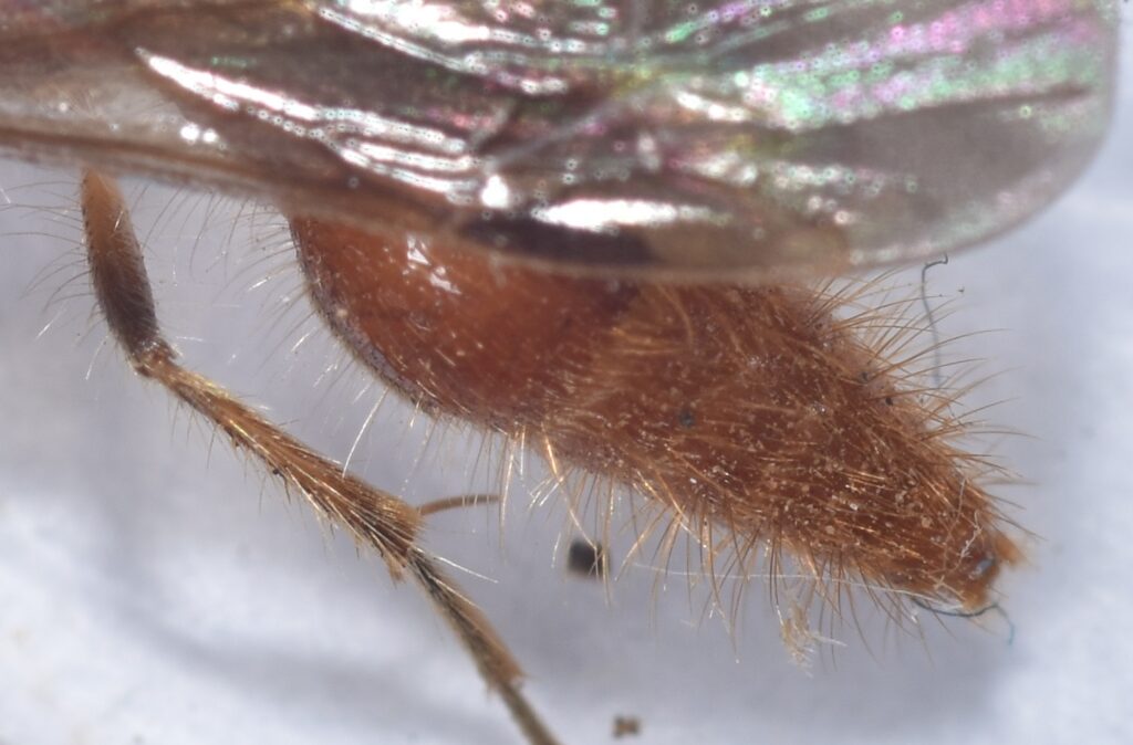
Conura is a genus in the family Chalcididae. Chalcididae is itself in the superfamily Chalcidoidea. Wasps in this superfamily are called chalcid wasps, and they often have a greatly enlarged hind femora (one of the leg segments), with a row of teeth on the lower margin. You can see this in the next photo, which shows one of the wasps that emerged from the cocoons.27

Most of you are probably familiar with aphids — those tiny, sedentary insects that spend their lives sucking sap from plants. Aphids have many enemies, but some of the most unsettling are the parasitoids. These wasps lay their eggs inside aphids. The larva develops inside and eventually pupates, usually still inside. At this point, the aphid is, mercifully, dead, and its body has been turned into a “mummy”. The body of a mummified aphid is swollen, papery, and brown, tan, or blackish in color, depending on the species of wasp that parasitized it. After the wasp metamorphoses into an adult, it chews its way out of the aphid, leaving a gaping hole in the aphid’s body.28

An aphid mummy
San Diego County 5/18/18
An aphid mummy
San Diego County 5/18/18

The wasp that parasitized these aphids was probably a braconid, but it wasn’t a braconid that came out. Instead, the parasite was itself parasitized by a wasp of the genus Pachyneuron in the family Pteromalidae.

A hyperparasitoid wasp (genus Pachyneuron, family Pteromalidae)
San Diego County 5/18/18
Another view

While we’re on the subject of parasitoids, I should mention what’s probably the weirdest type: adelphoparasitoids. These are tiny parasitic wasps in the family Aphelinidae. The females are parasitoids of whiteflies or scale insects, but the males of some species are hyperparasitoids of the females of their own species and sometimes even of other males!29

Fairyflies or fairy wasps (family Mymaridae) — Fairyflies are some of the most common wasps, but you’ve probably never seen one. They are unbelievably tiny. Most of them are between 0.5 and 1.0 mm (0.020 to 0.039 inches) long. All known fairyflies lay their eggs inside the eggs of other insects, which accounts for their small size. As you can see in the following photo, they have very strange, feathery wings.30

A fairyfly (Mymar sp.)
New Zealand
Author: S.E. Thorpe
License (as per the author): I, the copyright holder of this work, release this work into the public domain. This applies worldwide.
In some countries this may not be legally possible; if so:
I grant anyone the right to use this work for any purpose, without any conditions, unless such conditions are required by law.

Included within the fairyflies is the smallest known insect, Dicopomorpha echmepterygis. The males are blind and wingless and are about 40% smaller than the females (so it’s the male that’s the smallest insect). Their body length measures 186 μm, based on eight specimens. This makes them smaller than some species of Paramecium and amoeba. In other words, they are smaller than some single cells! 31

The smallest known insect — a male Dicopomorpha echmepterygis
Source: e
Huber J, Noyes J (2013). “A new genus and species of fairyfly, Tinkerbella nana (Hymenoptera, Mymaridae), with comments on its sister genus Kikiki , and discussion on small size limits in arthropods“. Journal of Hymenoptera Research 32: 17–44. Pensoft Publishers. DOI:10.3897/JHR.32.4663.
Authors: John T. Huber, John S. Noyes, J. Read
This file is licensed under the Creative Commons Attribution 3.0 Unported license.

The fairyflies also include the smallest known flying animals. These are wasps in the genus Kikiki. The genus includes a single species, K. huna, which is found in Costa Rica, the Hawaiian Islands, Trinidad and Tobago, Argentina, and India. It’s very likely that they’ve been spread by humans, probably by stowing away on boats. The females range from 158 to 190 μm. Very few males of the species have been studied. The one measurement of a male put it at 235 μm. It’s believed that the females of K. huna represent the smallest that an insect can get and still retain the ability to fly.32 33

The smallest known flying insect — Kikiki huna female
Source: Huber J, Noyes J (2013). “A new genus and species of fairyfly, Tinkerbella nana (Hymenoptera, Mymaridae), with comments on its sister genus Kikiki , and discussion on small size limits in arthropods“. Journal of Hymenoptera Research 32: 17–44. Pensoft Publishers. DOI:10.3897/JHR.32.4663.
Authors: Huber J, Noyes J
This file is licensed under the Creative Commons Attribution 3.0 Unported license.

Hyperhyperparasitoids? — If you’re like me, when you first learned about hyperparasitoids, your natural reaction was “So are there any hyperhyperparasitoids?” — that is, parasitoids of parasitoids of parasitoids? Apparently, the answer is yes. I don’t know if there are any parasitoids that parasitize only hyperparasitoids, but if the larvae of two hyperparasitoids happen to meet up within the body of a parasitoid host (which probably happens more often than you might expect), it’s quite possible that one will end up parasitizing the other.34

So are there any hyperhyperhyperparasitoids? Let’s just not go there.

Back to Stingers

So now we’ve got some insects stabbing their ovipositors into other animals. That seems like a good first step towards a stinger. But a yellowjacket doesn’t sting us in order to deposit an egg under our skin (which would presumably go on to hatch into a larva that would feed off our tissues and bodily fluids before bursting forth, xenomorph-style, from our body as a fully-formed adult.) Instead, it injects venom, whose sole purpose is to send us screaming. This seems like a bit of a leap evolution-wise. How do we get from injecting eggs to injecting venom?

I read one paper that described how a hypothetical enzyme could be transformed into a venom. The paper was focused more on snake venoms, but I think it still applies here. It pointed out that some venoms are related to immune system proteins that attack bodily invaders such as bacteria, and others are related to digestive enzymes. A subject that I’ve mentioned often in my blog posts is gene duplication. This is a common type of mutation in which the section of DNA that codes for a gene gets duplicated. At first, this just means that twice as much of an enzyme or other protein encoded by the gene may get produced. But having an extra copy of a gene means that one of the copies can evolve to perform a different function without the organism losing the ability to produce the original protein. So evolution is free to adapt the extra copy for a different purpose. Another type of mutation can change the signal that tells a gene where to become active. So, in the case of a snake, a mutation may tell a gene to make its protein in the mouth instead of the pancreas.35 In the case of an insect, a digestive enzyme could be redirected to the ovipositor.

So what would happen if such an enzyme were injected into another species? Hard to say. It could do nothing; it could make the target sick or even kill it; it could produce pain with or without actual damage or damage with or without pain. It could even have an effect on the target’s behavior (besides scaring it away or making it sick, I mean). After all, the substances manufactured by insects include hormones and neurotransmitters. In any event, a random foreign chemical injected into an organism will have a much better chance of having a negative effect on that organism than a positive one, especially if the chemical is, for example, a digestive enzyme. So it’s pretty easy to see how evolution could select for venoms that produce pain and/or damage.36

Some parasitoid wasps represent an intermediate step in the evolution from ovipositor to stinger. They use their stinger to inject an egg into the body of a host, but also inject venom along with it. The venom may paralyze the host or prepare it in some other way for consumption by the wasp larva. Eventually, some parasitoids developed a separate opening at the base of the stinger for depositing the egg, freeing up the stinger to became specialized for injecting venom.37

Some Stinging Wasps

In this section, we’ll cover some of the wasps that have full-fledged stingers. One thing you’ll notice when looking at the photos of these wasps is that, in most cases, the stingers are not visible. This is because they’re retractable — they are withdrawn inside the abdomen when not in use.

Umbrella wasp (Polistes dorsalis californicus) — Umbrella wasps are a type of paper wasp, which is a common name referring to any type of wasp that makes its nest out of chewed up wood fiber — paper, in other words.

It’s not surprising that paper wasps can sting. They have a nest to defend, after all. Surprisingly, though, the sting is generally described as only moderately painful.

Umbrella wasps (Polistes dorsalis californicus)
Mission Trails Regional Park, San Diego California 6/27/20

The use of wood pulp in paper making was probably inspired by paper wasps. The first paper was made out of recycled rags of hemp, linen, cotton, or other plant fibers. When Europe suffered a shortage of cotton and linen in the sixteenth century, manufacturers started looking for alternatives. The French naturalist René Antoine Ferchault de Réaumur is credited with introducing the use of wood fiber in 1719. One of Réaumur’s most important works is the Mémoires pour servir à l’histoire des insectes in 6 volumes. My French is not exactly fluent, but I think this means something like “Reports on the History of Insects”. And, again, it was in six volumes! I think it’s safe to say that Réaumur would have been familiar with the way paper wasps make their nests.38

Milde’s tarantula hawk wasp (Pepsis mildei) — If you haven’t read my earlier post on tarantula hawks, The Stuff of Nightmares, please do so. These are some beautiful, fascinating, and gruesome insects. A female tarantula hawk hunts tarantulas. When she locates one, she paralyzes it with a sting and drags the still living tarantula into a burrow. She then lays a single egg on the abdomen of the spider and seals the burrow. When the egg hatches, the wasp larva proceeds to feed off the tarantula, eating the non-vital organs first to keep it alive and fresh for as long as possible.

I’ve had the good fortune to see a tarantula hawk dragging a paralyzed tarantula on two occasions. Here’s a couple of photos from one of those encounters.

Milde’s Tarantula Hawk Wasp (Pepsis mildei)
Sweetwater River Trail 9/8/17
Milde’s Tarantula Hawk Wasp (Pepsis mildei)
Sweetwater River Trail 9/8/17

And here’s a video of the wasp dragging its prey.

When it comes to stings, tarantula hawks are something special. According to the Schmidt sting pain index (which I’ll be discussing later in the article), the tarantula hawk has the second most painful sting of any insect!39 And we have them right here in Southern California! Fortunately, since they don’t have nests to defend, tarantula hawk wasps will not normally attack humans. In other words, don’t bother them, and they won’t bother you.

Eastern cicada-killer wasp (Sphecius speciosus) — I recently spent a week in Virginia on the east coast of the U.S. As luck would have it, the 17-year periodic cicadas (Magicicada cassinii) were emerging.40 Cicadas are extraordinary insects. There are many different species, but M. cassinii is one of the most remarkable. It spends most of its life as a larva, living underground, where it feeds on the roots of plants. At the end of 17 years, all the cicadas emerge from the ground at the same time41 and molt one last time, metamorphosing into their adult forms.

As if this weren’t incredible enough, once they finish molting, the males all start calling to attract females. They do this by vibrating a couple of membranes on their abdomens, and the sound this makes is difficult to describe. It’s a loud buzzing — really loud. Collectively, the sound can reach 100 decibels. How loud is that, you ask? One website compares it to the sound of an “ambulance siren, inside driver window down”. It can actually cause hearing damage.42

17-year cicada (Magicicada cassinii)
Richmond, Virginia 7/27/21

17-year cicadas are large, meaty insects, so naturally, they have a lot of predators. One of those predators is the eastern cicada-killer wasp (Sphecius speciosus). As the name suggests, this wasp specializes in hunting cicadas. Just as a tarantula hawk wasp stings a tarantula to paralyze it and uses it to provision a burrow with food for its offspring, a cicada-killer wasp does the same thing with cicadas. Now, you might be wondering, how can it specialize in cicadas if they only emerge once every 17-years? I wondered this, too, and I haven’t found a definitive answer. My guess is that it hunts more than one type of cicada. (Most species of cicada are not ‘periodic’ — they spend much less time as larvae and emerge at random.) If you know the answer, please share your knowledge in the comments.

Once it stings a cicada, the wasp holds it underneath her as she flies off to a previously prepared nest cell. As already noted, 17-year cicadas are big insects, so the cicada-killer has to be big, too. It’s a large, scary-looking wasp with a wicked-looking stinger. Surprisingly though, its sting is actually quite mild — less painful than that of a honeybee.45

While in Virginia, I was fortunate enough to come across a dead cicada-killer wasp lying next to the path I was walking, so I was able to get some nice closeups.

A dead Eastern cicada-killer wasp (Sphecius speciosus)
Maymont Park, Richmond, Virginia 7/25/21
Another view
The stinger
A closeup
Notice how the sting protrudes from an opening in the abdomen. I would guess that, in life, it would be kept retracted inside.

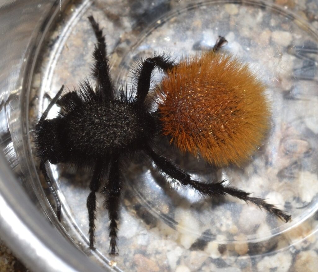
Velvet ant (family Mutilidae)46 — Velvet ants are actually wasps. The females are wingless and look a bit like large ants. Female velvet ants are parasitoids, usually of ground-dwelling bees or wasps. Because they have to spend a lot of time crawling around out in the open looking for the nests of their prey, they’ve had to evolve some serious defenses, one of which is an extremely painful sting. The sting of one species, Dasymutilla klugii, is rated three out of four on the Schmidt sting pain index. It also lasts up to 30 minutes.47 Check out my earlier blog post, Cow Killer, for more about velvet ants.

Velvet ant (Dasymutilla sp.)
Mission Trails Regional Park, San Diego County 9/16/19

Other Stinging Insects

While wasps (in the common, more restrictive, sense of the word) make up the vast majority of stinging insects, there are plenty of others among the ants and bees.

Harvester ants48 — Harvester ant is the common name for any kind of ant that collects seeds. This covers a lot of species and genera, so you might expect their stings to vary a great deal. But the following passage from Wikipedia seems to imply that they all have similar stings: “Harvester ants for their size have a rather potent venom. They inject it to [sic] their victim via sting by biting down and following up with a rapid sting from their abdomen, this causes 4-8 hours of sharp pain with effects similar to neurotoxicity such as piloerection49 and localized sweating around the area of the sting. ” If true, I suppose this is an example of similar lifestyles leading to convergent evolution.

Smooth harvester ants (Messor pergandei)
Anza Borrego Desert State Park, San Diego County 3/17/19
Smooth harvester ants (Messor pergandei) attacking a very unlucky caterpillar
Anza Borrego Desert State Park, San Diego County 3/17/19

Western or European honeybee (Apis mellifera) — Western honeybees are what most people think of when they think honeybee. They are, of course, highly social insects, and, since they have a hive to defend, one would expect them to have a fairly painful sting, and they do. On the Schmidt sting pain index, they rate a 2, which is moderately painful.50

Although I often got stung by wasps when I was a kid, as far as I can remember, I was never stung by a bee. That all changed on one of my recent hikes. A swarm of honeybees had built a hive inside the hollow of a tree. I used a friend’s phone to take a video of the swarm and was able to get quite close without being stung, resulting in a very nice video. Unfortunately, my friend’s phone malfunctioned shortly after this, and she lost the video. So the next time my hiking took me past the hive, I decided to try to get some more photos and videos.

I’m not sure why, but this time the bees seemed more aggressive. Maybe the first time, they were in the process of constructing the hive and didn’t have any eggs or young to defend, or maybe I was just too close for too long, but in any case, this time they were a lot more upset with me, and I got stung on the neck. Of course, my first thought was “What a great photo opportunity!”

I should explain. Honeybee stingers are barbed, which means that when they embed their stinger in your skin, they usually can’t pull it out again. When they fly away, the stinger is ripped out of their abdomen along with some of their guts. The bee, of course, dies from the injury. This may seem rather counterproductive, but it actually makes perfect sense from the point of view of evolution. The worker bees in a hive are all daughters of the same queen, so they share most of the same genes. As long as the hive survives, most of their genes will get passed down to future generations, regardless of the fate of any individual worker.

When the stinger is ripped out of a honeybee, the venom sac and the muscles connected to it are usually ripped out along with it. The muscles keep pumping for a while, insuring that a full dose of venom is injected into the target! So even if you swat the bee as soon as it starts to sting you, you’re still likely to suffer the full effect of its sting. Honeybees, by the way, are fairly unusual in only being able to sting once. Most stinging insects don’t leave their stingers behind and, thus, are able to sting repeatedly.51 52

So, anyway, back to the story. I left the stinger in my neck, venom muscles presumably still pumping away. (It actually didn’t hurt very much, so it wasn’t that much of a sacrifice.) When I got home, I awkwardly managed to take a few pics of the embedded stinger using my bathroom mirror to aim the camera. Here’s a couple of them. A nice photographic touch for an article on insect stings, if I do say so myself!

A honeybee sting embedded in my neck (Apis mellifera)
Mission Trails Regional Park, San Diego County 4/4/21
Another view

I didn’t think about it at the time, but I should have gotten one of the other hikers to take a video of it with their phone. Maybe it would have been possible to see the movements of the stinger as the muscles continued to pump venom. I also should have taken some photos of it after I scraped it off my neck. (I don’t know what I was thinking!) Here’s a photo someone else took of a honeybee stinger torn out of the insect’s body.

Honeybee stinger, torn from body of insect
Author:
Dejan Kreculj

This file is licensed under the Creative Commons Attribution-Share Alike 4.0 International license.

Finally (because I can’t seem to get enough of these ripped-out stingers), click here to view an amazing scanning electron microscope (SEM) image of a honeybee stinger.

Pseudo-stings

I mentioned at the beginning of the article that no male insects have stingers. That does not, however, prevent some of them from faking it. Many male insects have even evolved structures that look like stings and can be jabbed into an attacker, but that don’t inject venom. It’s an effective ruse. A quick jab from a pseudo-sting is often enough to startle even an experienced entomologist, giving the devious insect a chance to escape.53 54

The Schmidt Sting Pain Index55 56 57

No article about insect stings would be complete without discussing the Schmidt sting pain index. The toxicity of a sting can be determined experimentally, but toxicity is not closely related to pain. In fact, there are plenty of insect stings that are excruciatingly painful, but not at all toxic. Tarantula hawk wasps and velvet ants are two examples. The only way we currently have of finding out how painful an insect’s sting is is for someone to get stung by that insect and tell us. The entomologist Justin O. Schmidt set out to develop a sting pain index based on his personal experiences. The index he came up with rates stings from 0 to 4, with 0 being no pain at all and 4 being the most painful.

Many popular articles about Schmidt’s index suggest that Schmidt developed it by deliberately letting himself be stung. Schmidt himself denies this. His ratings are based on a lifetime of work in the field of entomology, during the course of which he estimates that he has (inadvertently) been stung by the majority of stinging hymenopterans. Whenever he got stung in the course of his work, he would make a note of how painful the sting was and how long the pain lasted. Obviously, the assignment of scores to stings is very subjective. That’s one reason the index only goes from 0 to 4. That way, different stings are only very broadly differentiated. If a species of wasp and a species of bee both have stings rated as 2, it doesn’t mean they’re the same or even very similar — it just means they produce roughly similar levels of pain.

Let’s take a look at some examples of Schmidt’s ratings.

Level 0. No pain in humans.

Level 1. Slight pain. Generally lasts five minutes or less. According to Schmidt, “almost pleasant, a lover just bit your earlobe a little too hard.” Examples of insects with level 1 stings are Western paper wasps, Southern fire ants, and most small bees.

Level 2. Average. Generally lasting between five and ten minutes. The majority of stinging insects have stings at this pain level. Western honeybees, yellowjackets, and bald-faced hornets have level 2 stings. Schmidt describes a yellowjacket sting as “hot and smoky, almost irreverent. Imagine W. C. Fields58 extinguishing a cigar on your tongue.”

Level 3. Significantly more painful than level 2. Lasting anywhere from one minute to half an hour. Klug’s velvet ant and the Maricopa harvester ant have stings in this category. Schmidt describes the latter’s sting as “After eight unrelenting hours of drilling into that ingrown toenail, you find the drill wedged into the toe.”

Level 4. The highest level in the index. A species of tarantula hawk wasp, Pepsis grossa, has a level 4 sting, which Schmidt deems to be “blinding, fierce [and] shockingly electric”. Another insect that gets a level 4 rating is the bullet ant (Paraponera clavata), found in Central and South America. According to Schmidt, this ant has the most painful sting of all insects. He describes it as “pure, intense, brilliant pain…like walking over flaming charcoal with a three-inch nail embedded in your heel.”

Schmidt’s descriptions of the various stings might give you the impression that his pain index isn’t meant to be taken seriously, but he actually did have a serious purpose in developing it. He wanted to collect evidence to either prove or disprove the hypothesis that the evolution of sociality in hymenopterans required the evolution of venoms that were both painful and toxic.

Pain is meant to act as a warning to an organism that it’s in danger of suffering damage — the pain of a burn lets you know that you need to take your hand off the hot plate. But since pain and toxicity aren’t closely related when it comes to stings, a painful sting can signal a danger that isn’t really there. Early hymenopterans were solitary, so just causing pain in an attacker would usually give the insect a chance to escape. Plus, solitary insects don’t provide large predators with much of a reward, so a little pain is usually enough to discourage them.

The evolution of sociality changed the equation. A large number of insects grouped together in a colony, along with their eggs and offspring, is a target much more worthy of predation. If a sting is painful, but causes no real damage, an intelligent predator will quickly realize that the meal is worth a little pain and learn to ignore the stings. So insects, the hypothesis goes, had to develop stings that were toxic, as well as painful. To study this hypothesis, Schmidt needed a way to relate the level of pain to the levels of toxicity and sociality. Thus, the Schmidt sting pain index was born.

Grad Students Are Nuts59

Another reason the Schmidt sting pain index has only four levels is that the figures represent an average over different locations on the body. For example, a honeybee sting to the back of the hand may only rate a 1.5, but a sting to the tongue from the same insect might be worthy of a 3. So, when assigning a level, Schmidt tried to estimate the average pain across a number of different body parts.

Michael Smith, a graduate student at Cornell University, decided to collect a bit more data. He proceeded to systematically test the pain levels of honeybee stings inflicted in random order to 25 different locations on his body. For six weeks, he received three test stings and two calibration stings a day until he had accumulated three stings to each location. The stings were rated on a scale from 1 to 10. The body locations chosen included “traditional” locations such as the upper arm, forearm, middle finger, thigh, upper lip, etc. Some of the more “non-traditional” locations included were the nipple, scrotum, and penis.

I would give you his results for all the various locations, but I know the only ones you really care about are the scrotum and penis. Unsurprisingly, both were “right up there near the top”.
Cow Killer

“An ant of velvet
On a dusty hiking trail
A wasp chirps softly” – Haiku14

Velvet ants are not really ants. They’re wasps. They’re called velvet ants because the females are wingless and look a bit like large, hairy ants. They’re also called cow killers. They earn this moniker by having excruciatingly painful stings.

You might be forgiven for thinking that the more painful a sting is, the more dangerous it is. In fact, the toxicity of a venom is unrelated to the level of pain it produces. The sting from a velvet ant is less toxic than that of a honey bee, but hurts way more. On the Schmidt sting pain index36, the velvet ant scores a 3. (The index only goes up to 4.) One of the few insects in Southern California with a more painful sting is the tarantula hawk wasp, which I covered in an earlier post. (See The Stuff of Nightmares.)

Dasymutilla magnifica – A beautiful desert species of velvet ant
Anza Borrego State Park, San Diego County, CA 6/22/19

I’ve actually been stung by a velvet ant. While hiking near where I live, I saw a Sacken’s velvet ant, a species whose long white fur makes it look kind of like the seed-head of a plant — if a seed-head had long black legs. I wanted to show it to my grandnephew, who’s a huge fan of bugs (can’t imagine where he gets that from), so I caught it in a small plastic container. The young woman I was hiking with was worried that it wouldn’t have enough air, so, to make her happy, I decided to transfer it to a larger container that I happened to have with me. When I got home, I discovered that the container had gotten squeezed in my pack and the lid had popped loose …and it was empty. It seemed unlikely that the velvet ant would be able to find its way out of the pack, so it stood to reason that it was still in there. Somewhere…

I started very gingerly pulling things out. Finally, I upended the pack and dumped out the remaining items. No velvet ant. I had just about concluded that it had escaped altogether, when I detected movement out of the corner of my eye. I jerked my hand back just as the wasp struck. I must have reacted just in time to prevent the insect from injecting more than a tiny amount of venom, because the sting was very mild — much less painful than a typical spider bite — and the pain faded completely in a few minutes. Obviously, I was very lucky.

Here’s a couple of photos of a velvet ant like the one that stung me (but not the same individual):

Sacken’s velvet ant (Dasymutilla sackenii)
Cuyamaca Rancho State Park, San Diego County, CA 8/8/20
Another view

Taxonomic Headaches44 45

Wasps are in the order Hymenoptera, which also includes bees, ants, and sawflies. All these insects are in many ways closely related. Bees have been described as vegan wasps (see my earlier blog post Vegan Wasps and Flying Mice) and ants could be thought of as wingless, social wasps.

Within the Hymenoptera, wasps form the suborder Apocrita and, within that, velvet ants make up the family Mutillidae. As noted, female mutillids are wingless. The males do have wings and often differ markedly in size, form, and coloration from the females. This presents a problem for entomologists. Velvet ants are solitary insects. The only time males and females hang out together are when they are actually in the process of mating. So, given any two velvet ants — one male and one female — how can you tell if they are the same species? Often the only way to know for sure is if you manage to catch them in flagrante. One entomologist who specializes in the group writes that, although he has frequently seen males in the vicinity of females (and actively searching for them), he’s only once seen them actually doing the deed. It seems that they prefer to conduct their affairs in private.

Obviously, this makes it very difficult for scientists to sort out the different species. Many times males and females of the same species have been given different species names. When the mistake is finally caught, one name (the first one given) becomes the official species name while the other becomes a synonym.47 Currently, there are around 150 species of velvet ant listed for North America. Of these, about a third are known from both males and females; another third are known only from males; and the final third are known only from females.

A male velvet ant in the subfamily Sphaeropthalminae
Mission Trails Regional Park, San Diego County, CA 8/13/18
A closeup
Note the three large ocelli on the top of the head. (See The Story of Eyes for an explanation of ocelli.)
A closeup of the abdomen

Dietary Habits

Adult velvet ants feed on nectar. I mentioned earlier that I caught a female to show to my grandnephew. Since hummingbirds also feed on nectar, but will accept sugar water as a substitute, I figured mutillids might do the same. So, we mixed up some sugar water, added a little red food coloring (this was more for the kid’s benefit than the bug’s — he thoroughly enjoyed making “nectar”), and put a few drops into the container with the wasp. The insect didn’t immediately go for it — she was probably distracted by all the unwanted attention — but later I saw her grazing on the sticky, drying droplets.

What the larvae of velvet ants eat is a bit more gruesome. Mutillid larvae are parasitoids — parasites that kill their hosts. With a few exceptions, they prey on the young of solitary, ground-nesting bees or wasps. After mating, the female velvet ant goes in search of a host nest. When she finds one, she invades the nest and lays an egg next to each larva or pupa. When an egg hatches, the mutillid larva begins feeding on the host larva/pupa. It takes only a week or two for the wasp to consume its host, after which, it metamorphoses into an adult and leaves the nest.

Naturally, a bee or wasp whose nest is invaded by a velvet ant defends it vigorously. And since the hosts are usually solitary, the nests tend to be scattered far and wide, so the female velvet ant has to spend a lot of time wandering around in the open searching for them. This means exposing herself to an increased risk of predation. As a result of these selection pressures, velvet ants have evolved a remarkable array of defenses. One of these is their extremely painful sting, which we’ve already discussed. I’ll cover the other defenses in the following sections.

Velvet ant (Dasymutilla sp.)
Mission Trails Regional Park, San Diego County 9/16/19

Body Armor60

The velvet ant’s exoskeleton is heavily reinforced. So much so, that collectors often resort to using tiny drill bits to drill holes for the pins, instead of trying to force the pins through the bug’s carapace. If an entomologist does try to push a pin through by brute force, when it finally breaks through, it will often continue on through the bottom of the carapace and into the collector’s finger. So, a drill bit is probably the way to go.

The top and sides of a velvet ant’s exoskeleton are covered with pits and ridges. This serves to strengthen the exoskeleton without adding a lot of weight. The ridges may also serve to redirect attacks. When a mandible or stinger catches on a ridge, it tends to slide along it towards an even thicker part of the armor.

Aposematic Coloration61 62

An aposematic signal is a warning directed toward potential predators. It’s a way of saying, “you really don’t want to mess with me”. Such signals often take the form of colorful, eye-catching patterns. Most velvet ants are covered in brightly colored fur. If a predator tries to eat one, it’s probably going to get a bad sting and, while the wasp in question may or may not survive the attack, the predator will quickly learn to avoid any similar-looking insects in the future. So, aposematic coloration helps to reduce predation on a species as a whole.

A defensive strategy often allied with aposematic coloration is Müllerian mimicry. This is when two or more species that share the same predators evolve very similar warning signals. If a predator learns to avoid members of one of the species, it will likely avoid members of all of the species, so sharing the same aposematic signals is mutually beneficial. There are many cases of velvet ants forming ‘Müllerian mimicry rings’ consisting of dozens of species. A study in “Current Biology” found that North American velvet ants form one of the largest known Müllerian mimicry complexes on the planet, consisting of eight extensive and distinct mimicry rings.63

A Chemical Alarm64

Aposematic coloration is a pretty good warning signal, but what if your attacker is color blind? Perhaps to fill that gap in its defenses, the velvet ant has evolved the ability to secrete an alarm chemical from a gland in its mandible. This chemical serves as an olfactory alarm signal. A predator quickly learns to avoid any insects that smell like it.

Stridulation65 66

Ok, but what if your attacker is color blind and has a head cold? Better to give him an auditory warning, too, just to be safe. Insects don’t have vocal cords, but many of them are able to produce sounds by rubbing together various body parts. This is called stridulation. Crickets, for example, are famous for their chirping, which they make by rubbing together their wings.67 When disturbed, a velvet ant emits a sound that has also been described as “chirping” or sometimes “squeaking”. It does this by rubbing together two of the segments of its abdomen.

When I showed that velvet ant to my grandnephew, we found that we could get it to stridulate by gently stroking its back with the cap from an ink pen. The sound was soft and difficult to hear, but then, it’s probably tuned to non-human ears. The vibration of the abdomen may also serve as a deterrent by provoking a startle response. In one experiment, a shrew was observed to drop a velvet ant as soon as it started stridulating, even though it hadn’t been stung.

I should note that stridulation may not be used exclusively for defense. According to one article, the female also uses it to attract males.68

In the following video, I’ve increased the volume to make the stridulation easier to hear. You can also clearly see the movement of the abdomen. Note: the velvet ant in the video looks a bit bedraggled. This is probably because of the time it spent loose in my backpack. It was not otherwise harmed and I later released it near where I found it. (It even got a nice meal of sugar water for its trouble. That’s the red splotches in the video.)

Scientists have actually studied the susceptibility of female mutillids to predation. In one study, entitled “The indestructible insect: Velvet ants from across the United States avoid predation by representatives from all major tetrapod clades”69, velvet ants from both the Eastern and Western United States were exposed to various potential predators that they would be likely to encounter in their native habitats. These included birds, shrews, lizards, toads, and one mole. (Apparently, moles were in short supply.) The results of the study can be summed up by the following excerpt from the paper: “…out of over 100 interactions between potential predators and various species of velvet ant, there were only 16 occasions where feeding strikes occurred. Only four velvet ants were eaten and three of the four were immediately regurgitated. Only one velvet ant was consumed and retained by a predator.”

The Mile High Club, Velvet Ant Style70 71 72

Female mutillids probably lost their wings as an adaptation to entering the burrows of their hosts. Wings would be easily damaged by attacking insects, so better to just do away with them. This presents a problem, though — it limits species dispersal — and this puts them at an evolutionary disadvantage, because if a species is only found in a small area, it will tend to adapt itself to the specific environment found in that area. If that environment changes drastically, the species could be wiped out.

Some mutillids have overcome this handicap by evolving phoretic copulation. Phoresy is a type of behaviour in which one animal hitches a ride on another animal. Some types of mites, for instance, will hitch rides on beetles or other larger animals. This way, they get more widely dispersed than would be possible if they just walked. In phoretic copulation, the male of a species carries a female to a new location either before or during mating. In the case of velvet ants, the male transports the female by flying. He actually carries her attached to his genitalia, so mating is done in mid-air. To facilitate this behavior, males of these species have evolved to be significantly larger than the females.

Rattlesnake!

“Everybody knew that you should never provoke a rattlesnake, much less tie it into a bow. But that didn’t stop Judd. What did stop him was the rattlesnake.” -Demetri Martin

It’s easy to forget how remarkable the rattlesnake is. We all learn about rattlesnakes as kids, so we’re used to them, even if we’ve never actually seen one in the wild. And, of course, many people are too busy being terrified of rattlers to think about them in any other way. But stop for a minute and really think about it — a snake with a rattle on its tail! If it didn’t actually exist, it would be a ridiculous idea. The evolution of the rattle will be one of the things I’ll be covering in this article, but first, we need to get the basics out of the way…

Rattlesnakes are native to North and South America with the greatest concentration in the southwestern United States and northern Mexico. There are two genera of rattlesnakes, Crotalus and Sistrurus. Sistrurus is the more basal genus, which is to say, Sistrurus evolved first and Crotalus branched off from it. The larger, more well-known of the rattlers are in Crotalus. Those in Sistrurus are smaller and have smaller, higher pitched rattles. There are 33 species of rattlesnakes altogether, only 2 of which are in Sistrurus.65

Rattlers are pit vipers (subfamily Crotalinae of family Viperidae). The “pit” part of the name refers to the pit organs, small heat-sensing pits located on each side of the head. These act like eyes, allowing a rattlesnake to detect warm-blooded prey even in pitch dark.66

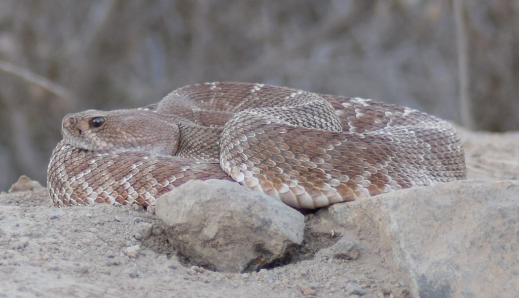
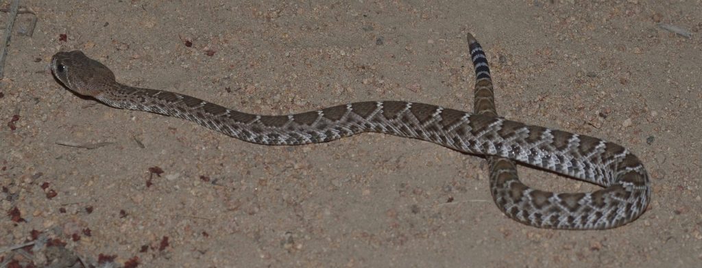
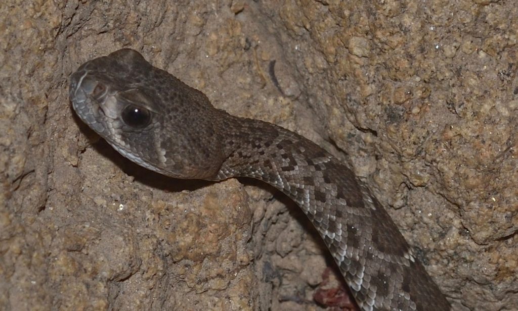
There are a number of Crotalus species here in southern California, but I’ve only had the uh… good fortune to encounter a couple of them. One is the red diamondback or C. ruber, which has a very pale, reddish coloration and a diamond pattern on its back. The other is the western diamondback or C. oreganus. This has a similar diamond pattern, but is usually dark gray or brown. Most of the western diamondbacks that I’ve seen have been in the subspecies C. oreganus helleri, which is also called the southern Pacific rattlesnake.69 73 74 (A third species, C. atrox, also goes by the common name western diamondback. Their range is supposed to include the southern tip of California, but, as far as I know, I haven’t encountered any.)75

I’ve run into rattlesnakes (almost literally a couple of times) in various places around San Diego County, but most of the encounters were in Mission Trails Regional Park, just west of San Diego. I don’t suppose they are any more common there than in most of the other places I hike; I just hike in Mission Trails a lot more often, since it’s close to where I live.

That said, I saw around 17 rattlers in 2018, almost all of them in that park. I came close to stepping on one twice. The second time was the scariest. I had just turned onto a trail. Overhanging branches of broom baccharis were blocking my view of the rattler, a huge southern Pacific. My foot landed right in front of the snake, which started rattling very loudly. Needless to say, I jumped back. I don’t know if the snake had already been coiled up when I almost stepped on it, but it was coiled up by the time I saw it. The snake partially uncoiled and, using some kind of beautiful, sinuous motion that I can’t begin to describe, propelled itself several yards away, deeper into the bushes, hissing and rattling the whole time. It stopped there and continued to rattle angrily as we took photos of it. Fortunately, there wasn’t much undergrowth, so I was able to get some great shots and a nice, long video. Those are posted below.

Southern Pacific Rattlsnake (C. oreganus helleri)
Mission Trails Regional Park, San Diego County 8/16/2018
This is the snake from the above video.
A closer look

In the video, you can hear one of the people I was hiking with calling to some women who were walking by on the main road, asking them if they wanted to see a rattlesnake. They declined. You can hear my friend responding “You’ll catch the movie…” Lol.

It was a fairly unnerving experience, since I’d already come close to stepping on a smaller rattler earlier that year. After that second encounter, I bought some snake chaps and started wearing them whenever I hiked in Mission Trails. I kept wearing them until the weather turned cooler, making rattlesnake encounters less likely. For whatever reason, I haven’t encountered as many rattlers this year (2019) and I haven’t felt the need to wear the chaps. You can bet I keep to the middle of the trail whenever I pass that particular overhanging bush, though.

Red diamondback (Cotalus ruber)
Mission Trails Regional Park, San Diego County 8/7/2018
A closer view

As hair-raising as that last story may have sounded, rattlesnake bites are actually very uncommon and, when they do occur, they’re rarely fatal. On average, there are 7000 to 8,000 venomous snake bites in the U.S. per year — that’s all types of venomous snakes, not just rattlers. And, on average, only 5 of those 7000 to 8000 snakebite victims die. Rattlesnakes don’t want to bite you. They will do so only if they feel threatened. If you step on one or try to catch it, then, sure, it’s going to bite. According to Wikipedia, “A majority of victims (about 72%) are males, often young and intoxicated”.76

One reason rattlesnake bites are rarely fatal is that the snakes have voluntary control over how much venom they inject. It takes energy for a snake to make venom, so it doesn’t want to waste it on something that it can’t eat. When a rattlesnake bites a person, it will often inject a smaller amount of venom than it would if it was attacking prey. In fact, around 20% of rattlesnake bites are dry bites — no venom at all!77

I do know one person who was bitten by a rattlesnake. A woman that I often hike with had just finished a hike. She went into the bushes for a call of nature and, as she was pulling her pants up, her foot broke through the ground and into a rattlesnake den. The snake started rattling and my friend felt a light tap on her calf. She said it felt like a tap on the shoulder. She thought maybe the snake’s rattle had hit her. After pulling her foot out (and presumably putting some distance between herself and the rattler), she examined her leg. She didn’t see any marks at first, but when she squeezed her leg, two tiny beads of blood oozed up from where the fangs had penetrated.

As I mentioned, this was at the end of the hike, so one of the other hikers was able to drive her directly to a hospital. This hiker was driving a Corvette and, in his words, “I’d been waiting for this my whole life!” To make a long story short, my friend managed to survive the drive to the hospital and the bite turned out to be dry. She never felt any effects from it at all. The poor snake was probably too surprised to even think about injecting venom.

Note: As soon as my friend was bitten, they called 911 and the ambulance met them as they were driving to the hospital. The paramedics checked her over quickly, but she wasn’t exhibiting any symptoms and, since the sports car was able to handle the twists and turns in the road better than the ambulance, the paramedics let her continue the trip to the hospital in that.

Rattlesnake bite
copyright © 2019 Hazell Belvin
Used with permission

So, to summarize, you’re unlikely to ever get bitten by a rattlesnake and, if you are, there’s a good chance that the rattlesnake will not inject a full dose of venom. But if you do get bitten, what should you do?

What To Do If Bitten

I was going to include list of things to do in case of snake bite, but I’m not a medical professional, so it’s probably better if I just direct you to a few websites that seem trustworthy. Click here for the Mayo Clinic’s snake bite page. Click here for a site that’s specific to rattlesnake bites. And finally, click here for a link to Johns Hopkins’s page.

Now that we’ve covered the basics, let’s take a closer look at the various things that go into the making of a rattlesnake.

The Pit Organ 78 79 80

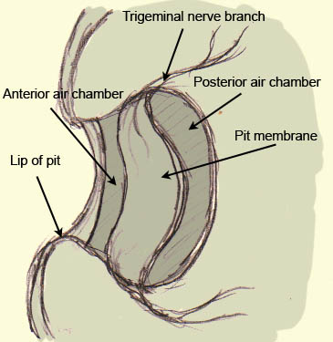
As mentioned earlier, the pit organs are small, heat-sensing pits on each side of the rattlesnake’s head. In the photo below, you can see one of the pits. It’s positioned between the eye and nostril, a little lower on the snout. The silvery oval inside the pit is the pit membrane. It may look like it’s swelling outward, but I think that’s an optical illusion. It’s actually curved inward, as depicted in the diagram below the photo.

Probably another southern Pacific rattler
The silvery oval is the pit membrane.
Mission Trails Regional Park, San Diego County 8/14/2017
A diagram of the pit organ
This image is in the public domain. It was downloaded from Wikipedia Commons at this url.

The pit organs function as crude eyes for detecting infrared light, a.k.a heat. For those of you who need a quick physics refresher, heat has several ways of traveling from place to place. One way is in the from of light waves in the infrared portion of the spectrum. The only difference between infrared light and red light is the wavelength. The wavelength of infrared light is longer than that of red light and cannot be detected by our eyes. As it happens, it can’t be detected by the rattlesnake’s eyes either. That’s why it needs pit organs.

In its regular eyes, the rattlesnake has special chemicals that are sensitive to light in the visible spectrum. The pit organ works a little differently. When infrared light from a warm object like a mouse hits a spot on the pit membrane, it heats up that spot. Infrared (IR) receptors in the form of heat-sensitive proteins detect the change in temperature and generate an electrical impulse that travels down nerves to the snake’s brain. The pit membrane is rich with blood vessels which quickly cool the spot back down to ambient temperature once it’s no longer being heated up.

Note that the opening that lets infrared light into the pit is smaller than the diameter of the pit. This means that the pit acts like an infrared version of a pinhole camera. The infrared light entering the pit forms a crude image in the form of a heat map on the surface of the membrane. So the pit membrane serves the same function as the retina in a regular eye.

The snake’s brain integrates the heat map from its pit organs with the visual images from its eyes. It’s kind of like how our brain integrates the 2-d images from each eye to form one 3-d image. So, what does this look like to the snake? I have no idea. I’m not a snake (despite what you may have heard). It’s like a person who’s been blind from birth trying to imagine what eyesight is like.

The heat image formed by the pits must be rather crude, but it’s good enough for the snake to detect and strike prey in total darkness. A BBC video that I found on YouTube shows some infrared images from a heat-sensitive camera developed by the military. According to the narrator, the rattlesnake’s pit organs are “30 times more powerful” than the camera. I think this probably means that the pit organs are able to detect a heat signature that is 30 times weaker; I doubt they form as sharp an image. It’s a great video, though. You can watch it here.

There is also evidence that the snake uses the pit organs for avoiding predators and for thermal regulation. In fact, it’s possible that the pit organs originally evolved for prey avoidance and/or thermal regulation and that rattlesnakes (or their ancestors) only later started using them for hunting.

So how could pit organs have evolved? I wasn’t able to find much about this, but one article mentioned that some snakes in the Boidae family have IR receptors in their labial scales (scales on the sides of the snout), without any specialized structures. Other snakes in the same family have IR receptors in the bottom of simple labial pits.81 These would seem to be examples of possible intermediate steps in the formation of a pit organ.

Those Eyes 82

Humans and many other animals have a small, central pit in the retina called a fovea consisting of tightly packed cone cells (the cells responsible for color vision). The fovea is where our vision is sharpest and it makes it possible for us to do things like reading and driving. A rattlesnake eye doesn’t have a fovea, so rattlesnakes are not able to see sharply defined images. (Never let a rattlesnake drive!) They rely mostly on the perception of movement and don’t appear to follow objects with their eyes.

Red diamondback (C. ruber)
Same one as in the earlier photos

Rattlesnakes are adapted for hunting at night, so they have lots of rod cells (the cells that are sensitive to dim light, but not color). They also have cone cells, though, so they have some color vision.83

Rattlesnakes have vertical pupils. That is, when the pupil is contracted, it forms a vertical slit. Not all snakes have vertical pupils. Studies have shown that it’s mostly ambush predators that have them. Computer simulations have shown that a vertical pupil allows a predator to better judge distance without moving the head — an obvious advantage, since moving the head could alert prey. (For a fascinating article about pupil shape, click here.)

Those Ears? 84

Yes, rattlesnakes have ears. They just don’t have ear openings. Or eardrums. What they do have are middle ears, but even those are not as specialized as those of mammals. In fact, mammals in general have much better hearing than reptiles because a couple of bones that form part of the jaw in reptiles (which are the ancestors of mammals) migrated into the middle ear and now form part of a mammal’s hearing apparatus — a remarkable and bizarre fact that I definitely need to do a blog post about one of these days.85

So rattlesnakes don’t have very good hearing. They do seem to be very sensitive to vibrations in the ground, though. These are transmitted to their ears through their bodies, especially their bones. So I suppose they do have pretty good hearing in that sense.

That Flicking Tongue 86

Snakes use their forked tongues for smelling. The forked tongue is not unique to rattlesnakes, of course — many reptiles have them — but it certainly adds to their mystique.

Snakes have a great sense of smell. In fact, it’s probably their most important sense. When a snake senses a change in its environment (through vibrations in the ground, for instance), it immediately starts flicking its tongue in and out, sampling the air. A groove, called a rostral groove, in the snake’s upper lip allows the tongue to move in and out without the snake having to open its mouth. When the tongue darts out, chemicals in the air stick to it. When the tongue is withdrawn into the mouth, the tines are inserted into two tubes in the roof of the mouth. These tubes form the Jacobson’s organ, also called the vomeronasal organ (VMN). This organ is stimulated by chemicals on the tongue to send nerve signals to the part of the brain that processes smell.

So why do snakes have forked tongues? This allows them to sense the direction from which a smell is coming. For instance, if more of a particular chemical sticks to the left tine than to the right, then the smell is probably wafting in from the left.

Snakes are also able to smell through their nostrils. I’m not sure how sensitive their noses are, but I get the impression that their tongues are far more sensitive. According to one article, a snake may pick up a scent through its nostrils and then start flicking its tongue to get a better sniff.87

I can’t resist mentioning that humans, as well as many other mammals, also have a Jacobson’s organ. It also consists of two tubes in the roof of the mouth. There’s a lot of debate about what purpose the Jacobson’s organ serves in humans. Some people think that it’s stimulated by sexual pheromones. Others disagree. There doesn’t seem to be any definitive evidence either way.

Those Venomous Fangs88

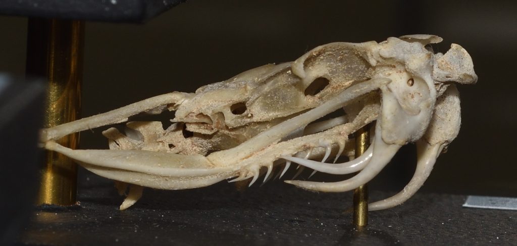
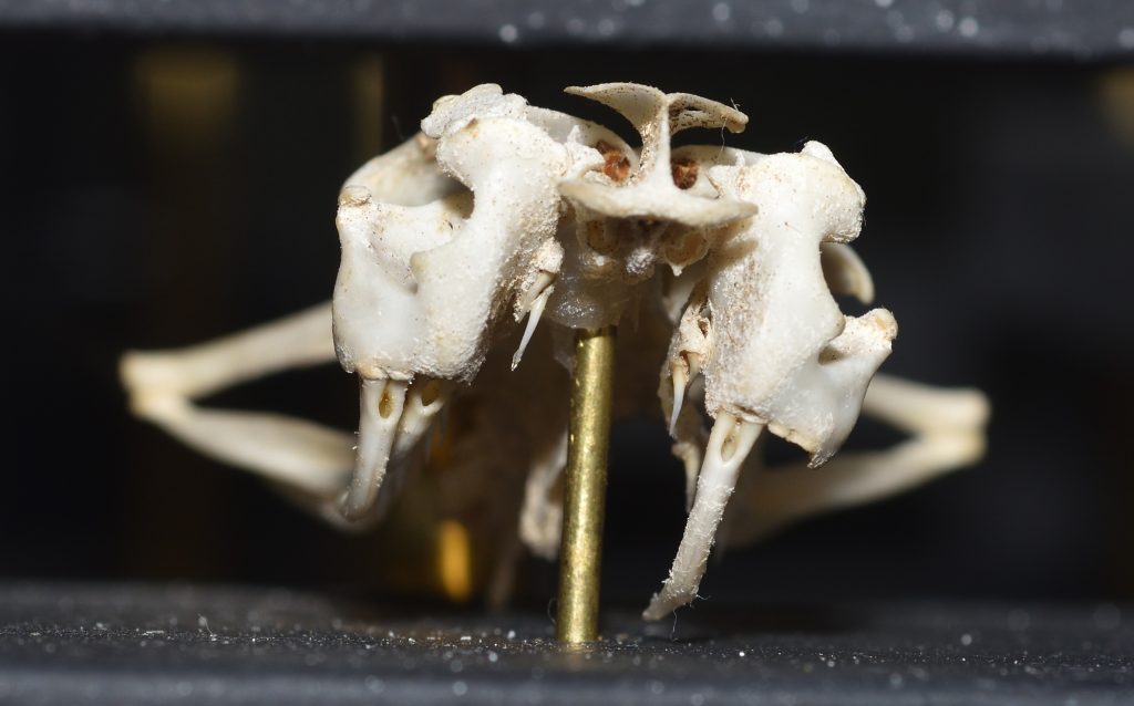
Rattlesnakes, in case you haven’t noticed, have two thin, curved fangs in the front of their mouths. These fangs are very much like hypodermic needles. They have a hollow core with an opening near the top where the venom enters the fang and a narrow slit on the front of the fang near the tip, through which the venom is ejected. Each fang is supplied with venom through a duct that connects to a large venom gland located near the back of the head. So there’s a separate venom gland for each fang. These glands are what give rattlesnake heads a triangular shape.89 When the snake bites, muscles on the sides of the glands squeeze the venom through the ducts into the fangs.

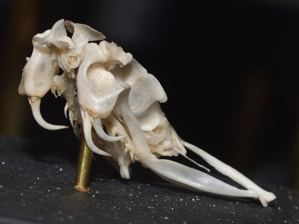
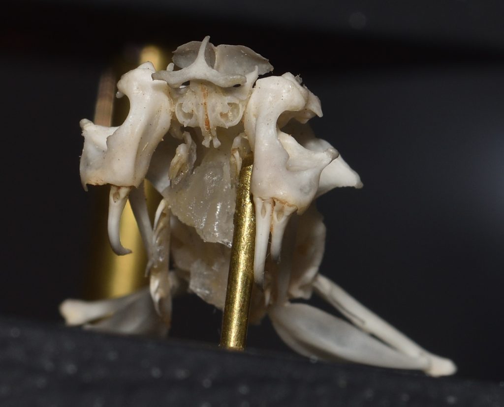
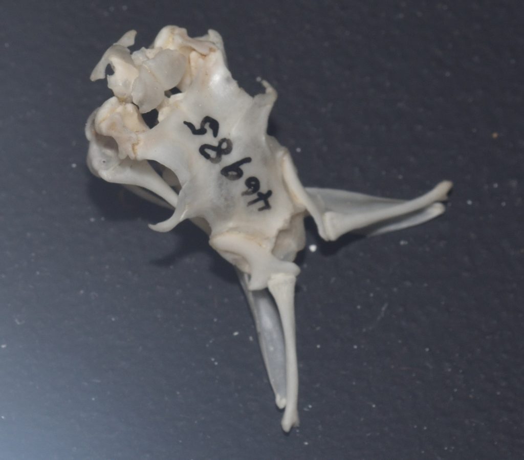
When the rattlesnake closes its mouth, the fangs fold back into the roof of the mouth, where they rest in a sheath of soft skin.90 Click here to see a video of a rattlesnake skull with the jaws rigged up to keep opening and closing. If you spend a little time watching this video, you can get a pretty good idea how the system works. Fangs are modified teeth, of course. They grow from the maxilla bone of the upper jaw. The other teeth, which are used for gripping the prey while the snake swallows it, grow from a separate bone, the pterygoid, that connects to the end of the maxilla and extends backwards toward the rear of the skull. The maxilla is connected at the other end to the rest of the upper jaw. The connections between all these bones are flexible, allowing them to rotate with respect to each other. When the rattlesnake opens its mouth, the pterygoid bones are pushed forward and the maxillas rotate, swinging the fangs into biting position.

Rattlesnakes shed their fangs periodically (once every 60 days or so) and they always have replacement fangs waiting in reserve. In fact, they have a series of 7 pairs of fangs in various stages of development behind their functional fangs! So even if a snake unexpectedly breaks a fang, it won’t have to wait long before it can deploy a new one.

So how did this whole remarkable system evolve? Let’s start with the venom. Venom is basically very strong saliva. This may raise a few eyebrows. How does saliva get magically transformed into venom? Well, it may surprise you to learn that ordinary human saliva injected under the skin of the hand can cause pain, redness, swelling, and partial loss of function.91

Obviously, human saliva did not evolve to be toxic. It just happens to contain some substances that don’t have very nice effects when injected under the skin. Actually, this isn’t too surprising given that the purpose of saliva is to aid in digestion by beginning to break down food in the mouth. So it makes sense that if it’s injected into the hand, it would begin to break down tissue. It’s also not hard to imagine evolution working with snake saliva to slowly increase its toxicity.

Venom — and this is true for almost all venomous creatures — is not a single substance. It’s a complex cocktail of chemicals and the different chemicals may serve very different purposes. For instance, some chemicals may act to actually kill the prey, while other chemicals only paralyze it. Still other chemicals may start the process of digestion. Different components of venom may also act on different systems. One chemical might break down muscle while another attacks the nervous system.

Different species of rattlesnake have evolved different mixtures of chemicals in their venoms to match their preferred prey. For instance, the venoms of C. ruber and C. oreganus are mostly necrotic — they break down tissue. The Mojave rattlesnake (C. scutulatus), on the other hand, has more neurotoxic components in its venom. Research has shown that the common ancestor of these three species had a more general purpose venom that contained both necrotic and neurotoxic elements. As this species differentiated into multiple species, its descendants modified the mix of chemicals in their venoms to reflect their presumably more specialized diets.92

That takes care of the venom, but how in the world did these snakes manage to evolve hypodermic needles with which to inject it? The first venomous snakes didn’t have fangs, but they did have teeth. These snakes probably caught their prey and held it in their mouths while the venom slowly seeped into the puncture marks made by the teeth. Chewing probably sped up the process. Eventually, some of the teeth evolved to be longer than the others and developed grooves that helped to channel the poison into the wounds. We still have these kinds of snakes today. These are the opisthoglyphous or “rear-fanged” snakes which make up the Colubridae family. (Technically, opisthoglyphous refers to the type of fang, not the group of snakes, but it’s often used informally to refer to the group.)

When I first read about rear-fanged snakes, I found the idea very confusing. How can a snake use fangs that are in the rear of its mouth? A picture of a snake called a boomslang on Andrew Durso’s excellent (and excellently-named) blog “Life Is Short, But Snakes Are Long” cleared things up for me. Click here to see the photo I’m talking about. It’s around the middle of the page. In this photo, you can see that the fangs aren’t actually in the rear of the mouth. They’re just at the rear of the maxilla bones. The pterygoid bones continue on beyond the fangs. (In these snakes, the maxilla and pterygoid bones are not able to rotate relative to each other.) So the fangs are really more toward the middle of the mouth. These snakes bite down on their prey and “chew” the venom into it.

The fangs of rattlesnakes — the kind that are in the front of the mouth and fold back when not in use — are called solenoglyphous fangs. These types of fangs evolved to inject large amounts of venom quickly and deeply. When a rattlesnake strikes a prey animal, it doesn’t hold on to it. After injecting venom into the prey, the snake immediately releases it. It then uses its excellent sense of smell to track the prey to where it finally collapses. This way, the snake doesn’t risk being injured by a struggling animal.

There’s another group of snakes that have what’s called proteroglyphous fangs. These fangs are also in the front of the mouth, but they don’t fold back. They tend to be about a third as long as rattlesnake fangs and they are also hollow. Snakes that have this type of fang are all in the Elapidae family and they include such infamous serpents as cobras, mambas, and death adders. Elapids bite their prey and hold on while they wait for the venom to take effect.

Scientists believe that solenoglyphous and proteroglyphous snakes both evolved from opisthoglyphous ancestors. So the distant ancestors of rattlesnakes would have had grooved fangs near the middle of the mouth. Over time, the ridges of the grooves folded over to form tubes and the fangs migrated to the front of the mouth. Evidence for this last part can be found in embryology. In the rattlesnake embryo, the developing fangs are located in the middle of the mouth in the same place as in rear-fanged snakes. As the embryo grows, the short piece the maxilla behind the fang grows much faster than the rest of the maxilla, pushing the fang forward in the mouth. You can find a nice set of photos illustrating this on the same blog page that I mentioned earlier, right under the picture of the boomslang. Click here to go back to that web page.

Those Insanely Flexible Jaws

Click here to see a video that explains how a snake swallows its prey. (It’s not a rattlesnake, but the principal is the same.) Snakes have more bones in their jaws than mammals do and these bones are not as tightly bound together. They have ligaments that stretch, allowing the bones of the jaw to spread apart, making room to swallow the prey whole.

You may be wondering how a snake goes about pulling a prey animal down its throat. I haven’t found any explicit description of this, but I’m guessing the snake clamps down harder with one side of the jaw than the other (remember how much more flexible those jaws are than ours!) and then twists its head so that the other side of the jaw moves a little farther up on the animal. It then quickly opens its mouth and snaps down again to get a new grip and repeats the process, this time clamping down with the other side of the jaw and twisting the head in the other direction.

It’s also possible, and this is entirely speculation on my part, that the pterygoid bone can be drawn back a little farther into the mouth to pull the prey a little farther in. Then the snake releases its grip long enough for the pterygoid to spring forward a little before it clamps down again. As always, if anyone out there has more info, please leave a comment. Once the prey gets down into the throat, the muscles of the throat and body can take over.

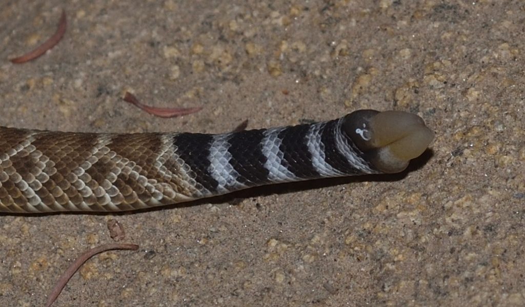
And Finally, That Rattle 93

The rattle consists of a series of hollow, interlocking segments. The segments are made of keratin — the same material that makes up our hair and fingernails and the scales of reptiles. In fact, the rattle segments are modified scales. Rattlesnakes are not born with rattles. Instead, the new-born rattler has a pre-button or first button on the end of its tail, which is a single segment of keratin. The pre-button is lost after a few days when the baby rattlesnake sheds its skin for the first time. The pre-button is replaced with the first rattle segment (labeled d in the diagram below). Obviously, this segment was developing before the snake molted, so it must have contained living tissue. By the time it’s ready to replace the pre-button, though, it has become a hollow shell of dead material and it has a second, living segment forming behind and partially within it. By the time the snake is ready to molt again, the living material in this second segment will have died, leaving a hollow keratin shell, and a third segment will have started growing behind it. In this way, the snake adds a new segment to its rattle every time it sheds its skin.

The parts of a rattle
This photo is in the public domain. It was downloaded from here.

The series of segments at the bottom of the above diagram represents a disassembled rattle. Notice how each segment bulges out in two places. The front bulge of a segment fits loosely inside the rear bulge of the segment in front of it. So the segments remain connected to each other, but are loose enough to rattle against each other when the snake shakes its tail. 94

The pre-button on the tail of a baby red diamondback (C. ruber)
Mission Trails Regional Park, San Diego County 10/14/19

The YouTube channel What’s Inside? has an entertaining video in which the creator of the channel cuts open a rattle to see, well… what’s inside. As you can hopefully guess by now, he found that there was basically nothing inside — just the interlocking walls of the segments. You can watch that video by clicking here.

This is the baby rattlesnake from the previous photo.
Close up

The muscles that shake the rattle are some of the fastest muscles known — comparable to those that power a hummingbird’s wings. They fire an average of 50 times per second and can keep firing for hours.

A rattlesnake sheds its skin several times a year, depending on the availability of food, so the number of segments in the rattle cannot be used to determine the age of a snake. Also, a rattlesnake will often lose segments from the end of the rattle in battles with other rattlers, when they are bitten off by predators, and just from general wear and tear. Usually, the rattle of a full-grown snake will have no more than 7 or 8 segments. (Note, the rattlesnake shown in the video near the beginning of this article — the one I almost stepped on — had a rattle with 12 segments! That was one big, healthy snake!)

So how did this amazing device evolve? Obviously, the rattle of modern rattlesnakes is a warning device (an aposematic device if you want to be technical). A rattle could not have just appeared full-blown in the rattlesnake lineage, though — it had to have evolved gradually. But it’s hard to imagine how a very early, proto-rattle could have been of much use as a warning device. So how could it have come about?

Actually, many modern snakes that don’t have rattles still shake their tails. They do this for a variety of reasons. Some snakes appear to shake their tails as a distraction. If a predator is trying to eat it, a snake may shake it’s tail to try to attract the predator to that end of its body. Obviously, the snake has a better chance of surviving the encounter if the predator is attacking its tail rather than its head.

Other snakes shake their tails to attract prey. A small, writhing tail may look like a worm or insect larva to a hungry bird. Some snakes actually have tails that have evolved to resemble specific kinds of insect larvae or other animals.95 Without a doubt, the most remarkable example of this is the Iranian spider-tailed horned viper which, as the name suggests, has evolved a structure on the end of its tail that resembles a spider!96 Click here to watch a video of this stunning creature.

Finally, a rapidly shaking tail, even without a rattle, may serve as a warning to predators that have learned to avoid such snakes.

In one study, researchers compared the tail shaking of 56 species of snake that didn’t have rattles to the tail shaking of rattlesnakes. They found that the more closely a species was related to rattlesnakes, the more similar their tail shaking was to that of rattlers.97

So the rattle probably started with a shaking tail. Over time, various modifications were added for various reasons. Eventually, in one lineage of snakes, these modifications led to the evolution of the rattles we see today.

In Conclusion

There’s so much more I could write about rattlesnakes. I should at least mention that rattlesnakes are ovoviviparous, meaning that the mother snake incubates her eggs inside her body and the babies are born live. Then there are rattlesnake roundups and the story of antivenin and dens full of hibernating rattlesnakes and on and on and on… but this is supposed to be an article, not a book, and it’s already much longer than I planned. If you made it this far, I hope you enjoyed reading it. And, as always, if you have any interesting facts or stories to add, feel free to put them in a comment. I’ll leave you with a few photos of rattlesnake skulls that I took at the San Diego Natural History Museum:

Red diamondback (C. rubers) 9/13/2019
It looks like a replacement fang is coming in for the right fang. It’s not unusual for the original fang and the replacement to both be present for a time before the original finally works loose.
Red diamondback (C. rubers) 9/13/2019
Front view of previous skull
In this photo, you can see that the replacement fang is coming in to the outside of the function fang. It will shift into the inner position, displacing the old fang.
Western diamondback (C. oreganus) 9/13/2019
This snake also appears to have had a replacement fang coming in.
Western diamondback (C. oreganus) 9/13/2019
Front view of previous skull
Note the orifices in the bases of the fangs where the venom would enter them.
Western diamondback (C. oreganus) 9/13/2019
Top view of previous skull