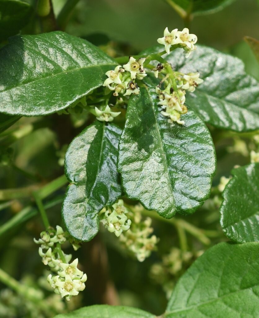
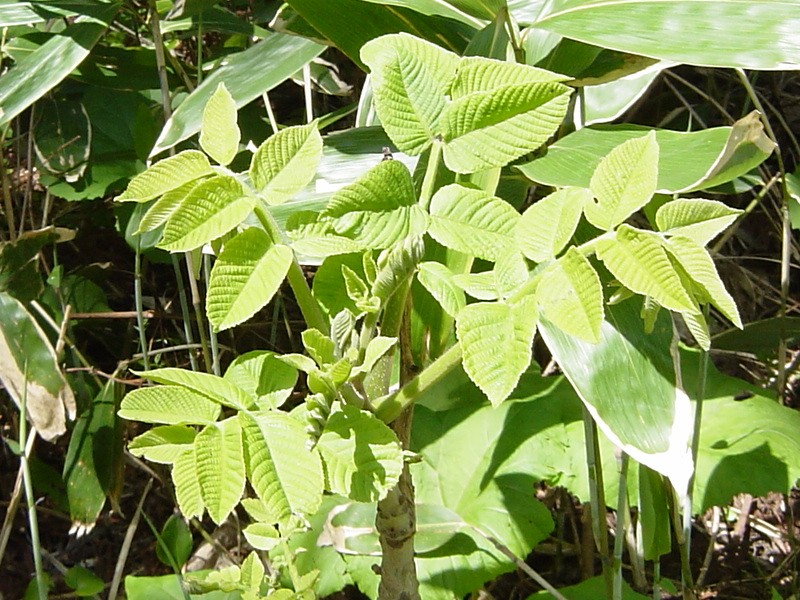
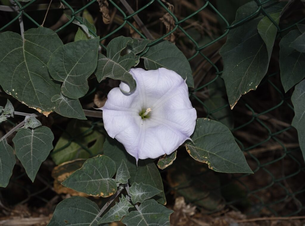
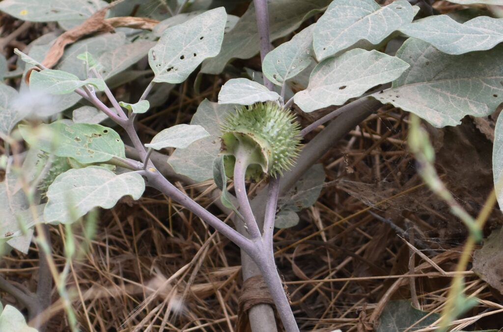
“In spring, the ivory flowers bloom on the sunny hill or in sheltered glade, in summer its fine green leaves contrast refreshingly with dried and tawny grassland, in autumn its colors flame more brilliantly than in any other native, but one great fault, its poisonous juice, nullifies its every other virtue and renders this beautiful shrub the most disparaged of all within our region.” – John Howell1
Most of the people I hike with aren’t especially cognizant of the botanical life around them. If a flower is unusually pretty they might notice. They might even comment on it. For the most part, though, they hike for the scenery, the exercise, and the companionship.
One plant that does get a lot of attention, however, is poison oak. Whenever a patch is spotted, someone is sure to deliver a warning to the rest of the group. “Watch out, poison oak!” is a common refrain. I can’t really blame them. A poison oak rash is no fun. I have a habit of lying down on the ground in order to get a better shot of whatever small creature I’m photographing. I also have a habit of focusing so intently on my subject that I don’t pay attention to what I’m lying in. After one such ill-considered photo session, I came down with the worst case of poison oak in my life. It was on both arms and both legs. (A couple of people asked me why I didn’t have it on the rest of my body. I explained that I don’t normally hike naked.) It itched like crazy, and it lasted for three or four weeks. Calamine lotion, for all its sterling reputation, didn’t make much of a dent in my suffering. It was a long few weeks.
It’s a pity poison oak and its close relative, poison ivy, have such a “great fault”, because they’re actually very interesting and attractive plants. The poison ivy on the east coast of the United States is especially beautiful in the Autumn, when the leaves turn a bright red. As one floral designer put it, “It would be a great fall accent—if you could use it. The trouble is, you couldn’t count on re-orders.”2
The Genus
Both poison oak and poison ivy are in the genus Toxicodendron, which includes a number of other “poisonous” plants such as poison sumac (T. vernix) and the lacquer tree (T. vernicifluum), which grows in Asia.
There are a number of species of poison oak and poison ivy:3
Western poison oak (Toxicodendron diversilobum) – very common in western North America. This is the one I normally see in Southern California. In fact, it’s the most common woody shrub in California.
Atlantic poison oak (Toxicodendron pubescens) – grows in eastern parts of the U.S., mostly in sandy soils.
Poison ivy (Toxicodendron radicans) – grows in all states east of the Rockies. It’s very common in my home state of Virginia. It also grows in Central America.
Western poison ivy (Toxicodendron rydbergii) – grows in northern parts of the eastern United States. It can also be found in the western U.S. and Canada, but it’s much less common than western poison oak.
Asian poison ivy (Toxicodendron orientale) – grows throughout east Asia. It could be a variety of western poison ivy (T. rydbergii), rather than a separate species.
The other member of the genus that most Americans will have heard of is poison sumac. The only species in North America is Toxicodendron vernix. It has leaves with 7 to 13 leaflets, as opposed to the usual three leaflets characteristic of poison oak and poison ivy. It grows as a small tree or a tall shrub. When I lived in Virginia, I often used to see what I thought was poison sumac, but, according to my reading, it’s not nearly as common as most people think, and what is often identified as poison sumac really isn’t. So maybe it was some other type of sumac I was seeing. According to one source, the best way to find true poison sumac is to find a bog, trip, and catch yourself. The plant you catch yourself on will be poison sumac.45
The lacquer tree (Toxicodendron vernicifluum), as the name suggests, is a tree. I’ll have more to say about it in a later section.
Leaves of Three
What English speaker hasn’t heard the rhyme “Leaves of three, let it be?” Unlike many such popular bits of folklore, this one is pretty accurate. Both poison oak and poison ivy tend to have their leaves (technically leaflets) arranged in groups of three. Certainly, there are other plants that have this characteristic, but not as many as you might think, so if you avoid all plants with “leaves of three”, you’ll probably be safe.
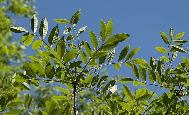
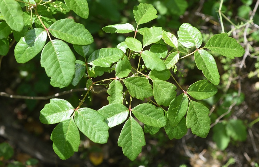
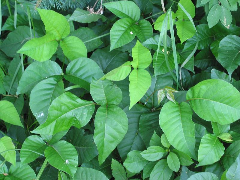
So how can you tell if what you’re looking at is poison oak or poison ivy? Well, where you’re at is your first clue. If you’re on the east coast of the U.S., chances are it’s poison ivy. If you’re here in the west, it’s probably poison oak. This isn’t always the case, though. A more reliable clue is the shape of the leaves. Poison ivy leaves are pointed at the tip. The leaves of poison oak have scalloped, toothed, or lobed edges and have blunt tips.
Western poison oak (Toxicodendron diversilobum) San Diego County 4/4/21
Poison ivy (Toxicodendron radicans) Author: Esculapio This work has been released into the public domain by its author, Esculapio. This applies worldwide. In some countries this may not be legally possible; if so: Esculapio grants anyone the right to use this work for any purpose, without any conditions, unless such conditions are required by law.
I should mention that both poison oak and poison ivy drop their leaves for part of the year. So how do you identify one of these plants if they don’t have any leaves? Well, you could try rubbing one of the sticks on your arm, and see if you get a rash. Otherwise, I’m not sure. Probably, you should just try to remember where you’ve seen the plants growing in the past and avoid that area. (And, yes, you can definitely get a rash from the leafless twigs.)
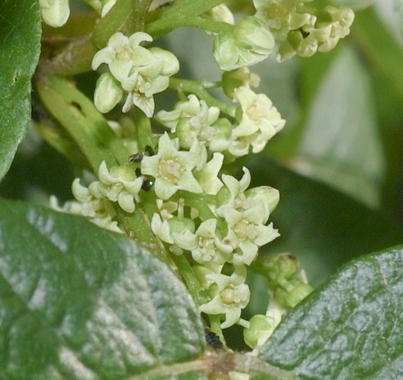
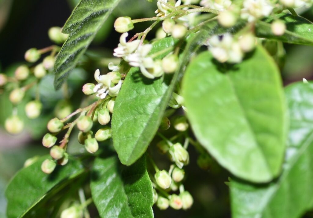
As noted in the quote from John Howell at the beginning of the post, T. diversilobum blooms in the spring with ivory or greenish-white flowers. The flowers of T. radicans are similar.
Western poison oak (Toxicodendron diversilobum) in bloom Mission Trails Regional Park, San Diego County 4/1/21
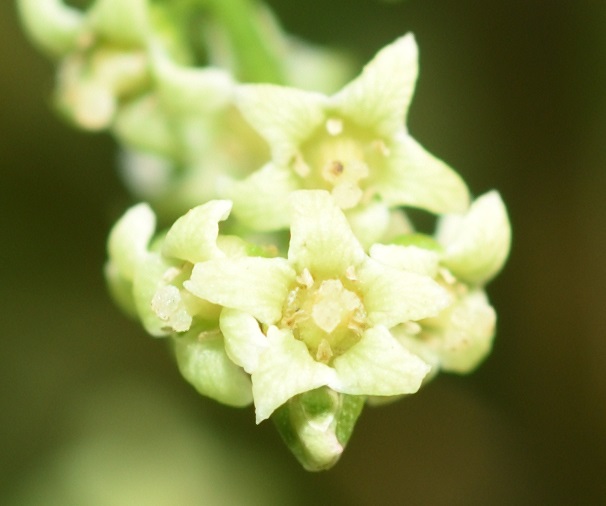
Western poison oak (Toxicodendron diversilobum) flowers Mission Trails Regional Park, San Diego County 4/1/21
@A closeup
When fertilized, they develop into greenish-white or tan berries. Technically, the berries are drupes. A drupe is a fruit that consists of a fleshy outer part surrounding a hard pit, which contains the seed. Poison oak, by the way, is one of the few plants in California with white (or nearly white) berries. Another one is snowberry (genus Symphoricarpos), whose berries are very white and are considered poisonous to humans.7 So another good rule to follow, at least in California, is to not mess with anything that has white berries.
The small, off-white berries of poison oak (Toxicodendron diversilobum) San Diego County 4/16/21
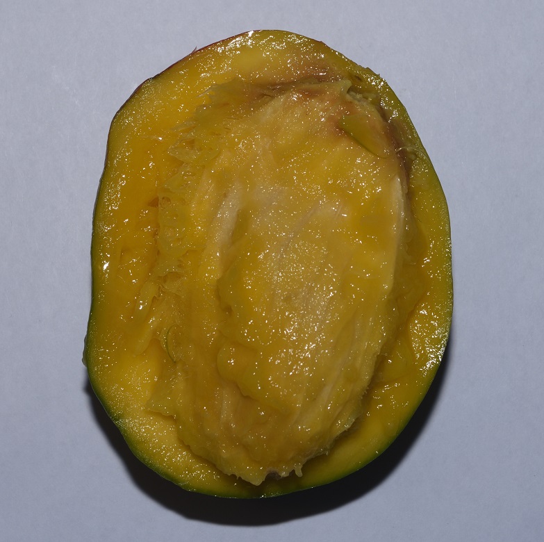
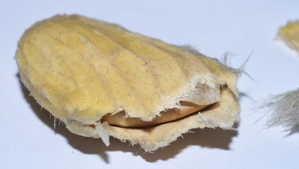
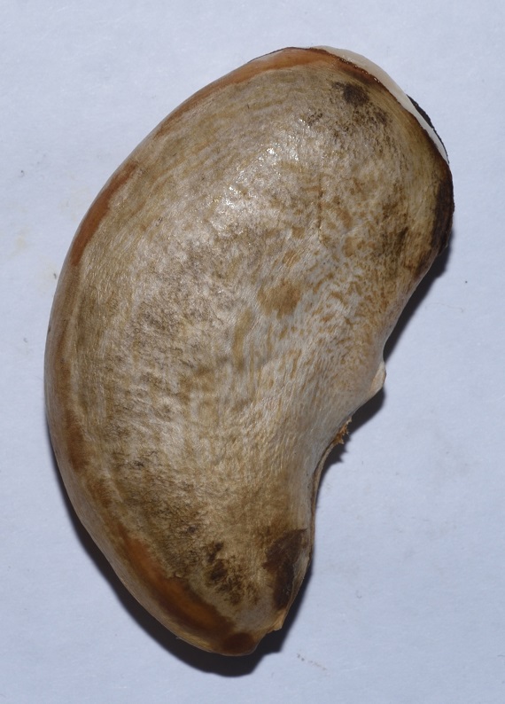
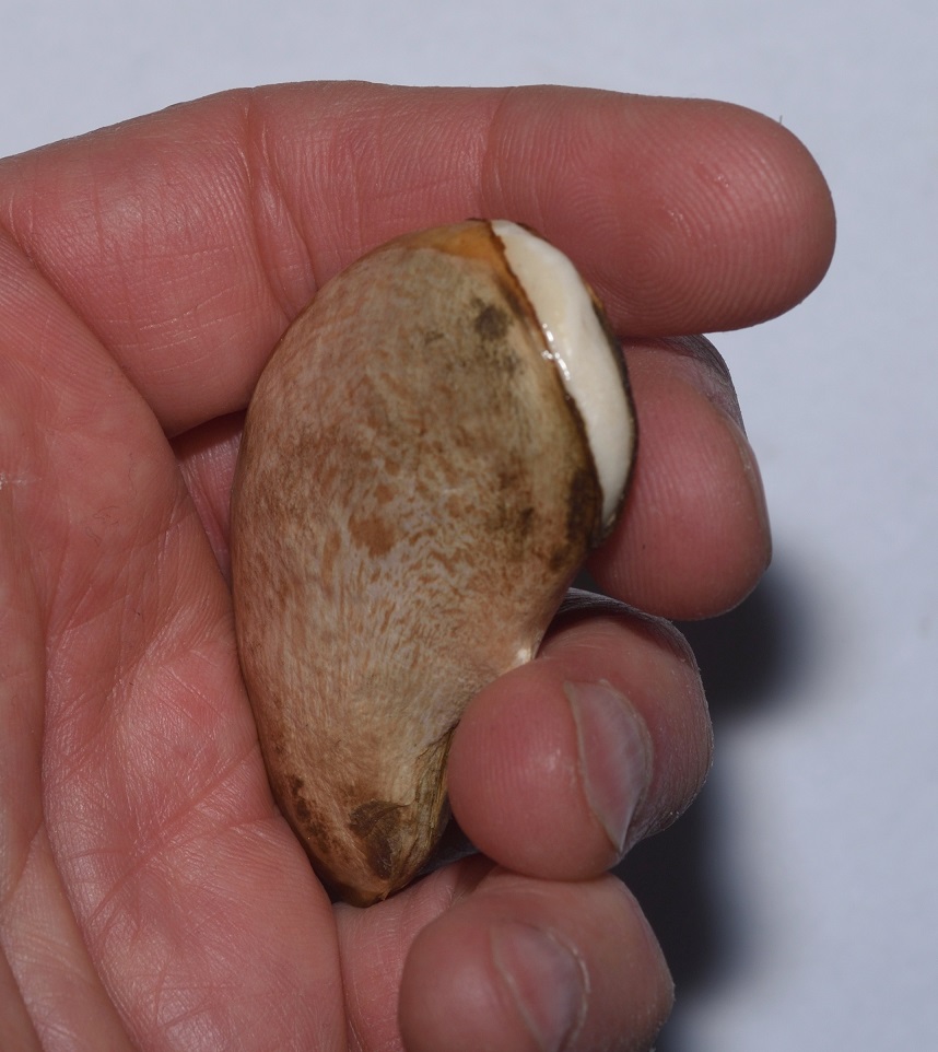
Surprisingly, the genus Toxicodendron, with all its not-so-popular plants, is in a very popular family: Anacardiaceae or the cashew family. Besides cashews (Anacardium occidentale), Anacardiaceae also includes the pistachio tree (Pistacia vera) and the mango tree (Mangifera indica). So, that’s kind of weird, but cashews, pistachios, and mangos obviously don’t contain whatever substance is found in poison oak that causes rashes, right? Well, actually, they do. The substance is an oil called urushiol. In the case of mangos, it’s found in the skin of the fruit, not the meat. If you eat a mango, make sure you peel it first. Also, wash it after you peel it. Your knife can transfer some of the urushiol from the skin to the fruit. Apparently, some people are able to eat mangos with the skin intact without suffering any negative consequences. Presumably, those people would also be immune to the effects of poison oak. But immunity to the effects of urushiol often wears off with repeated exposure. You can be immune to it for years, and then suddenly find yourself reacting. Many people who unwisely eat the skin of a mango will find themselves with a painful rash on their lips. This is known as ‘mango mouth’.910
So, what about cashews and pistachios? Most people seem to be able to eat these without any problem, but, yes, some people are allergic to them. Often, if you’re allergic to one of the three (cashews, pistachios, or mangos), you’ll be allergic to all three, although that’s not always the case.1112 Just as most of the urushiol found in a mango is in the skin and not the meat of the fruit, most of the urushiol in cashews and pistachios is in the shell. Even so, cashews and pistachios are not eaten raw. (If you buy these nuts, they may be described as raw on the packaging, but they’ve probably been roasted at least enough to break down the urushiol.)13
More on Urushiol
I guess it’s a bit late in the game to be mentioning this, but poison oak and its relatives aren’t technically poisonous. Urushiol is an allergen, not a toxin. In other words, it doesn’t directly attack your body. Instead, it causes an immune reaction. This is why you can be seemingly immune to poison oak for years and then suddenly develop an allergy to it. Repeated exposure sensitizes your immune system to the substance, until it finally starts reacting to it.
It’s also worth noting that if you’re already sensitive to urushiol, you can’t build up an immunity to it by repeatedly exposing yourself to it in small amounts. It doesn’t work that way. In fact, you would just be increasing your sensitivity to it.
Why does our immune system react to urushiol? No one knows for sure. I could describe how urushiol “attaches to certain proteins of the skin, where it acts as a hapten, leading to a type IV hypersensitive reaction”, blah, blah, blah, but the fact is, in most animals, this doesn’t happen.14 Remarkably, humans and maybe a few other primates seem to be the only animals that have an immune reaction to the compound! Your dog can wallow in poison oak or poison ivy all day, and it’ll be just fine. You, not so much. The oil dries on the dog’s fur, and it can be transferred to you — over and over again — until you get around to giving the dog a bath.
The name ‘urushiol’ comes from ‘urushi’ — the Japanese word for the Chinese lacquer tree (Toxicodendron vernicifluum). The sap of this tree is tapped to produce lacquer. The sap is filtered, heat-treated, or colored before it’s applied, and the applied sap must then be cured by drying it in a warm, humid environment for 12 to 24 hours. Curing causes the urushiol to polymerize, forming a clear, hard surface. In its liquid state, the sap can cause extreme rashes, even from the fumes. Once cured, it may still cause rashes in some people, but this is uncommon.15
It can be a bit difficult to figure out what to believe when it comes to poison oak and poison ivy, since there’s a lot of misinformation out there. One thing I’ve read is that if you think you’ve been exposed to it, there’s no rush. It takes hours for the oil to penetrate the skin and, as long as you scrub the affected area thoroughly with soap and water when you get home, you should be okay. This doesn’t seem to be correct. The oil actually penetrates very quickly, and if you want to avoid a reaction, you need to scrub it off pretty quickly after coming into contact.
In theory, simply coming into contact with the plant is not enough to get a rash. The oil is contained within the tissues, not on the surface, so, in order to be exposed to it, you have to rupture the tissue containing it. Of course, you have no way of knowing if a plant you come in contact with has already been bruised or injured, in which case, the oil will be there waiting for you. Plus, some people are very sensitive to exposure, so better safe than sorry.
Urushiol is very sticky and, once released, it will dry onto surfaces and can take a very long time to degrade. (According to one account, the oil may stay active for one to five years!16) So if you’ve come down with a rash, it’s best to thoroughly wash everything that might still have some of the oil on it, such as the clothes you were wearing at the time.
Another misconception is that scratching the rash will spread it. The blisters do not contain urushiol, so breaking them doesn’t make the rash worse. You also can’t get it from someone else, unless they still have some of the oil on their skin or clothes.17
A popular belief that is correct is that you can get a rash from burning poison ivy, oak, or sumac and breathing in the smoke. Inhaling the fumes can cause a systemic reaction, as opposed to the usual contact reaction, and a systemic reaction can be much more serious. Symptoms may include headache, nausea, vomiting, fever, swollen lymph nodes, and difficulty breathing. Swelling in the respiratory system may even lead to death.
Calamine lotion can help make the symptoms of a rash more endurable, but it doesn’t make the rash go away any faster. It also won’t do anything for a systemic reaction. In that case, you should go to the doctor as soon as possible.18
Why Urushiol?
So why would a plant produce all that allergenic oil? Usually, when a plant produces a toxic substance, it’s to protect against herbivores. But, as I’ve already mentioned, urushiol is neither toxic nor allergenic to most animals. In fact, birds, deer, goats, and many other creatures happily dine on the urushiol-laden berries of poison oak and its relatives.
We don’t really know why some plants produce urushiol. It’s been suggested that it may protect from bacterial or fungal infection when the plant is injured, and the stickiness of the resin may help to seal wounds. This is all just speculation, though.1920
What of the Pollen?
Some sources will tell you that every part of a poison ivy or poison oak plant contains urushiol. Apparently, this isn’t entirely true, because much of the honey produced in the U.S. contains pollen from one or the other of these plants. Honey bees are quite happy to visit the flowers of poison ivy and poison oak, and a study of honey produced in East Texas found that poison ivy pollen occurred in 83% of the samples. Granted, it wasn’t found in large amounts, so I suppose it could be that the low concentration makes it safe to eat.21 On the other hand, the book “Nectar and Pollen Plants of Oregon and the Pacific Northwest” (Burgett et al. 1989) describes poison oak as a “producer of light honey in areas where it grows in sufficient abundance”. In fact, you can buy “poison oak honey” online.22 (Click here if you want to order some.) So maybe the pollen really is free of urushiol.
“In the summer, in the College garden, the woolly foxglove, Digitalis lanata, is visited by little bees which become stuporose and lie upside down in the flowers, seeming unable to fly away when disturbed.” – Dr Henry Oakeley, Garden Fellow at the Royal College of Physicians (RCP) Medicinal Garden.
As long as we’re on the subject, I would be remiss if I didn’t mention that, although pollen from poison oak and poison ivy seem to occur fairly regularly in honey without causing any ill effects, there is such thing as poisonous honey. Some plants produce nectar or pollen that is poisonous to humans and sometimes even to bees! If enough of this is incorporated into a batch of honey, then it could become toxic.
So why don’t we ever hear of people getting sick or dying from eating bad honey? Two reasons: First, bees usually visit a variety of flowers, so that no one plant species contributes the bulk of the pollen or nectar. When a problem does occur, it’s usually because of a lack of species diversity, forcing the bees to visit flowers that they might normally avoid. Second, when honey does become tainted, it often has a strong, unpleasant taste, so most people would naturally avoid it.
That said, there have been cases of poisoning by honey. Most often, the plant responsible is rhododendron. Rhododendron and other plants in the family Ericaceae produce chemicals called grayanotoxins, which are toxic to both bees and humans. Honey that becomes tainted with these chemicals can cause a reaction called ‘rhododendron poisoning’ or ‘honey intoxication’. I want to emphasize again that this happens very rarely, but it does happen. And sometimes it’s even changed the course of history.
Rhododendron poisoning is said to have caused Xenophon’s Greek army to have retreated from Persia in 399 bc. Later, during the Third Mithridatic War in 65 bc, the Roman general Pompey’s soldiers reportedly suffered casualties, some of them lethal, from eating rhododendron-tainted honey deliberately left behind by the enemy forces. According to historical accounts, consuming such honey would lead to a “feeling of drunkeness, to vomiting and purging, and madness that lasted for days” (Collins).2425
The botanist Frank Kingdon-Ward and his companions experienced rhododendron poisoning while traveling towards Tibet through northern Burma. He describes the symptoms as similar to acute alcohol poisoning. The local Tibetans had apparently built up a tolerance for the toxins, as they were able to eat the honey without suffering any ill effects. (Collins) 26
In 2005, the British writer, Piers Moore Ede, published a travel book, “Honey and Dust: Travels in Search of Sweetness”, in which he describes traveling the world in search of the last of the tribes that still hunt wild honey. In the book, Ede tells of eating the “wondrous toxic honey” collected by the honey hunters of Nepal. This honey, too, was tainted by rhododendron. According to Ede, the effects of honey intoxication “resembled drunkenness at first, but then became visual, like a magic mushroom trip I remembered from university. Painted dots were dripping across my irises like technicolor rain. My body felt light and tingly, filled with warm rushes and heat-bursts. It was wild and strangely wonderful.”2728
“The poisonous weed, being in shape but little different from our English ivie; but being touched causeth reddness, itchings, and lastly blysters, the which howsoever, after a while they pass away of themselves without further harme; yet because for the time they are somewhat painefull, and in aspect dangerous, it hath gotten itselfe an ill name, although questionless of noe very ill nature.” — Captain John Smith31
The term ‘poison ivy’ was coined by none other than our own Captain John Smith of Jamestown fame. Poison ivy is a native of North America, and its first mention in the literature is the above-mentioned quote from Smith. (Poison oak, of course, is so-named because of the resemblance of its leaves to those of oak trees, although the two plants are not closely related.)
European collectors were always looking to buy “exotic” new plants from the New World, and they were willing to overlook whatever disagreeable aspects some of those plants might have presented. In fact, some of those disagreeable aspects might have been regarded as a plus. What uses might medicine make of new substances that had such strong effects on the body? In those days, most medicines were plant-based, so it’s not surprising that physicians would have looked to poison ivy and its relatives for potential new treatments. Throughout the 18th and 19th centuries, scientists experimented with poison ivy as a medicine.
One physician-botanist, André-Ignace-Joseph Dufresnoy, was in the habit of giving botany lectures that were open to the public. During one such lecture, he displayed some leaves of poison ivy and described the ill effects of the plant. A young florist in the audience was skeptical. After the lecture, he approached Dufresnoy and proceeded to test the truth of his assertions by rubbing the leaves on his hands. No doubt the florist came to regret his impulsiveness, but when the rash finally cleared, he discovered that an old sore that he had had on his wrist had completely healed!
When Dufresnoy learned of this miraculous cure, he quickly concluded that poison ivy offered great potential as a medicine. He boiled some leaves in hopes of making an infusion that was safe for internal use. Apparently, this mostly worked, because Dufresnoy tested the concoction on himself and reported only mild side effects. Sweating and urination increased, and he experienced some slight stomach upset. He went on to prescribe such extracts to patients with various skin maladies and to at least one patient with paralysis of the legs. Reportedly, he had some positive results.
Dufresnoy, by the way, nearly went to the guillotine because of his infatuation with poison ivy. During the French Revolution, he sent some young plants to a physician friend. At the time, poison ivy was in the genus Rhus, instead of Toxicodendron, so Dufresnoy later wrote to his friend, asking “How are our dear Rhus? How I long to see them!” The word for Russians in French is ‘Russes’, and the Russians werethreatening to join the wartime coalition against France. Dufresnoy’s letter was intercepted, and the revolutionaries interpreted ‘Rhus‘ as Russians (Russes). (People weren’t particularly consistent with their spelling back then.) Fortunately, the harsh judge who was originally due to hear the case was deposed, and Dufresnoy was able to explain to the authorities that he was just writing about plants.
In the 20th Century, botanists moved the “poisonous” members of the Rhus genus into a separate genus, Toxicodendron, which means “poison tree”. The chemist who identified urushiol as the allergen in poison ivy and figured out its chemical structure was a Japanese scientist named Rikō Majima. Japanese scientists at the time had a hard time competing with Western scientists, who were far ahead of them technologically, so Majima decided to focus on local natural products that western scientists were unlikely to spend much time on. He studied the lacquer tree from 1907 to 1922, eventually extracting the allergen and identifying it as a mixture of closely related products called alkyl catechols.
Poison Ivy in Popular Culture
For whatever reason, poison oak doesn’t receive a lot of attention in popular culture. Poison ivy is another matter. Something about the plant just seems to capture the public’s imagination. Here’s a few examples.
In 1959, the Coasters were the first group to record the song “Poison Ivy”, which was written and composed by Jerry Leiber and Mike Stoller, respectively. Ostensibly, the song is about a woman who “comes on like a rose” and is “pretty as a daisy,” but will “really do you in if you let her get under your skin.” In reality, it’s about the clap (gonorrhea), a subject that didn’t normally get a lot of air time in 1959.
Another example, of course, is the Batman supervillain Poison Ivy. Dr. Pamela Lillian Isley is a botanist and biochemist, who survives a poisoning attempt, only to find that she has acquired an immunity to plant poisons and (perhaps more remarkably) the ability to control plants.32
Poison Ivy is also the name of a popular movie starring Drew Barrymore as a psychopathic teenager. The movie tagline is “What Ivy wants, Ivy gets”. It spawned several sequels.
Finally, Toxicodendron species have played starring roles in many a “poison garden”. As I’ve noted in other blog posts, plants routinely produce toxins to protect themselves from animals that would like to eat them. Animals, such as us, that do eat them, find or evolve ways to reduce the toxicity to a safe level. As a result, the line between toxic and non-toxic is blurred. Nevertheless, some botanical gardens have leveraged our natural proclivity towards sensationalism by fencing off an area which they designate as their “poison garden” and, of course, charging extra to see it. It’s silly, but mostly harmless, fun. If you’re ever in Northumberland, England, you can visit the “Deadliest Garden in the World” at Alnwick Castle;33 Ireland has a poison garden at Blarney Castle;34 and then there was the Medici Poison Garden in Florence, Italy, which I’m not sure ever actually existed, but was supposed to have supplied Catherine de Medici with the poisons that went into her various potions. (She had something of a reputation as a sorceress.)35
Plants breathe carbon dioxide. This means that, in the short term, some plants will actually benefit from the increased CO2 levels associated with global warming. In a six-year study conducted by researchers in North Carolina, an experimental forest was subjected to increased levels of carbon dioxide. The researchers found that the greater availability of CO2 improved poison ivy’s ability to conduct photosynthesis and manage water usage, allowing it to grow larger and more abundant. Not only that, but the increase was greater for poison ivy than it was for similar woody plant species, meaning that, in the future, poison ivy could out-compete other plants and become even more common than it already is. And the bad news doesn’t end there. A higher carbon dioxide level also influences the production of urushiol, leading to a more potent form of the compound.
So, yeah, the future is looking very hot and very itchy.
I can tell you it’s a very disturbing experience to take a drag on your cigarette and find out you don’t have one.. Or to talk to your brother about himself thinking he’s someone else… or having your friends come round to see how you are, having a long chat with them only to realize they are not there. I cannot remember it all as it was only three days ago and I can’t operate properly (it’s taken 2 hrs so far to write this because I can’t see).
I do remember trying to smoke my belt, throwing my cat in the bathtub, thinking my mum was Spike Milligan. The list goes on.
What a long, strange trip it’s been researching this topic. I knew the plant Datura had a reputation as a psychedelic — and a dangerous one at that — but… wow. The above quote is one of the less disturbing ones I found. Many of them contained stuff that I didn’t feel comfortable including in a blog that’s supposed to be more or less family friendly. Of course, Datura didn’t evolve in order to send humans on bad trips, and there’s more to the plant than it’s psychoactive properties.
Datura is a small genus containing nine species, all with similar features. It’s in the family Solanaceae or the nightshades. Solanaceae is an unusual family. It contains many of our most important crops, including tomatoes, potatoes, eggplants, and peppers. Yet, it also contains many famously poisonous plants, such as nightshade, belladonna, and henbane.
It may seem counterintuitive that poisonous plants and food crops should be so closely related, but, in fact, it’s not really that strange. Just as animals tend to defend themselves when attacked, so do plants. And since plants can’t run or bite or sting, they have to rely on more passive methods of defense, such as toxins. If humans are able to eat a particular plant, it’s not necessarily because the plant doesn’t contain any toxins. Our bodies can naturally detoxify some foods, as long as we’re in good health and the toxin levels aren’t too high. Sometimes we only eat the less poisonous parts of the plant and avoid the other parts. We may also process a plant in such a way that most of the toxins are removed. Toxins in some dried beans are removed by soaking and boiling, for example.26
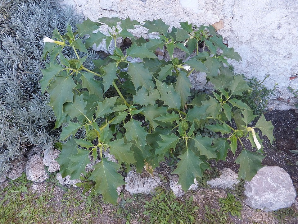
Some of the common names for members of Datura are raving nightshade (my personal favorite), thorn apple, stinkweed, Devil’s apple, jimsonweed, Devil’s weed, Devil’s trumpet, toloache, and moonflower. The most common species here in Southern California is Datura wrightii. This particular species is sometimes called sacred datura, due to its use in Native American religious rituals.2729D. wrightii is native to the Southwestern United States and northwestern Mexico. It’s also widespread in Central California, although the concentration is much higher in the south.37
Sacred datura (Datura wrightii) Datura is one of those plants that’s easy to recognize even when it’s not in flower. San Diego County 4/19/17
Sacred datura (Datura wrightii) Mission Trails Regional Park, San Diego County 5/2/21
Sacred datura (Datura wrightii) Mission Trails Regional Park, San Diego County 5/2/21
Sacred datura (Datura wrightii) with a seed pod Los Peñasquitos Canyon Preserve, San Diego County 9/16/17
Another Datura species, D. stramonium, is found scattered throughout California. According to Calflora38, it’s not native to California, although a number of other sources call it a native of North America, without being more specific.394041D. stramonium is a rather important member of the genus, since it very early on managed to spread to the Old World. It now grows all over the world, wherever the climate is warm and temperate.
D. stramonium looks a lot like D. wrightii, but its leaf margins have much deeper lobes.
Although the name ‘jimsonweed’ or ‘jimson weed’ is often used for other members of the genus, the name originated with D. stramonium. It’s actually a corruption of the name ‘Jamestown weed’. The story goes that some English soldiers were camped out in Jamestown, Virginia while trying to suppress Bacon’s Rebellion.42 They foolishly used the leaves of the plant in a salad and spent the next eleven days tripping out. Here’s a quote from one of the people who witnessed it.43
The James-Town Weed (which resembles the Thorny Apple of Peru, and I take to be the Plant so call’d) is supposed to be one of the greatest Coolers in the World. This being an early Plant, was gather’d very young for a boil’d Salad, by some of the Soldiers sent thither, to pacific the Troubles of Bacon; and some of them eat plentifully of it, the Effect of which was a very pleasant Comedy; for they turn’d natural Fools upon it for several Days: One would blow up a Feather in the Air; another wou’d dart Straws at it with much Fury; and another stark naked was sitting up in a Corner, like a Monkey, grinning and making Mows at them; a Fourth would fondly kiss, and paw his Companions, and snear in their Faces, with a Countenance more antick, than any in a Dutch Droll. In this frantick Condition they were confined, lest they should in their Folly destroy themselves; though it was observed, that all their Actions were full of Innocence and good Nature. Indeed, they were not very cleanly; for they would have wallow’d in their own Excrements, if they had not been prevented. A Thousand such simple Tricks they play’d, and after Eleven Days, return’d to themselves again, not remembring any thing that had pass’d.44
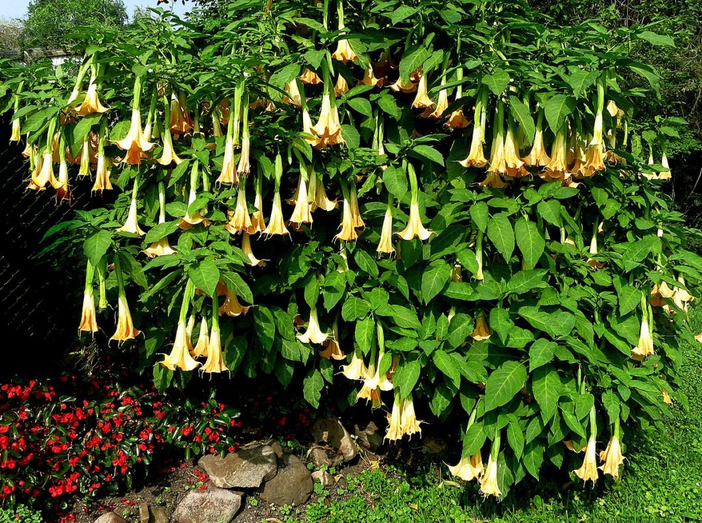
Another genus of plants in the nightshade family is Brugmania. This genus contains seven species and is closely related to Datura. The common name for members of Brugmania is angel’s trumpets. I’m not sure why these plants deserve such a positive rating, while Datura is stuck with Devil’s trumpet and Devil’s weed. It’s certainly not due to their being any less dangerous, as the members of Brugmania are among the most toxic of ornamental plants. Maybe it’s just because angel’s trumpets are woody shrubs or small trees, and their large, trumpet-shaped flowers hang down in such a distinctive manner, making them more popular as ornamentals. Datura is very attractive in it’s own right, but it’s a much smaller plant and the flowers are displayed in a more traditional, upright manner. 45
An interesting thing about angel’s trumpets is that they are only known from cultivars. The genus appears to be extinct in the wild, and it’s suspected that its extinction may be linked to the extinction of whatever animal was responsible for the dispersal of its seeds. If this is the case, then angel’s trumpets only survive today because humans decided to grow them as ornamentals. This would make them evolutionary anachronisms. (See my earlier post Strange Fruit and Other Anachronisms.)
Soon after I moved to California, I saw an angel’s trumpet growing alongside a street in San Diego. It was a hard plant to miss, with its huge, hanging flowers. I’ve got a photo of it somewhere. Until such time as I can dig it up, here’s one I found on Wiki Commons.
On a trip to South Korea in 2012, I visited Jeju Island, a large volcanic island just off the southern tip of the Korean Peninsula. It’s a beautiful place with lots to see, including some of the largest larva tubes in the world. A popular hiking destination is the crater and lake at the top of the central volcano, Mount Halla.46
At this period in my life, I was into oil painting, not hiking, so I wasn’t in any shape to hike all the way to the top of the mountain. Instead, I contented myself with hiking maybe a third of the way. There, I set up an easel and did a small painting. I had just squeezed out a large glob of titanium white paint onto my palette, when a large hawk moth suddenly appeared and hovered over my palette for a second before dipping its proboscis into the glob of paint. It didn’t take the moth long to realize its mistake, and it flew off with a bit of white paint stuck to the tip. It was a memorable experience, although I felt bad for the moth, which may have died as a result of the encounter — oil paint doesn’t come off easily.
So why would a moth do such a thing?
Attracting Pollinators
For millions of years, flowering plants and insects have been evolving mutualistic relationships. The plants provide insects with food in the form of pollen or nectar, and the insects pollinate the plants. When an insect visits a flower, it often gets some of the pollen stuck to its body. When it later visits the flower of another plant of the same species, some of this pollen may rub off onto the pistil of the flower, fertilizing the plant.47 This may sound like a simple, straightforward relationship, but it’s not. There are all kinds of complicating factors. Some insects want to eat other parts of the plant, such as the leaves, that the plant doesn’t want eaten, so plants have to figure out how to protect themselves from the “bad” insects while attracting the “good” ones. Some insects cheat by chewing through the petals to get to the nectar, bypassing the reproductive parts of the flower and doing nothing to pollinate the plant. And then, of course, there’s competition between plants to attract pollinators. As a result of this competition, many plants have evolved to specialize in one particular species or group of insects.
You can often make an educated guess what kind of bug or other animal pollinates a plant based on the color of its flowers. If the flowers are bright blue or violet, there’s a good chance the plant is pollinated by bees. If they’re bright red, hummingbirds may be the target pollinators. If a plant has white flowers, and, moreover, if those flowers open at night, they are probably evolved to attract moths or even bats.48
And therein lies our answer. Bright, white flowers are easier to see at night, so many plants have evolved white flowers to attract moths. And moths, in turn, have evolved to associate large, splotches of white color with a meal. That unlucky Korean moth mistook my blob of white paint for a flower! (In case you’re wondering, no, I wasn’t painting at night. Hawk moths tend to be nocturnal, but many fly during the day, as well.)
Datura flowers tend towards white, although they can also be pale yellow, pink, or pale purple. They also open at night. And they’re big. So, as you’ve probably guessed by now, they are pollinated primarily by big moths, namely hawk moths.
Sacred datura (Datura wrightii) with my hand for scale Mission Trails Regional Park, San Diego County 5/23/21
Sacred datura (Datura wrightii) Mission Trails Regional Park, San Diego County 5/23/21
Sacred datura (Datura wrightii) Mission Trails Regional Park, San Diego County 5/23/21
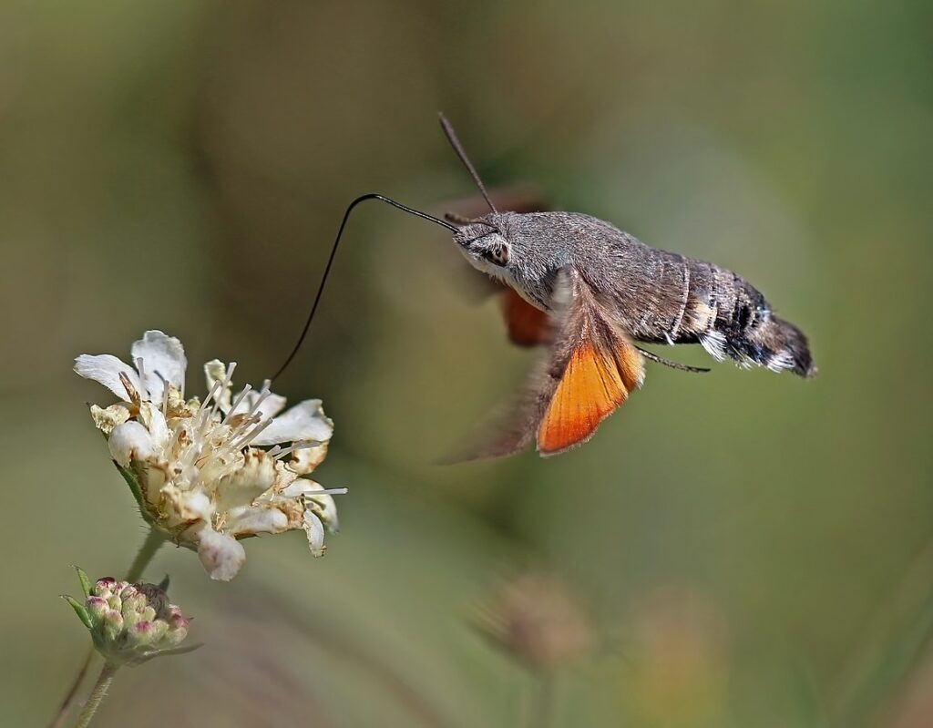
Hawk or sphynx moths make up the family Sphingidae. They are also called hummingbird moths, because from a distance they look remarkably like hummingbirds. They’re about the same size, and they hover in front of flowers to feed just like hummingbirds. The moth I saw in Korea was definitely a hummingbird moth.
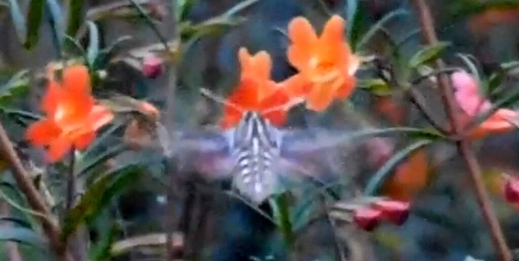
I see plenty of hawk moths here in San Diego County, which isn’t surprising given how much Datura we have. Of course, they feed on other flowers, too. Here’s a bad photo of one feeding from a monkey flower.
A hawk moth sipping nectar from a sticky monkey flower (Diplacus aurantiacus) San Diego County 4/24/17
And here’s a much better photo someone took of one in Bulgaria.
Surprisingly, hawk moths don’t seem to be immune to the psychoactive properties of Datura and are said to sometimes become intoxicated on the nectar.51 As to what form this “intoxication” takes, I read somewhere that they kind of stumble around unsteadily. Unfortunately, I haven’t been able to locate the article I read this in.
One species of hawk moth, Manduca rustica, is especially fond of sacred datura.52 I suspect that many of the moths I see in my area belong to this species.
I did have one other close encounter with a hawk moth. I was hiking at night in Mission Trails Regional Park, and one landed on the back of my wrist. Naturally, it was my right wrist, and my phone was in the right pocket of my jeans, so I had to use my left hand to fumble around in my right pocket trying to get it out. And, naturally, the moth took off the moment I was ready to shoot.
Datura has become a very important model organism for genetic research, largely due to the botanist Albert Blakeslee.54 A normal Datura plant has twelve pairs of chromosomes, but, apparently, it’s relatively easy to create mutant strains that have extra chromosomes. Blakeslee used the chemical colchicine to induce such mutations. He was able to create trisomic plants — plants with three copies of a particular chromosome, instead of a pair.
Time for some terminology. In genetics, the letter n is used as a placeholder for the number of different chromosomes in an organism’s genome. A cell is haploid if it contains only one set of n chromosomes. Most germ cells (reproductive cells) are haploid. If a cell contains 2n chromosomes or two copies of each, it is said to be diploid. In most multicellular organisms, the somatic cells — that is, anything that isn’t a germ cell — are diploid. If a cell has more than 2 sets of chromosomes — that is, 3n or greater — then it’s said to be polyploid. These terms are also applied to organisms as a whole. For instance, if an organism’s somatic cells are polypoid, then the organism is also said to be polyploid.55
Aneuploidy is when an organism’s chromosome number differs from the wild type (i.e. the unmutated type) by part of a chromosome set. For example, an organism’s cells could have two copies of most chromosomes, but three copies of one of them. Such an organism is said to have 2n + 1 chromosomes in its genome. An organism could also have two copies of most chromosomes, but only one copy of one of them. Then, it’s said to have 2n – 1 chromosomes. If an otherwise diploid organism is missing one pair of chromosomes entirely, then it has 2n – 2 chromosomes. This type of mutation is usually lethal.56
Most organisms that have nuclei in their cells (that is, organisms that aren’t bacteria or Archeae) are diploid. We humans have 23 different chromosomes, but most of our cells have two copies of each one for a total of 46 or 23 pairs. So humans have 2n chromosomes, where n = 23.
Using colchicine, Blakeslee was able to produce Datura plants with a chromosome number of 2n + 1. He was even able to produce one plant that was haploid — it only had one set of chromosomes. “This was the first haploid to be found in vascular plants and created something of a sensation. Haploid plants, one thought, should be gamophytes, but here was a little plant, rather weak and nearly sterile, that was clearly a sporophyte, but that had only one set of chromosomes in its cells.”57
Blakeslee’s results inspired a great deal of research into polypoidy. Eventually, a total of six stages in the polyploidy series of Datura were identified (1n, 2n, 3n, 4n, 6n, and 8n). It’s not clear from my reading if Blakeslee himself identified any of the polypoids or if he just produced aneuploids and a haploid, but there is no doubt that his pioneering work was of vast importance to the field of genetics. It eventually led to the concept of genetic balance: “…an organism that has complete extra sets of all of its genes is not very different from a normal organism of its type, whereas an organism that has a single extra chromosome, giving it an extra ‘dose’ of the genes carried on this chromosome and thus changing the ratio of these genes to those borne on the other chromosomes, has a profoundly changed appearance.”58
All species of Datura contain tropane alkaloids such as scopolamine and atropine, which are both toxins and hallucinogens. These chemicals are mostly found in the flowers and seeds. In some species, such as D. wrightii, there may also be high concentrations in the roots.
Within hallucinogens, Datura is classified as a deliriant. Most deliriants, including Datura, are anticholinergic drugs, meaning they inhibit the neurotransmitter acetylcholine. The state of delirium produced is generally characterized by stupor, agitation, restlessness, confusion, and realistic visual hallucinations. Scopolamine and atropine, in particular, are known for their tendency to produce dream-like hallucinations and these hallucinations tend to be disturbing and unpleasant. People who take Datura often lose control of their actions and have little or no memory of the experience.
The real danger presented by Datura, however, is not so much its toxicity or the often disturbing nature of the hallucinations it produces. The real danger is its unpredictability. As already noted, the amount of toxin varies according to the part of the plant, but it also varies according to its stage of development and the environment in which grows. The concentration of toxins decreases during the day and increases at night. It’s also more toxic after a rainy period than a dry period. All this makes it extremely difficult to estimate what a safe dose would be.61
Despite its dangers and the often dark nature of the “trips” it produces, Datura has long been used in folk medicine and religious rituals around the world. What follows are a very few examples of such use.
The Chumash were a Native American tribe who lived in the coastal regions of Central and Southern California, a bit north of where I live.65 They enter this story because, at some point, someone among them used red-ochre to paint an image of a 5-armed pinwheel on the ceiling of a cave. The cave is called Pinwheel Cave and it’s located in the Wind Wolves Preserve of the San Emigdio Mountains. The pinwheel is believed to represent a Datura blossom as it unwinds at dusk. The photo below is one I took of a real Datura blossom unwinding. I don’t have permission to show you any of the photos of the cave painting, but you can click here to see an article that contains a couple.
An unopened blossom of sacred datura (Datura wrightii) Mission Trails Regional Park, San Diego County 5/23/21
The idea that the pinwheel represents a Datura blossom is bolstered by the fact that around 50 clumps of chewed Datura have been found tucked into crevices in the stone alongside the painting. This strongly suggests that the chumash people were using the plant ritually at the time the painting was done around 400 years ago. (Give or take. Radiocarbon dating indicates that the site was occupied from around A.D. 1530 to 1890. I’m not sure how, based on that, it was decided that the painting must be 400 years old. You’ll have to take that up with the authors of the referenced articles.)
Archaeologists have long speculated that some of the more abstract, “psychedelic” art found in rock paintings might have been related to the ritualistic use of hallucinogens of one type or another, but this is the first time that traces of hallucinogens have actually been found associated with cave art. One popular theory is that such caves were the haunt of individual shamans whose artwork represented images from their “vision quests”, and that the caves were avoided by other members of the tribe. However, the number of chewed “quids” of Datura found in Pinwheel Cave, as well as the number of tools and other artifacts littering the cave floor, suggest that the site may have been used communally — maybe in initiation ceremonies or in rituals conducted before a hunt.
Besides the pinwheel, a number of other, less distinct blobs and smears are painted in red-ochre on the cave ceiling. Researchers have interpreted one of the figures as an insect, possibly a hawk moth.
Huskanawing
The Algonquin Indians of Virginia used Datura stramonium in an initiation ritual called huskanawing. The following is based on a description in Beverly’s “The History and Present State of Virginia, In Four Parts”.66
The ceremony took place every fourteen or sixteen years or oftener, depending on the ages of the young men in the tribe. A group of these young men would be selected based on “treasures” they had acquired in the course of their traveling and hunting. They would be taken into the woods, where medicine men carefully fed them a plant mixture called wysoccan, which contained D. stramonium. They would be kept confined there in isolation for several months “giving them no other sustenance, but the Infusion, or Decoction”.67 (This part may sound a bit hard to believe, but I suppose whatever plant matter was in the “Infusion” could have been enough to keep them alive for a few months.)
This steady dose of wysoccan would cause the boys to become “stark, raving mad” for eighteen to twenty days. Not only that, but they would supposedly lose all memory of their lives, their parents, and even their language. The dose would then be gradually reduced, but, before the effects had worn off, they would be brought back into the village, where they were carefully observed to see if they retained any memories of their former lives. If one of them showed any sign of remembering, then he would have to undergo the entire ritual again, only this time with even more severity, and he would be unlikely to survive it.
Once back in their village, the initiates were expected to learn everything over again, even their language, so that they could begin life again as men, having forgotten their previous lives as boys. Obviously, it’s likely that the young men only pretended to have forgotten everything and were very careful not to acknowledge that they recognized anyone in the village, not wanting to have to repeat the ritual (and probably die in the process). On the other hand, given some of the stories of modern people who’ve experimented with the plant, a total, if temporary, loss of memory may not be all that farfetched.
Witches and Werewolves
The medieval belief that witches flew around on broomsticks may have been the direct result of the said witches tripping out on D. stramonium. Supposedly, “flying ointments” were compounded out of the seeds and roots of Datura and applied to the body, producing states of “extraordinary derangement and delusion”. As for the broomsticks, the handles were used to apply the ointment to the “sensitive vaginal membranes “.
Experiments on these banned religious practices were actually carried out by Andres Laguna, a physician to Pope Julius III, and Giovanni Battista Porta, a colleague of Galileo. The results showed that the potions did take the women on “journeys”, but only in their dreams. It was also reported that men would drink potions of Datura and then put on wolf skins and run about on all fours.68
So, did D. stramonium really shape our legends about witches and werewolves? I don’t know, but, given the plant’s documented effects on people, it doesn’t seem like too much of a stretch.
The “Safe” Use of Datura
If Datura is really so dangerous and unpredictable, how is it that so many cultures have apparently learned to use it safely? First, tribal cultures are much closer to nature than we are. They are in everyday contact with the plants and animals they live among, and their knowledge of the attributes and uses of various plants is formidable. Second, the use of plants like Datura would likely have been controlled by shamans (a.k.a. medicine men), who had spent much of their lives learning under what conditions to harvest the plants and how high a dosage to use. And third, in most tribal cultures, Datura was limited to rituals. It probably wasn’t used as a recreational drug.
I hope I don’t come off sounding preachy by emphasizing how dangerous Datura can be. I think some hallucinogens have great potential in medicine and therapy. Some may even be safe to use recreationally, as long as you know what you’re doing. But I don’t think Datura is one of them. It’s just too dangerous. And why would you want to knowingly take a substance that has a reputation for bad trips?
There once was a small frog named the Pacific chorus frog. To scientists, it was known as Hyla regilla, and it had an impressive range, extending from southern Alaska all the way to Baja California. In 1986, the species was moved from genus Hyla to Pseudacris, and it became known as Pseudacris regilla.
There things stood until 2006, when some pesky molecular biologists came along and determined that P. regilla should be split into three species — the northern species would retain the scientific name, P. regilla, as well as the common name, Pacific chorus frog; the central California species would, henceforth, be called P. sierra or the Sierran tree frog; and the southern species, which ranges from southern California into Baja California, was to be rechristened P. hypochondriaca or the Baja California tree frog.
Actually, that’s not quite true. The authors of the 2006 paper used incorrect names for two of the species, and they had to fix their mistake in a subsequent paper. Eventually though, we did end up with those three separate species, and that remains the case to this day. (Well… except that their conclusions are still controversial and some people disagree with the whole thing. Taxonomy is hard.)53
Anyway, I’ll be using the three-species breakdown in this article. Specifically, I’ll be focusing on the southern species, Pseudacris hypochondriaca, simply because that’s the one that I see in my area.
The Baja California Tree Frog (Pseudacris hypochondriaca)636970
Just to avoid confusion — well, more confusion — I should mention that the Baja California tree frog is also called the Baja California chorus frog. The latter name is actually more accurate, since P. hypochondriaca is rarely found in trees. Nevertheless, Baja California tree frog seems to be the more popular name, so I’ll go with that one for this article.
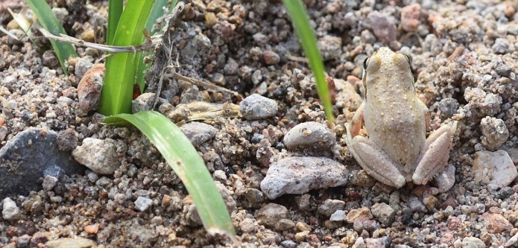
The adult frogs measure around 0.75 to 2 inches (44 to 50 mm) from snout to vent. Their color varies quite a lot, depending on the environment. Usually, they come in green, grey, tan, or brown. They can go from light to dark or vice versa in a matter of minutes, and they can change color entirely over a period of hours or weeks. Often, there are darker markings on the back and legs, which vary in intensity.
Baja California tree frog (Pseudacris hypochondriaca) San Diego County. 4/1/21
One feature that’s pretty constant is the dark stripe that extends from their nostrils to their shoulders and sometimes beyond, passing through the eye along the way.
Baja California tree frog (Pseudacris hypochondriaca) San Diego County. 4/1/21
This photo may give you a better idea of its size. San Diego County. 4/1/21
Here’s a video of a Baja California tree frog. As you can see, this one is a darker gray color than the one in the previous photos, and it has much darker markings on the back and legs.
Here’s another specimen with similar, but not identical, markings to the one in the video.
Baja California tree frog (Pseudacris hypochondriaca) Mission Trails Regional Park, San Diego County 11/4/17
Note the yellow-orange coloration on the bottom of the legs of the frog in the following photo. This is the usual coloration found on the ventral (bottom) surface of their bodies.
Baja California tree frog (Pseudacris hypochondriaca) Mission Trails Regional Park, San Diego County 6/9/17
Baja California tree frogs have sticky pads on their toes, which allow them to cling to surfaces. They don’t usually climb trees, but that’s because they don’t want to, not because they can’t. I put one in a small plastic terrarium, and it was all I could do to keep it from scampering up the side of the container and escaping whenever I opened the lid. It had no trouble at all climbing the almost vertical walls of the terrarium.
Like most frogs, P. hypochondriaca is a powerful jumper. Here’s a slow-motion video of a jump. (The odd sound in the background is my voice slowed waaay down.)
And here’s the same jump at normal speed.
Baja California tree frogs eat a variety of bugs. Like other frogs, they have a sticky tongue that they use to snare their prey. Contrary to popular belief (and every cartoon frog you’ve ever seen), frogs don’t shoot out long, thin tongues to pick off bugs at a distance. In fact, their tongues are large and thick and attach near the front of the mouth, so they kind of roll out of the mouth. Click here to view a video of a tree frog eating.
The tadpoles are suspension feeders71, eating things like algae, bacteria, protozoa, and debris.
When a trait is vitally important to an organism’s survival, you can bet that evolution will come up with some tricks to enhance that trait. The frog’s tongue is no exception. If you watched the video of a frog eating, you might have gotten the impression that the tongue just flops out of the frog’s mouth. In fact, it whips out so fast that it hits the prey with a force of around five times the force of gravity. And it’s not just sticky — it’s coated with “reversible saliva”.
Some materials react very differently according to the type of force being applied to them. Silly Putty is the classic example. It’s soft and pliable in your hands, but when you drop a ball of it, the ball bounces as if it were made of rubber. Ketchup is another example. When you shake a bottle of ketchup, you’re actually changing the state of the ketchup, making it more watery and easier to pour.
Frog saliva reacts to shear forces in a similar way to ketchup, but in reverse. The saliva on the tongue is normally thin and watery. This allows it to seep into the cracks and crevices of the prey when the tongue strikes it. But when the tongue starts to retract, the shear forces produced change the state of the saliva so that it instantly becomes thick and sticky. In fact, it becomes more viscous than honey. It turns watery again when the tongue is back at rest inside the mouth.
A frog’s tongue is ten times softer than a human tongue, allowing it to mold itself around the prey, increasing the contact area. It’s stretchiness causes it to act like a bungee cord, pulling the captured bug toward it and “storing the intense applied forces in its stretchy tissue and dissipating them in its internal damping.”
Hollywood is in southern California, and the most common frog in southern California is, by far, the Baja California tree frog. So, when movie makers needed the sound of frogs croaking in the background, they used the first frog they found, which was, unsurprisingly, P. hypochondriaca. (Well, actually, it could have been the central species, P. sierran, whose range extends close to that area. All three species of Pseudacris seem to have identical calls.)75
It was a good choice. We expect bullfrogs to be loud — bullfrogs are big, after all — but P. hypochondriaca is less than 2 inches long. How loud could it be? Incredibly loud, as it turns out. Their croaks can be heard almost a mile away.
The croak of the Baja California tree frog has been used in so many Hollywood movies, that it’s become the “sound a frog makes” in the popular imagination all over the world. (Spoiler: when you watch Rambo running through the Vietnamese jungle, the frogs croaking in the background are not Vietnamese frogs.) Many languages have invented onomatopoetic76 words for the sound a frog makes. In English, it’s “ribbit”. If you learned as a child that “Frog says ribbit”, you have P. hypochondiaca to thank.
Here’s a video of some Baja California tree frogs ribbiting.
The frogs in the video are croaking over each other, so it might be a little hard to single any one of them out. You can click here for the sound of one frog croaking.
Only the males croak, and they do it, of course, to attract females. One way to tell a male from a female is the loose, darker-colored flap of skin under its jaw. When it’s croaking, the flap inflates with air, becoming a resonance chamber. Here’s a photo of a male in full ribbit.
Males make their calls even more noticeable by coordinating their croaks. The first male to start calling is the “chorus master”, and nearby males gradually join in, creating a “frog chorus”.
Mating in Baja California tree frogs takes place in the water, using a behavior called amplexus. The male crawls onto the female’s back and grasps her with his front legs. Fertilization takes place externally — the female releases her eggs into the water, and the male releases his sperm to mix with them.
Females lay their eggs in small clusters of ten to eighty, which they attach to sticks, stems, or grass in shallow areas. Altogether, a female will lay between 400 and 750 eggs. The eggs hatch into tadpoles, which take two to two and a half months to metamorphose into frogs.
Here’s some photos of P. hypochondriaca tadpoles.
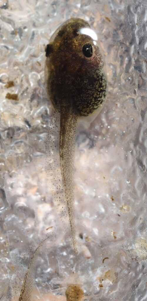
Baja California tree frog tadpole (Pseudacris hypochondriaca) San Diego County. 4/9/21
Baja California tree frog tadpole (Pseudacris hypochondriaca) San Diego County. 4/9/21
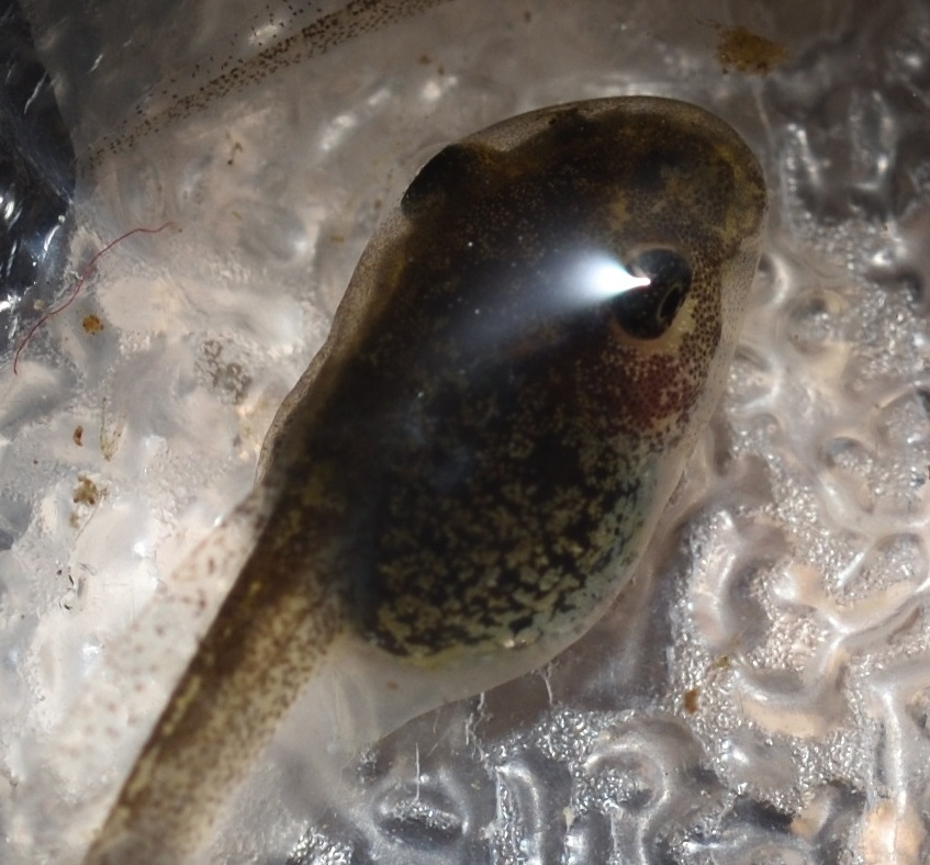
Baja California tree frog tadpoles (Pseudacris hypochondriaca) San Diego County. 4/9/21
Here’s a view of the ventral (bottom) surface. You can see that the mouth is positioned at the bottom of the head, so it can scrape material such as algae from the substrate.
Ventral surface
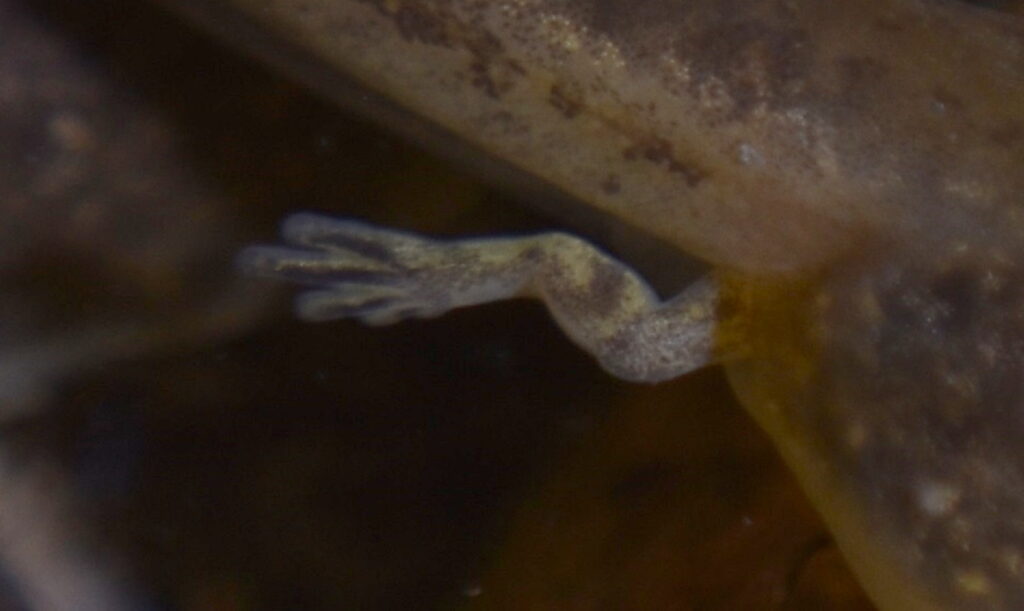
A closeup of a newly formed hind leg on a tadpole. Mission Trails Regional Park, San Diego County 11/4/17
Frogs were not the first amphibians to evolve, but they’ve been around for a very long time. Molecular studies suggest that their origins may extend as far back as the Permian, 265 million years ago, before the rise of the dinosaurs.
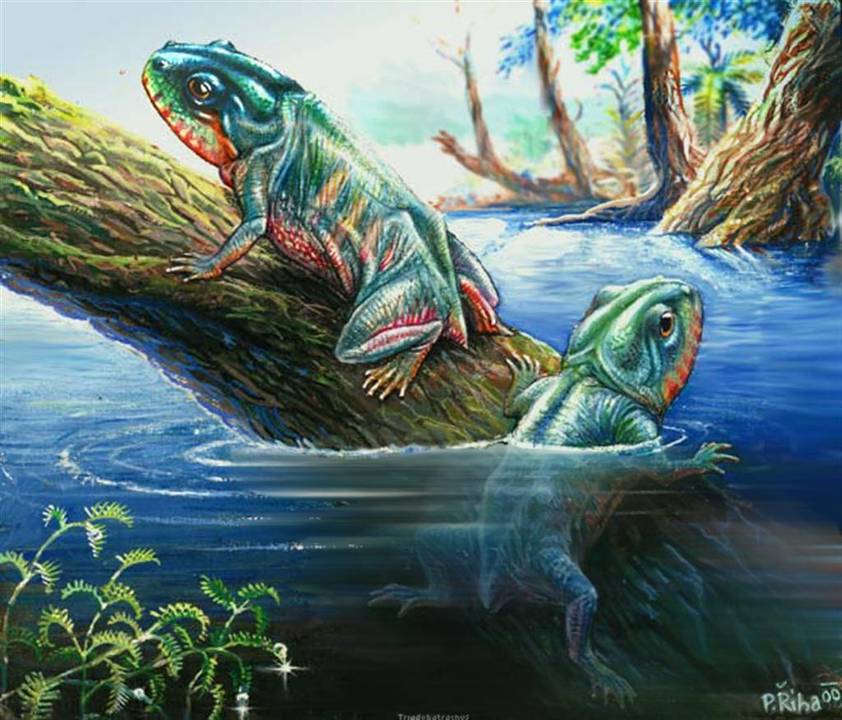
The oldest actual fossil we have in the frog lineage is a “proto-frog” called Triadobatrachus. It lived around 250 million years ago in what is now Madagascar, and it’s a nice example of a transitional fossil — something partway between a true frog and its earlier, less frog-like ancestors. It had a large skull consisting of thin bones, similar to modern frogs, but it couldn’t jump, and it had at least 26 vertebrae. (Modern frogs only have four to nine.) And it may have retained its tail as an adult.7980
Despite their early start, frogs don’t seem to have really come into their own until after the asteroid wiped out the non-avian dinosaurs,81 around 66 million years ago. This left lots of niches vacant, and frogs were only too happy to fill their share of them. Frogs suffered too, of course — an estimated three-quarters of life on Earth died in the aftermath of that cataclysm — but as many as ten types of frogs managed to make it through, and three of those types went on to evolve into most of the frogs we know today. So, how did frogs manage to survive an extinction event that killed off so many other groups? We don’t know for sure, but it could be that, like many modern frogs, they had the ability to spend long periods of time underground — a trait that would have been very useful when everything was going to heck above ground.
Two of those major surviving frog lineages were in what is now Africa and make up the groups Microhylidae and Natatanura. The third lineage, the superfamily Hyloidea, originated in South America, and it is from that lineage that our little Baja California tree frog eventually sprang.
When a bird eats a berry, it digests the pulp and poops out the seed. But not right away. It takes a while for the seed to work its way through the bird’s digestive tract, and by the time it reaches the exit, it’s likely to have traveled a long way from the plant that produced it. In this way, the plant is dispersed.
Every species must have some means of dispersal. Animals can walk or swim or fly to new locations, but plants don’t have that luxury. They have to find more indirect methods of dispersal, and one of the most common is reliance on animals. Usually, this means enclosing the seed in a tough coating that can withstand acidic digestive juices, grinding gizzards, and mashing molars, then wrapping the whole thing in tasty, nutritious pulp that some animal will find irresistible. If the resulting fruit is a small berry, it’ll probably be a bird that’s recruited to disperse it. But many fruits are much larger than your typical berry,77 and larger fruit likely evolved to attract larger, more earth-bound animals.
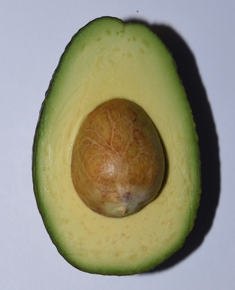
This leads to a mystery. Some plants produce very large fruit that have very tough rinds. And sometimes the seed or seeds inside are also intimidatingly large. Think of an avocado. I’ll help you out — here’s a photo of one.
That is a big pit. What kind of animal could possibly swallow an avocado pit whole? Sure, lot’s of small animals could just eat around it, but that doesn’t help the plant disperse its seed.
Now, I’ll grant you, the avocados of today are the result of thousands of years of domestication, so it’s quite possible that larger fruit have been selected for. But why would the seed also increase in size? In fact, there is evidence that the original version of the fruit had less flesh than today’s avocado. But the pit was actually larger. A larger pit and less pulp — the mystery deepens.
Avocado plants are native to South America, and there is one South American animal that may sometimes eat avocados whole. Surprisingly, that animal is the jaguar! Yes, jaguars are carnivores. Nevertheless, they do occasionally eat avocados, but probably not that often. Also, being carnivores, jaguars are very uncommon. Why? Simple math. A carnivore has to eat lots of other animals during its lifetime, so its prey animals have to greatly outnumber it. For this reason, it’s unlikely that avocados evolved to be dispersed by jaguars — there just aren’t enough of them.83
Maybe you’ve already guessed where I’m going with this. If not, I won’t keep you in suspense any longer. The reason you won’t find any animals in South America large enough to be the dispersal agents for avocados is that they’re all extinct, and they went extinct thousands of years ago!84
The Americas in the Pleistocene
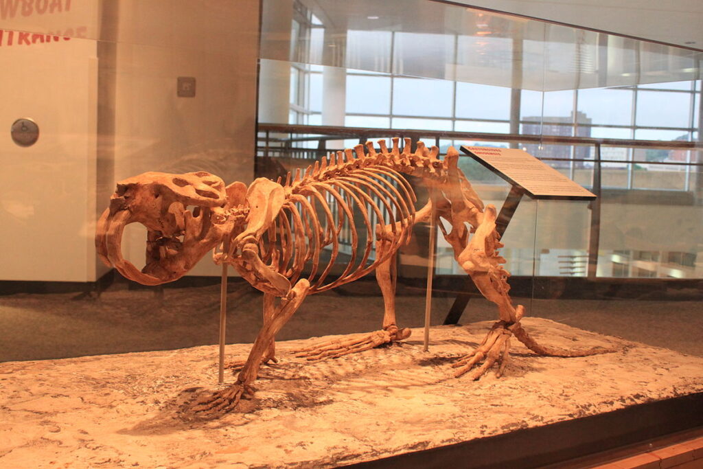
According to current estimates, humans first colonized the Americas around 15,000 years ago. This was toward the end of the Pleistocene epoch, which lasted from 2,580,000 to 11,700 years ago.85 The Americas in the Pleistocene were very, very different from the Americas we know today. And I don’t just mean no cars or roads or white picket fences. I mean, there were some seriously large animals roaming the landscape. 15,000 years ago, North America was home to mammoths, mastodons, giant ground sloths, lions, camels, wild horses, short-faced bears, saber-toothed cats, a beaver the size of a bear, and lots of other beasts you’ve probably never heard of.
Giant beaver (Castoroides ohioensis) Minnesota Science Museum: Mississippi River Gallery Author: Ryan Somma from Occoquan, USA This file is licensed under the Creative CommonsAttribution 2.0 Generic license.
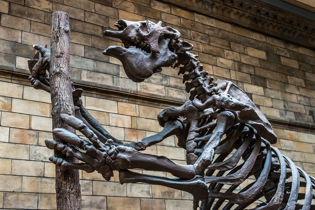
In South America, the situation was much the same, although the selection of large mammals was different. North and South America were separated until around 2.8 million years ago when the Isthmus of Panama formed, so the southern continent evolved its own set of mammals.86 Giant ground sloths actually evolved in South America before crossing the isthmus into the north, so South America did have them.
If you’re not familiar with giant ground sloths, you should look them up. The largest one, Megatherium americanum, was around 20 feet long, 4 tons, and about the size of a modern elephant. On its hind legs, M. americanum would have stood around 12 feet (3.5 meters) tall!8788
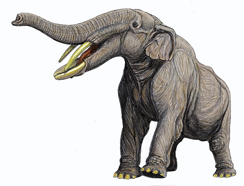
South America didn’t have any mammoths or mastodons, but it had gomphotheres, a family of elephant-relatives, many of which easily held their own in the behemoth department.89
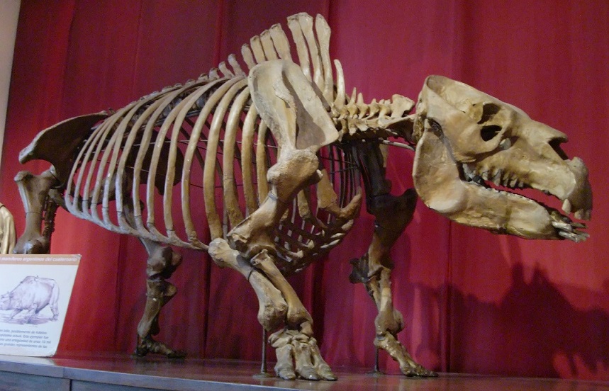
Another large South American mammal of the Pleistocene was the toxodon. Actually, Toxodon is an entire genus nested within an order of extinct hoofed animals called Notoungulata. The most well-known species of toxodon is Toxodon platensis. It’s estimated to have weighed over a ton and was around the size of an American bison or an African black rhino.90
Yet another group of large extinct mammals from South America are the glyptodonts, members of the suborder Glyptodontinae. They were closely related to modern armadillos and had a similar armored body plan. In fact, molecular studies have found that the glyptodonts actually were armadillos — Glyptodontinae is nested within the family Chlamyphoridae, which includes most modern armadillos.
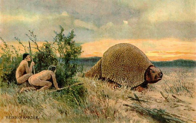
The best-known of the glyptodonts belong to the genus Glyptodon. These were about the size and weight of a Volkswagon Beetle.919293 Here’s a very old (probably 1920) drawing of a glyptodon.
This is just a small sampling of the large mammals that inhabited the Americas of the Pleistocene epoch. Note that all the mammals mentioned here are actually genera or even families. In other words, there wasn’t just one type of gomphothere or one type of glyptodont. These were groups of animals that contained many, many variations. The New World had an incredibly diverse collection of very large mammals .
Scientists use the term megafauna to mean very large animals. How large varies somewhat — the term is a bit loose. Some people define megafauna as including any animal with a mass as great or greater than a human being. Others apply the term only to really big animals — those weighing in over a ton. For the purposes of this article, I’ll stick with the first definition.94
Today, most of the megafauna are gone from North and South America. In fact, most of the megafauna are gone from everywhere except Africa (and we’re working on that). As mentioned, the megafauna were mostly extinct in the New World by 13,000 years ago. (A few literal die-hards, like the giant sloth, managed to hold on until about 11,000 years ago). So what happened to them? Well, humans showed up in North America around 15,000 years ago. 2,000 years later, most of the big mammals were extinct. Seems like a no-brainer.
Actually, some pretty good arguments have been made that humans couldn’t have driven all those animals to extinction in such a short period of time just by over-hunting. It’s possible that other factors, such as climate change, were involved. Still, it’s highly suspicious that most of the megafauna went extinct so soon after humans appeared. Maybe it was a combination of human over-hunting and some other factors. I don’t know. The important thing is, by about 13,000 years ago, most of the really big mammals were gone.
In modern Africa, large herbivores such as elephants routinely take large fruit into their mouths whole, mash the soft pulp, and swallow the seeds whole, along with the pulp. Later, they poop out the seeds in another location, along with a generous helping of natural fertilizer. Many African plants have evolved large fruit specifically in response to the presence of large megafauna such as elephants, rhinos, giraffes, and so on.9697
There’s no reason to think that things were any different in the Pleistocene of North and South America. Many plants, including the avocado, must have evolved to rely on the large megafauna for dispersal. When these animals went extinct, the plants were left without their primary dispersal agents, which means it suddenly became much harder for them to spread their seeds. Now, 13,000 years later, these plants are still producing ridiculously large fruit — as if they were still expecting a giant sloth or gomphothere to show up. And, in a sense, they are. 13,000 years is a blip in evolutionary time, especially for plants, which tend to evolve more slowly than animals. It’s as if the plants haven’t yet “noticed” that the megafauna are gone!
When a fruit evolved to be eaten by an animal or group of animals that are no longer around, botanists refer to such a fruit as an evolutionary anachronism. An anachronism, in the general sense, is something that is out of place in time. If you’re reading a story that’s supposed to be taking place in the Middle Ages, and one of the characters uses a slang term from the 21st Century, that’s an example of a literary anachronism. Botanists and evolutionary scientists have adopted the term to describe these strangely out of step fruit.
But strange fruit are not the only examples of evolutionary anachronisms. In general, anytime a species preserves intact a physical or behavioral trait that evolved in response to evolutionary pressures that no longer exist, the trait is said to be an example of an evolutionary anachronism.
So, maybe you’d like to see an example of an evolutionary anachronism that doesn’t involve fruit…
The American pronghorn is sometimes called the American antelope. It’s not a true antelope, although it does resemble one. In scientific terms, it’s a member of the order Artiodactyla, the even-toed ungulates. This is a large order, which includes deer, pigs, true antelopes, giraffes, and (surprisingly) hippos.
The pronghorn is the fastest land mammal in the New World. It’s been clocked at 55 mph for 0.5 mi (88.5 km/h for 0.8 km). The interesting thing about that figure is that it’s too fast! Nothing alive today in North America is anywhere near fast enough to catch a pronghorn. Why would an animal evolve to be so much faster than it needs to be?
The answer, of course, is that it wouldn’t. There’s nothing alive in North America today that can catch a pronghorn, but what about back in the Pleistocene? When I was listing the various megafauna that lived in North America during the Pleistocene, I forgot to mention cheetahs. That’s right, cheetahs used to live in what’s now the United States! Other predators, such as hyenas, short-faced bears, and lions, might also have posed a threat to pronghorns.
So the American pronghorn is likely an example of an evolutionary anachronism — a prey animal that’s still ready to run super-fast at the first glimpse of a cheetah or whatever other scarily-fast predator might have been around all those years ago.
When I defined the term ‘evolutionary anachronism’, I specified that it had to be an ‘intact’ trait. In other words, the trait cannot have noticeably devolved. Let me give you an example of what I mean.
Many animals that live their entire lives in caves have lost their eyesight. In some cases, they’ve lost their eyes entirely. In other cases, they still have eyes, but those eyes no longer function. In other words, they’re vestigial. If, for whatever reason, a species with vestigial eyes were to migrate out of its cave and re-colonize the surrounding area, the environmental condition that caused animals to evolve eyes in the first place — namely, the presence of light — would reappear. But its vestigial eyes would not start functioning again. By this time, the species has presumably been blind for many thousands of years. So, it’s too late — its eyes are permanently broken.
On the other hand, the pulp and seed of an avocado are still functional. If an animal were to come along tomorrow that liked the taste of the pulp and was big enough to swallow the fruit whole, it could easily fill the role of disperser for the plant. Ergo, the avocado is an anachronistic fruit, not a vestigial one.
So, it’s likely that the fruit of avocados evolved to be swallowed whole by animals like gomphotheres and giant sloths. Such animals were very common for most of the Pleistocene, and, sure enough, avocados had a much larger natural range back then than they do today. But, if the animals that were responsible for dispersing them have been extinct for all this time, why haven’t avocados also gone extinct?
Well, note my use of the phrase ‘natural range’ in the last paragraph. Most of the avocado’s megafaunal dispersers went extinct towards the end of the Pleistocene, but not quite all of them. There was one survivor: humans. Fortunately for the avocado, people like how they taste. And people can be very good dispersers when they want to be. So, it may be that the avocados would have gone extinct if humans hadn’t decided to fill in for the sloths and gomphotheres.
Then again, maybe avocados could have held on for a long time without humans. Just because their primary dispersers went extinct, that doesn’t mean that nothing would ever disperse them. I already mentioned the possibility that jaguars might occasionally disperse an avocado seed. It’s very unlikely that jaguars could have kept the avocados going indefinitely, but they might have enabled them to hold on for a little while, if only by the skin of their non-existent teeth.
Most fruits float. They didn’t evolve for floating. It’s merely a side-effect of the way they’re constructed — most fruits are light enough and waterproof enough to float for a while. For that reason, many plants that appear to be anachronistic can still be found alongside rivers. When the river floods, some of the fruit get washed downstream, and, with luck, some of them may come to rest in a place where they can take root.
Even animals that don’t routinely disperse a plant’s seeds may occasionally disperse them. For example, an animal may be in the habit of eating the pulp of a fruit and spitting out the seeds or eating around them. This doesn’t help the plant because it doesn’t disperse the seeds. Unless, of course, the animal decides to carry the fruit someplace else before it eats it. In that case, the discarded seeds may still be able to find a suitable new location in which to germinate.
If a seed has evolved to survive passage through the digestive tract of an animal, it has necessarily evolved a thick coating. This could make it very hard for the seed to absorb water and germinate. For this reason, many plants have evolved seed coatings that are just barely tough enough to make it through the target animal’s guts. In fact, many won’t make it through, but those that do should be scarified enough by the passage that water will be able to penetrate the coating. The seed absorbs the water, swells up, and germinates. Thus, many plants have evolved seeds that almost have to pass through an animal’s gut if they are ever to germinate.
It’s a tricky balancing act. A seed that hasn’t passed through a digestive tract may eventually germinate, but it may take months or even years. So, if a plant’s primary dispersers are gone, it may still be able to hold on for a long time thanks to that very occasional seed that doesn’t pass though an animal’s gut, but manages to germinate anyway.
But many plants may not be able to hold on without their primary dispersers. Plants rarely fossilize, so we have no way of knowing how many plants went extinct along with their megafaunal dispersers. It seems likely that at least some did, especially the ones for which humans didn’t have any use.
More Strange Fruit
I’d like to spend the rest of the article showcasing a few interesting, and very strange, fruit that are likely anachronistic. It turns out that grocery stores are great places to go hunting for the “ghosts” of extinct megafauna.
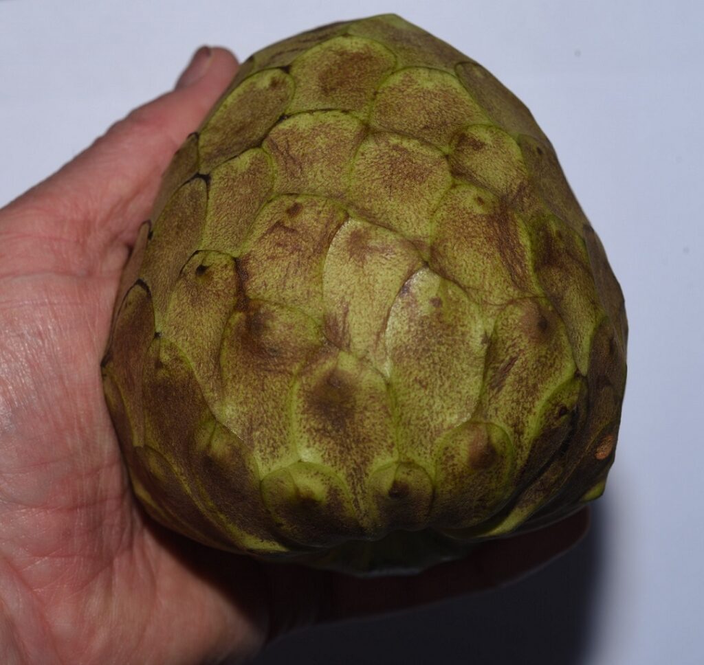
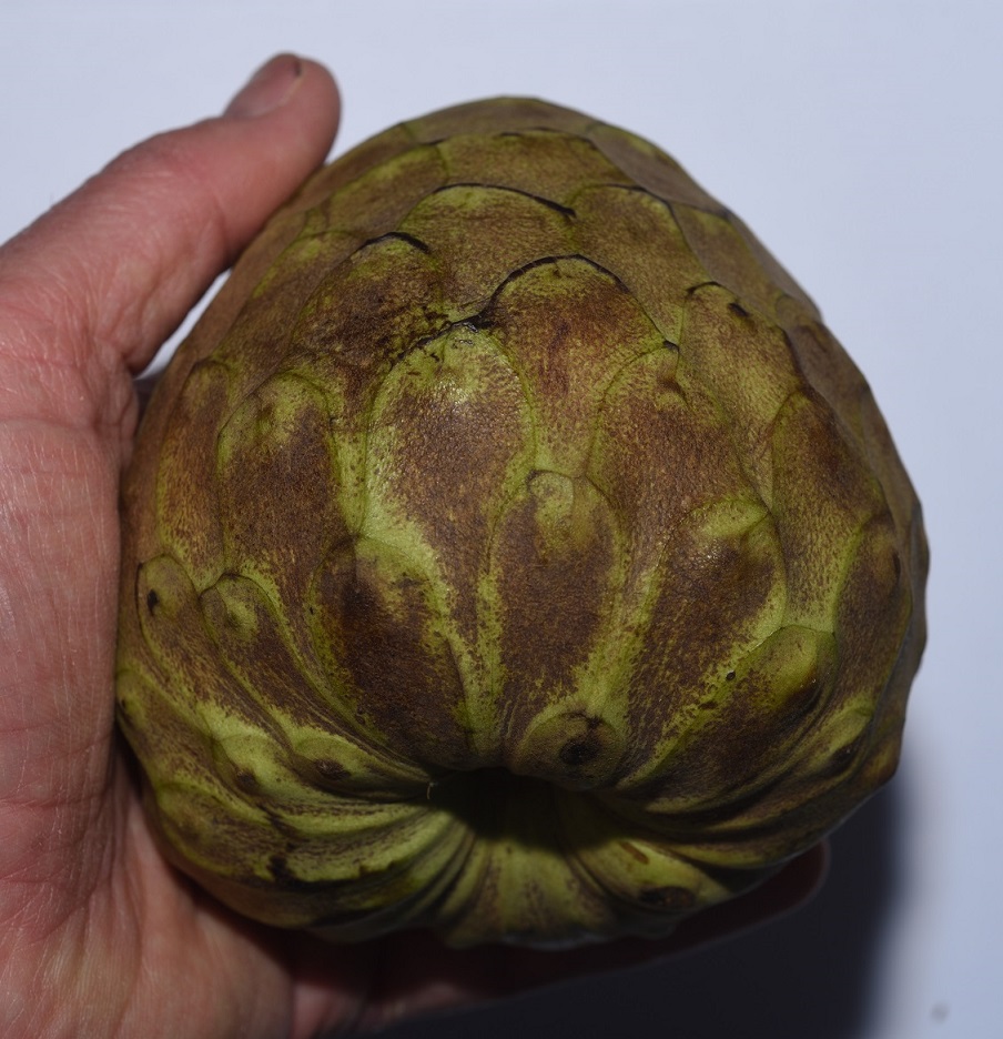
That’s what Mark Twain called the fruit of cherimoya(Annona cherimola), a plant that grows from central Mexico, through Central America, and down the west coast of South America, almost to its very tip. I won’t argue with Twain’s assessment. The one I tried was exquisite, delectable, scrumptious… just seriously good. Its taste is complemented by the texture of the flesh, which is similar to custard and gives it the other common name it goes by — custard apple.
My only complaint is that the seeds are numerous and evenly distributed throughout the pulp, so that you have to spit them out with every mouthful. This makes eating one a bit of a hassle. If someone could come up with a seedless variety, I expect it would take the world by storm.
Cherimoya
I googled “seedless cherimoya” just to see if, by chance, anyone had come up with one. I found a number of articles referencing work done at UC Davis that isolated a mutant gene in a closely related fruit — the sugar apple — that caused it to be seedless. It seems like it would be a simple matter to knock out the same gene in cherimoya, thereby producing a seedless variety. But the articles I saw were from 2011, and I haven’t found any follow up articles, so I guess it’s not that easy.103
Cherimoya
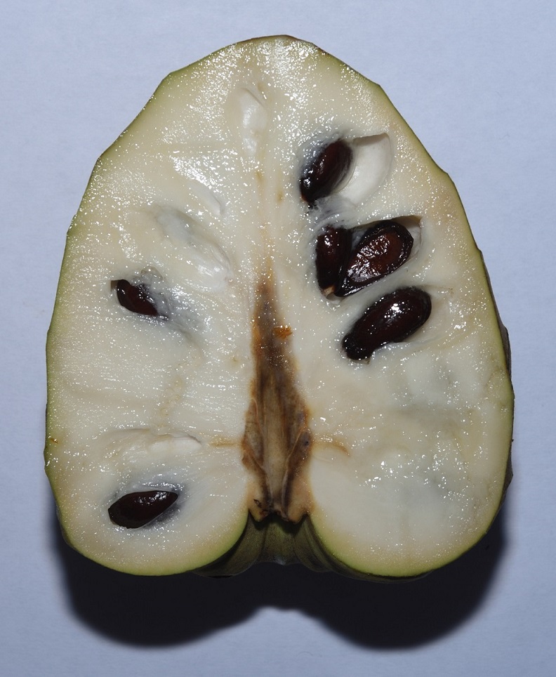
A cherimoya fruit sliced down the middle
The best evidence that cherimoya is anachronistic is its large size. The seeds are also fairly large and not super easy to separate from the pulp, so it seems likely that the fruit evolved to be eaten by large mammals that would mash the fruit and swallow the pulp along with the seeds.
Sliced for eating
A cherimoya seed
A cherimoya seed dissected
There are at least two other edible fruits produced by plants in the Annona genus that may be anachronistic: soursop (Annona muricata)104 and sweetsop or sugar apple (Annona squamosa).105. Both are similar in appearance to cherimoya, but I haven’t tried them, so I don’t know how they compare taste-wise.
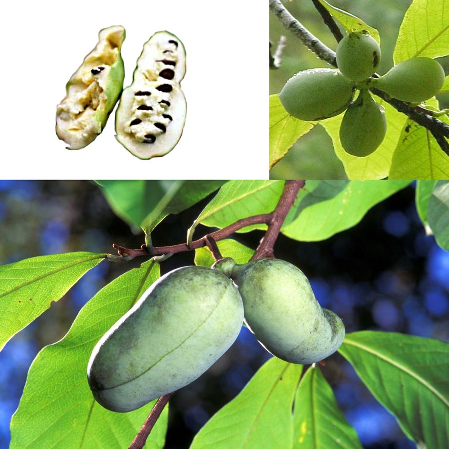
I’ve never eaten a pawpaw (Asimina triloba). Chances are, you haven’t either. In fact, it’s possible your only clue to the existence of this fruit is the lyrics of that old folksong. (If you’re not familiar with the song, well now… you should just click here and have a listen.)
The plant is a small deciduous tree that’s native to the eastern United States and Canada. It’s in the same family, Annonaceae, as the aforementioned cherimoya, and is quite closely related. Supposedly, the fruit has a “sweet, custardish flavor somewhat similar to banana, mango, and pineapple”.108 The fruit has a shelf life of only about three days, which explains why you don’t find them in supermarkets. If you’re lucky, you might find some in a roadside stand. Otherwise, unless you happen to live way down yonder near a pawpaw patch, you probably won’t run across them.
Pawpaws (Asimina triloba) Author: Agnieszka Kwiecień (Nova) Permission is granted to copy, distribute and/or modify this document under the terms of the GNU Free Documentation License, Version 1.2 or any later version published by the Free Software Foundation; with no Invariant Sections, no Front-Cover Texts, and no Back-Cover Texts. A copy of the license is included in the section entitled GNU Free Documentation License.
Several things point to pawpaws being anachronistic. First, of course, is their large size. The seeds are also fairly large, although not ridiculously so. Second, the fruits tend to fall from the tree within a day of ripening, and then they rot very quickly. This suggests that they are targeted towards animals large enough to pick them directly from the tree.
Third, pawpaw patches are rare, except where indigenous people routinely eat them. This suggests that their only remaining primary dispersers may be humans. Finally, wild pawpaw trees usually only grow in floodplains, and yet, they grow just fine when they’re planted in uplands. This, again, suggests that people are their only remaining primary dispersers.
The jackfruit (Artocarpus heterophyllus) has the distinction of being the largest fruit of all trees. Here’s a photo of a jackfruit that was found in Kerala, India.
The one I bought in an Asian market wasn’t quite that big, but it wasn’t exactly small, either. Here it is on the cutting board.
Jackfruit
And here’s one of my grandnephews for scale.
Mother, son, and jackfruit
I don’t know if anyone has ever been killed by a jackfruit falling from a tree, but it wouldn’t surprise me. Then again, maybe people just instinctively avoid walking under something that looks like this.
When I cut my specimen open, I was surprised how tough the rind was. It took some effort to cut through it. I was also surprised by how sticky the insides were. The pulp stuck to my hands like glue. It took a lot of soap and water to get it off. It was also kind of slimy. My grandnephew (the older one, who’s four) refused to eat it because it was “too slimy”. (This is the same kid who routinely handles earthworms and thinks they’re “cute”!)
A jackfruit cut open
The fruit is very good. According to Wikipedia, it has a “subtle pineapple- or banana-like flavor”.111 Personally, I thought it tasted more like cantaloupe.
A closeup of the pulp and a few seeds
My grandnephew holding a couple of seeds
The seeds can also be eaten. I boiled and ate a bunch of them. After 10 minutes of boiling, the water was stained a dark reddish-brown from all the tannins. After boiling, the seeds have to be peeled, which is no fun, but they do have a nice, sweet taste to them.
So is jackfruit anachronistic? It’s important to realize that, like the avocado, and like most fruit consumed by people, the jackfruit has been domesticated. It’s probably quite a bit larger than the original, wild variety. But also like the avocado, the original fruit was probably still pretty big, and the seeds likely haven’t changed significantly.
So the huge size, tough rind, and large seeds strongly suggest a fruit that evolved to be mashed in the mouths of megafauna and the seeds swallowed whole without grinding. The presence of high levels of tannins in the seeds would be a deterrent to insects, but it would also make them distasteful to any large animal that bit into them or ground them between its molars.
So what kind of megafauna are we talking about? It’s not known exactly where jackfruit originated, but they’re believed to be indigenous to the rainforests of the Western Ghats in southern India. This area is a hotspot of biodiversity. Among the many large animals found there is the Indian elephant (Elephas maximus indicus) — an obvious candidate seed disperser for the ancestral jackfruit. Turn the clock back around 50,000 years, and India was home to, not one, but two giant elephants — Palaeoloxodon namadicus and Stegodon namadicus. P. namadicus, in particular, may have been the largest land mammal that ever lived. According to one estimate, it weighed around 24 tons (21,772 kg) and stood 16 feet (4.8 m) tall at the shoulder.112 The largest mammal and the largest fruit? Seems like a good match.113
Mango
The mango (Mangifera sp.) is another fruit that probably originated on the Indian subcontinent. The most common variety, appropriately named the common mango, is Mangifera indica. I’m not sure what variety I got when I visited the local grocery, but here it is.
A mango (Mangifera sp.)
And here it is cut in half. Notice how the pulp clings to the pit.
A mango sliced in half
In the next photo, I’ve extracted the pit. Notice how large it is compared to the fruit as a whole. I actually used a scouring pad to remove most of the pulp from the pit and I still couldn’t get it all off — that’s how tightly the pulp is bound to it. This is no accident. A large animal eating the fruit isn’t going to spit out the pit — that would be a waste of pulp — it’s going to mash the fruit and swallow both pulp and pit.
…and with the pit extracted.
I continued my dissection, extracting the seed from the pit.
Papayas (Carica papaya) are sometimes called pawpaws, but they are unrelated to the pawpaw (Asimina triloba) discussed earlier. They have the usual large size, suggestive of an affinity for megafauna, but the seeds are a surprise.
A papaya (Carica papaya)
Instead of one huge seed or a few dozen moderately large ones, the papaya has lots of very small seeds concentrated in a large central cavity. This is an interesting variation. The large cavity reduces the weight of the fruit, so the stalk attaching it to the tree doesn’t have to be quite as thick. The large number of seeds increases the chances that one of them will successfully germinate and grow to maturity.
A papaya cut in half
The seeds themselves are fascinating. They’re wrapped in some sort of gelatinous pouch. Here’s a photo of a whole seed (right) and a seed with its pouch removed (left).
A papaya seed with its covering removed (left) and with its covering intact (right)
I dried this one off as best I could…
…before slicing it open.
Papaya seeds are edible and taste a bit like horseradish. I would love to know more about that gelatinous pouch. I’m not familiar with any other type of seed that has such a covering. I’ve been searching for more info online, but all I’ve found so far is some stuff about papaya seeds’ reputed health benefits and a lot of rather dubious articles extoling their value for detoxing and parasite cleansing.
Papaya is thought to be native to Mexico, Central America, and northern South America. Several members of the papaya family can be found growing wild in the tropical valleys of the Andes. If papaya originated in South America, then, like the avocado, it was probably dispersed by South American giants such as ground sloths, gomphotheres, and toxodons.
The osage-orange or hedge-apple (Maclura pomifera) is a small tree or large shrub, which has a small natural range in parts of Texas, Oklahoma, and Arkansas. The fruit is neither an orange nor an apple. It’s technically edible, but no humans actually eat it, because it contains an unpalatable milky latex. Even horses don’t much care for it, although one of its many names is horse-apple.
In fact, nothing much eats it. When the fruits ripen, they drop to the ground. There, unless they roll downhill or get dispersed by a flood, they rot underneath the tree that produced them. This is, itself, a clear sign of anachronism. Nothing today eats them, but something must have eaten them at one time — something that’s no longer around. Molecular analysis indicates that M. pomifera probably diverged from other members of Maclura during the Oligocene, around the same time that some groups of mammoths, mastodons, and ground sloths first appear in the evolutionary record. This suggests that these mammals could have been the original seed dispersers for the plant.
You might be wondering, if the latex contained in the osage-orange is distasteful to horses and people, why would these larger mammals be any different? The fact is, many plants that people do eat produce chemicals that are toxic to some degree. Peppers, for instance, are eaten by no other animal other than humans. Large mammals, such as us, are able to handle limited amounts of mild toxins, and much larger mammals, such as elephants, are able to eat some fruits that would be toxic or unpalatable to us. Partly, this is because their slower digestion gives them more time to break down the toxins. Bad-taste is often an indication that something is not safe to eat. If something is safe for an animal to eat, chances are it won’t taste bad to that animal. So, to a mammoth or ground sloth, an osage-orange might have tasted pretty good.
Although we don’t eat the fruit, people have managed to find other uses for osage-orange. The wood was prized by Native Americans as the best wood for making bows (from which it gets another one of its common names, bois d’arc, which is French for bow wood.) They prized it so much, in fact, that they would travel hundreds of miles to get it.
The common name ‘hedge-apple’ refers to its use by European settlers as living fences or hedges around their fields to keep out free-range livestock. Pruning the plants would cause them to sprout adventitious roots from their bases. These would interweave, forming a dense, thorny barrier.
The use of osage-orange as hedges allowed it to become naturalized far beyond its limited natural range. The invention of barbed wire eventually ended its use by farmers, but, by that time, it had become well-established in much of the U.S. It’s still occasionally used as an ornamental.
I’ve concentrated on fruits in this article because they represent the most obvious examples of anachronisms, and because most of the research has been focused on them. This is probably only scratching the surface of the subject, though. One thing I haven’t mentioned is the presence on some plants of ridiculously large thorns — thorns that seem unnecessarily large to fend off the tiny herbivores the plants have to deal with in their modern environment. In the case of trees, these thorns often go far up the trunk — far beyond the reach of any modern animal — and then stop. Plants don’t waste resources any more than they have to. If nothing can reach beyond a certain height, then there’s no point in growing thorns beyond that height.119 There are many more examples of evolutionary anachronisms that I haven’t covered here, and as more research is done on the subject, many more examples will undoubtedly be found.
One final thing I should point out is that whether or not a particular trait is anachronistic rarely, if ever, has a simple yes or no answer. The person responsible for originating the concept of evolutionary anachronisms is a scientist named Dan Janzen, and as Janzen points out “…each trait is to some degree anachronistic.” The reason for this is simple. Evolution is slow. When the environment changes, it takes time for poorly adapted organisms to die out, leaving better adapted ones to carry on. This means that organisms are not really adapted to their current environment — they are adapted to their past environment.
Another important point is that the fruits I’ve been discussing are probably not adapted to a particular species of megafauna, or even a particular genus or family. As has already been pointed out, plants tend to evolve more slowly than animals. If most of a plant’s original dispersers have gone extinct, chances are, another set of dispersers will have evolved to take their place. (At least, that was the case until humans came along and upset the apple cart.)
All this leads to the concept of degrees of anachronism. Some organisms represent extreme anachronisms and are probably on their way to extinction (unless, of course, humans have stepped in and taken the place of their former dispersers.) Other organisms may have evolved traits in response to environmental conditions that have long since changed, but they still manage to get by pretty well, and they will probably have time to adapt to the new conditions. As in all things biological, it’s complicated.
Evolution has become something of a unifying theme for these blog posts. It didn’t start out that way. My original plan was just to write short pieces about the subjects of some of my photos. As time passed, however, the articles grew longer and more detailed, and since, as the evolutionary biologist Theodosius Dobzhansky wrote, “Nothing in biology makes sense except in the light of evolution”, 108 I inevitably found myself writing more and more about the evolutionary history of my subjects.
Of course, evolutionary history is sometimes hard to come by. The fossilization process favors organisms with hard parts, living in specific kinds of environments. As a result, the fossil record for many organisms is very sparse — sometimes even nonexistent. Fortunately, these days we can infer a lot about evolution from molecular studies. Unfortunately, funding for such studies is limited, so they’ve only been done for a small fraction of taxonomic groups.
For this post, I thought I’d write about a spider. I first considered writing about the western black widow (Latrodectus hesperus). Another possibility I considered was the green lynx spider (Peucetia viridans). Both are visually striking, and I have some very nice photos of both species. There’s also a fair amount of info online about them. The problem was, I couldn’t find much about their evolution.
Then I had an idea… L. hesperus and P. viridans are markedly different, both visually and behaviorally, yet they are both members of the suborder115Araneomorphae, the most widespread and “modern” of living spiders. So I thought, why not write about both in the same article? This would allow me to compare and contrast them, while speculating about the evolutionary forces that led to their remarkably divergent traits.
As I turned the idea over in my head, I thought of the many other spiders I’ve photographed over the years. One of my favorites is the spotted orbweaver (Neoscona crucifera). It, too, is in Araneomorphae, and although it lacks the striking coloration of L. hesperus and P. viridans, it makes up for it by having a body completely covered in spiky bristles. In the right light, this makes for some great photos.
And then there’s the silver argiope (pronounced are-guy-oh-pee) or Argiope argentata, also in Araneomorphae. This large spider has a body that looks like it’s made of polished silver!
So, in the end, I decided I would write a single blog post that covered all four spiders. It’s a bit of an experiment, but we’ll see how it goes.
A female western black widow (Lactrodectus hesperus) Mission Trails Regional Park, San Diego County 10/3/18
You’ve almost certainly heard of black widows. You’ve probably seen more than a few of them. Actually, there are several species that are commonly called black widows. All are members of the genus Latrodectus — the “true” widows. (There are also “false” widows, such as the noble false widow, which is very common in Southern California. It doesn’t look much like the true widows, in my opinion, but I guess most people don’t look all that closely.) Not all widows are black, though. There’s also a brown widow (Latrodectus geometricus), which is found around the world, but probably originated in South America.121 There’s even a red widow (Latrodectus bishopi), which is found in Florida.
There are three species of Latrodectus in North America that are known as black widows. The southeast has the southern black widow, L. mactans. That’s the one I used to see in Virginia, before I moved to the West Coast.122 Then, there’s the northern black widow, L. variolus, which is most common in New Jersey, Delaware, and Maryland.123 Finally, there’s the one that’s native to the western U.S. — the western black widow or L. hesperus.124
A female western black widow (Lactrodectus hesperus) Mission Trails Regional Park, San Diego County 10/3/18
Adult females of L. hesperus are usually shiny black with an hourglass-shaped red mark on the bottom of their abdomen. The hourglass can sometimes be yellow or, very rarely, white. Males are about half the length of the females and are usually tan colored.
Black widow males are rarely seen, as they are much shorter lived than the females. As with humans, the male’s only real purpose in life is to find a female and mate with her. If he’s lucky, he may get to mate several times before he dies. The term “widow”, by the way, is taken from the supposed habit of the female to eat the male after copulation. While this does occur, it probably happens more frequently in captivity than it does in the wild. (It’s harder to make a hasty retreat when you’re doing it in cage). Cannibalism seems to be even less common among western black widows than it is among some other members of the genus.
Latrodectus is in the cobweb family, Theridiidae. Like many cobweb spiders, L. hesperus spins messy, tangled webs. After constructing the top part of the web, the spider constructs “gumlines” running from the web to the substrate beneath. These gumlines are stretched taught, and they’re coated near the bottom with sticky silk. If a bug walks into one of the these lines, the sticky silk clings to it. When the insect struggles to free itself, the gumline breaks free from the substrate and snaps up, propelling the unlucky bug into the tangled web above.
While we’re on the subject of webs, the male black widow has an interesting habit. When he finds a female he wants to mate with, he starts by trashing her web. A male will cut out large sections of a female’s web and bundle them up in his own silk. Why, you ask? Researchers have found that this makes the web less attractive to rival males. Male spiders find females by following the scent of their pheromones. One theory is that when a male cuts up a female’s web, he’s selectively cutting up the parts where the female’s pheromones are the most concentrated. He doesn’t destroy the whole web — that would only cause the female to get to work building a new one. Typically, he’ll destroy about half of it — just enough, presumably, to deter other males, increasing the chances that it will be his sperm the female will use to fertilize her eggs. 125
Black widows enclose their eggs in round, pale, cream colored sacs. The newly hatched spiderlings have totally different colorations from the adults, as you can see in the following photos.
Western black widow spiderling (Latrodectus hesperus) fresh out of the egg sac. Mission Trails Regional Park, San Diego County 10/3/18Another angle
I can’t possibly leave the subject of black widows without discussing their bite. Black widows are probably the most feared spiders in North America because of the supposed danger their venom poses to humans. And, to be fair, black widow venom is reputed to be 15 times stronger than that of the prairie rattlesnake!126 Of course, that’s by volume, and a spider is going to inject a much smaller volume of venom than a rattlesnake (assuming the rattlesnake injects any venom at all, which it often doesn’t. See my earlier blog post: Rattlesnake!) 127
So, how dangerous is their bite? Not all that dangerous, actually. It is painful, and it can cause nausea, muscle aches, and paralysis of the diaphragm, which might make it hard to breathe. Nevertheless, most bite victims suffer no lasting damage. That said, a bite can be fatal to small children, the elderly, or people in poor health.128
Given that very few people actually die from black widow bites, it’s a bit surprising that they have such a bad reputation. Perhaps it’s because that shiny black body with its bright red hourglass just makes them look kind of evil. Then again, maybe it’s because, before indoor plumbing, most black widow bites happened in outhouses.
You see, outhouses tend to smell. And those kinds of smells attracts flies. Spiders eat flies. Probably for this reason, black widows like to hang out under the toilet seats of outdoor privies. And, definitely for this reason, most black widow bites are on penises. To quote from the article where I found this lovely bit of trivia: “A 1944 case study published in the Annals of Surgery noted that, of 24 black widow bite cases reviewed, eleven bites were on the penis, one was on the scrotum, and four were on the buttocks. A full 16 of the 24 victims were bitten while sitting on the toilet.”129
Green lynx spider (Peucetia viridans) with a captured honey bee San Diego County 10/3/17
The green lynx spider is a beautiful spider. Yeah, yeah, I know… No spider is beautiful! They’re horrid, ugly creatures that should be wiped from the face of the earth! So, why are you reading this post again? I suspect you don’t really believe that. More likely, like me, you know how to appreciate a beautiful spider when you see one. And this is one beautiful spider.
A lynx spider, in general, is any member of the family Oxyopidae. Most lynx spiders are ambush hunters. Despite this, they tend to be fast runners and jumpers, and have excellent eyesight. Apparently, their rapid movements and agility reminded someone of a lynx; hence, the common name. Most species, including P. viridans, have large, stiff bristles on their legs, which act as spines, helping them hold on to their prey.
Another view of the green lynx spider from the last photo with its captured honey bee San Diego County 10/3/17
Lynx spiders tend to be drably colored and small to medium in size. Members of the genus Peucetia, however, tend to be larger, and they’re often a vivid green. This helps them blend in with the green shrubbery where they’re usually found.
A male green lynx spider (Peucetia viridans) Mission Trails Regional Park, San Diego County 7/7/21
A closeup of the pedipalps — the two appendages on the front of the head. The complex structures on the ends are the palpal bulbs. The spider loads these organs with sperm and uses them to transfer the sperm to the female’s receptacle during mating.132
A view from above
A view of the distinctive eye pattern of a lynx spider. We’re looking toward the front of the head from a little bit above the body. So the front of the head is on the right in the photo.
Like black widows, P. viridans constructs a roughly globular egg sac. However, the lynx spider’s sac is covered with large protrusions. I don’t know what’s inside the sac that causes the protrusions. When I get a chance, I’ll dissect one and post what I find.
Green lynx spider (Peucetia viridans) with egg sac Los Penasquitos, San Diego County 9/16/17
A green lynx spider tends to flee when approached by a human. Unless, that is, it’s a mother spider guarding its eggs or offspring. In that case, she not only stays put, she may even spit venom into the intruder’s eyes! This behavior was first reported in the 1940s by a WWII soldier who suffered impaired vision and conjunctivitis (pink eye) when a green lynx spider sprayed him. The behavior was confirmed 40 years later by Linda Fink, a researcher who was sprayed on 15 different occasions by mother spiders guarding their egg sacs. (I assume she was wearing eye protection.)133
The green lynx spider very rarely bites humans, and, unlike the black widow, it’s bite is not dangerous to people. (It also doesn’t typically hang out under toilet seats.)
Spotted orbweaver (Neoscona crucifera) Batiquitos Lagoon, San Diego County 10/7/18
Note: I should probably mention that Wikipedia and at least one other website I found described N. crucifera’s range in the U.S. as extending west only as far as Arizona. This appears to be incorrect, as the iNaturalist website shows a large number of observations in California, including the San Diego area, where I live. My own photos of what I believe to be spotted orbweavers have been identified as such, both on iNaturalist and on BugGuide. I’m not an expert on spider identification, though, so if anyone wants to set me straight, feel free to post a comment.
The spotted orbweaver, as the name suggests, builds orb webs. These are flat, vertically-oriented, wheel-shaped webs — the kind you typically think of when you think ‘spider web’. They tend to build them more or less at eye level, so I’ve gotten many a spotted orbweaver web in my face while hiking at night, when they’re most active. The spiders usually build their webs at dusk and take them down before sunrise. The webs are quite large, which contributes to the frequency with which I blunder into them.
In constructing orb webs, N. crucifera is very different from both lynx spiders, which don’t build webs for prey capture, and black widows, which build cobwebs. Another thing to notice when comparing these three spiders is that both black widows and spotted orbweavers have very tiny eyes. The lynx spider, on the other hand, has large eyes — especially the front two — and has excellent vision for a spider.
This makes sense. L. hesperus and N. crucifera both use webs to trap their prey. All they have to do is sit in their webs and wait. When a bug gets caught, the vibrations it sets up in the strands of silk let the spider know exactly where it is. The spider doesn’t really need to see at all when it comes to finding prey.
The lynx spider, however, is a hunter, not a trapper. Granted, it often hunts by ambush, sitting still and waiting for something to wander by, but since it doesn’t use a web, it’s important for it to be able to see what it’s ambushing. Plus, lynx spiders don’t always wait in ambush. Sometimes they actively stalk prey.
Spotted orbweaver (Neoscona crucifera) Mission Trails Regional Park, San Diego County 8/10/18Another viewA closeup of the abdomen
The bite of N. crucifera is not dangerous to humans, and the spider is not aggressive. When it does bite, it’s comparable to a bee sting.135
Silver argiope (Argiope argentata) Mission Trails Regional Park, San Diego County 7/26/18
Maybe I shouldn’t have raved quite so much about the green lynx spider. The silver argiope certainly gives it a run for its money in the beauty department. That silvery metallic sheen makes it look a little like a (maybe slightly morbid?) Christmas ornament.
Like N. crucifera, the silver argiope is an orbweaver. Both the spider and the web are large and conspicuous. Unlike the spotted orbweaver, A. argentata is diurnal (active during the day). And, since it tends to build its webs on prickly pear cacti, it don’t have to worry too much about running into them. (And if I did run into one, I’d probably be more focused on the cactus than the web.)
Silver argiope (Argiope argentata) Mission Trails Regional Park, San Diego County 7/26/18
As if its large size and striking coloration weren’t eye-catching enough, the silver argiope builds decorations called stabilimenta into its web. In mature spiders, these stabilimenta take the form of zigzag patterns like the ones visible in the above photo. Often, the spider will build four zigzag patterns and arrange its front and back legs to line up with them. It’s not known if these decorations are for defense or for luring prey. Arguments have been put forth in support of both possibilities. One suggestion is that they make the web more visible to birds, so they’re less likely to fly into them.
Silver argiope (Argiope argentata) Lake Murray, San Diego County 7/22/18
Many other spiders build stabilimenta into their webs (although N. crucifera is not one of them), and the decorations vary in design from species to species. I’ve seen some very impressive ones in photos online. In Southern California, though, I haven’t seen anything that compares to the web decorations of A. argentata.
Spiders make up the order Araneae, which is subdivided into three suborders. As mentioned, all four of the spiders discussed here are in one of those suborders, Araneomorphae. Araneomorphae was the last of the three suborders to evolve. And, since the araneomorphs represent, by far, the most widespread and successful of the spiders, we’re probably justified in thinking of them as the most “modern” spiders.
The araneomorphs have a number of features that tend to give them advantages over other spiders, but probably the most important are their innovations in silk production. Araneomorphs have evolved entirely new forms of silk, which allow them to build webs capable of catching flying insects. This has greatly expanded their ability to find prey. Eventually, one group of araneomorphs developed major ampulate silk or MA silk — the toughest of the silks — and this, together with the development of flagelliform silk and aggregate silk protein glue, has allowed them to create the beautiful orb webs for which they are famous.
Two of the spiders in this article weave orb webs: the spotted orbweaver and the silver argiope. This might lead you to assume that they’re more “evolved” than the western black widow and the green lynx spider. Surprisingly, this is not the case. These latter two spiders likely evolved from orb weavers. In fact, the largest clade141 of araneomorphs — the RTA142 clade — for the most part, don’t use silk to capture prey!
It’s strange to think that after evolving all these sophisticated varieties of silk, so many spiders would then abandon building webs for prey capture, but having a large repertoire of new silks allowed spiders to explore new methods of prey capture that were not possible with the earlier silks. For example, bolas spiders target male moths, which they attract by giving off pheromones that mimic the pheromones of female moths. The spider hangs by a line of MA silk, and dangles another line below it, which ends in a droplet of sticky liquid silk. When the spider detects a moth close by, it swings the droplet at the insect. The liquid silk is able to soak past the dusty coating of scales and stick to the moth’s wings or body, and the spider reels it in. This technique was not possible until the araneomorphs developed the sticky protein glue used in the droplet. It also would not be possible without the strength of MA silk.143
A lynx spider doesn’t use silk at all in prey capture, but it hunts above the ground, in bushes or trees, and it’s likely that it never would have gotten off the ground if it hadn’t evolved from web weaving ancestors.
As far as black widows and other cobweb spiders are concerned, transitioning from an orb web to a cobweb might seem like a step backward, but that’s just our human tendency to think of cobwebs as less advanced because they’re less aesthetically pleasing. In fact, the cobweb has some advantages over orb webs. Perhaps the most important one is that it surrounds the spider, protecting it from predatory wasps. And, as we’ve seen, a cobweb can be supplemented with gumlines to increase its effectiveness at prey capture.
When you look at insects, you find a dizzying variety of forms and lifestyles. Most have wings, but not all. Most have six legs, but some have none. They include predators, herbivores, and detritivores. Spiders, by comparison, are more conservative. All spiders (with the exception of one herbivorous species) are carnivores. All have the same general body plan, although they’ve refined that body plan in countless ways: enlarging, shrinking, and stretching parts, and adding colors, spines, and sensory hairs.
The reason spiders are more conservative when it comes to form and lifestyle can probably be attributed to the essential part that silk production plays in their lives. No insect produces more than one variety of silk. A single spider, on the other hand, may produce half a dozen varieties, which it uses for many different purposes. Tinkering with the composition and use of silk is largely responsible for the great variety of spiders we see today.
Still, silk isn’t the only thing that evolution has tinkered with in spiders. The lynx spider, which is mostly an ambush predator, has evolved to blend in with its environment. The black widow, which has unusually potent venom and doesn’t need to blend in, has taken the exact opposite approach — developing a pitch black coloration with a bright red hourglass mark on its abdomen. These two features act as a warning to would-be predators that they don’t want to mess with it. (It’s not always an effective warning, as some wasps, such as dirt daubers, routinely prey on black widows.)
The reason for the polished silver coloration of the silver argiope is not clear, but it’s known that it reflects UV light. Many flowers reflect UV light to make them stand out to pollinators, so maybe the silver color makes the spider look like a flower to approaching insects.
So, why are some spiders covered in hairs, while others… not so much? First, I should mention that they aren’t technically hairs. Hair is a mammal thing. The bristles on spiders evolved independently of mammal hair. Nevertheless, for convenience, I’ll continue to refer to them as hairs.
So, why are some spiders so hairy? In the case of lynx spiders, the bristles are mostly on the legs, and they’re stiff and sharp — an obvious adaption to the spider’s hunting style. In many other spiders, the hairs are mostly sensory in nature. Some types detect chemicals. Those are used for smelling and tasting. Other are tactile in nature. Some of these provide the spider with a sense of touch. Others are used to detect the movement of air currents, which may alert the spider to approaching predators.
The spotted orbweaver builds its webs more or less out in the open, and it builds them at night. I would speculate that having lots of sensory hairs allows these spiders to keep a lookout for things that might want to eat them, even in the dark. A black widow also builds webs, but it builds them in more enclosed, out of the way places, where it’s less exposed. As a result, the widow has less need for sensory hairs to warn it of predators. The silver argiope also builds its webs out in the open, and it’s diurnal, so you would expect it to have lots of sensory hairs. It does have a lot of hairs on its legs, although it’s body appears to be mostly hairless. Maybe that’s because it needs to keep its body bear in order to use that silver coloration most effectively.
I should emphasize that this is speculation on my part. I haven’t read any articles that explain why some spiders have more hairs than others.
Hopefully, this article has given you some appreciation of the many forces that have shaped spider evolution. If you’d like to learn more, please check out my earlier post on Silk.
Nested within the sunflower family of flowering plants, known to botanists as the Asteraceae, is the tribe Madieae — the tarweeds and their relatives. Tarweed or tarplant is the common name for a number of different plants in this tribe. (There are a few plants outside of Madieae that are also sometimes called tarweeds, but we won’t concern ourselves with them here.)
Tarweeds are native to the Western United States.128 Two species I commonly see in Southern California are graceful tarplant (Holocarpha virgata ssp. elongata) and clustered tarweed (Deinandra fasciculata). Graceful tarweed is the more attractive of the two, in my opinion. (I suppose that’s why it’s called ‘graceful’.) Here’s a photo of a flower head of this species.
Graceful Tarplant (Holocarpha virgata ssp. elongata) Mission Trails Regional Park, San Diego County. 7/19/19
Note the dark brown on the stamens. This is a common characteristic of the tarweeds, and it’s what initially attracted me to these plants. The flower heads are not large — maybe half an inch across — but the dark specks against a yellow background really make them stand out when you see them in the field. Here’s a view from the side.
Graceful Tarplant (Holocarpha virgata ssp. elongata) Mission Trails Regional Park, San Diego County. 7/19/19
And, for the sake of completeness, here’s what the leaves look like.
Graceful tarplant (Holocarpha virgata ssp. elongata) Mission Trails Regional Park, San Diego County. 7/19/19
Clustered tarweed, as its name implies, has its flower heads in clusters.
Clustered tarweed (Deinandra fasciculata) Dictionary Hill, San Diego County. 5/18/19
Here’s a closeup of one of those flower heads.
Clustered tarweed (Deinandra fasciculata) Mission Trails Regional Park, San Diego County. 7/24/19
As already mentioned, Madieae is a tribe in Asteraceae. This is the sunflower or aster family. If you know even a little bit about botany, then you probably know about the Asteraceae. If not, here’s a quick introduction.
With the possible exception of the orchids (family Orchidaceae), Asteraceae is the most diverse family of flowering plants. Their most distinctive featureis the flower head or capitulum. I say ‘flower head’, rather than flower, because what looks like a single flower is really a composite of many smaller flowers. For this reason, the family used to be called the Compositae. In fact, it’s often still called that, although it’s not technically correct. At some point, taxonomists decided that the scientific names for plant families should all end in -aceae. Hence, the name change. But Compositae has been used for so long, and it’s so (relatively) easy to pronounce, that it’s still used a lot, even in scientific papers.
In most Asteraceae, the capitulum is a composite of ray flowers and disk flowers. The disk flowers are clustered in the center. Surrounding them are the ray flowers. Each ray flower has a single petal that grows out, away from the central disk flowers. In the photo below, I’ve labeled the ray and disk flowers for a tarweed capitulum.
Capitulum (flower head) of a graceful tarplant (Holocarpha virgata ssp. elongata) Mission Trails Regional Park, San Diego County. 7/19/19
In some plants, the capitulum consists of only disk flowers or only ray flowers. When it does have both, often the ray flowers will be lacking pistils and stamens, or, if they do have them, they’ll be sterile. Only the disk flowers will have functional reproductive parts. Sometimes, it’s the other way around — the disk flowers will be sterile and the ray flowers will handle the reproductive duties.
Wikipedia lists seven genera of plants in the Madieae tribe that are commonly called tarweeds or tarplants. I won’t repeat that list here. Check out the Wikipedia article by clicking here, if you’re interested. They all share very similar characteristics. In all but one genus, Blepharizonia, the petals are yellow. In Blepharizonia, they’re white. Another genus, Hemizonella, is distinguished by having tiny flower heads, and the leaves and stems are very hairy.
Other Madieae
There are numerous genera in Madieae that don’t usually go by the common name tarweed. I should point out, though, that botanists will sometimes refer to any member of the tarweed tribe as a tarweed. (Similarly, you’ll sometimes hear a member of the sunflower family referred to as “a sunflower”, even though it’s obviously not literally a sunflower. It’s usually clear from the context that the person just means it’s in that family.)
Anyway, the only examples of these ‘not tarweed’ tarweeds that I’m familiar with are yarrows. Here’s some photos of golden or yellow yarrow (Eriophyllum confertiflorum).
Golden yarrow (Eriophyllum confertiflorum) Volcan Mountain, San Diego County. 5/21/17Golden yarrow (Eriophyllum confertiflorum) Volcan Mountain, San Diego County. 5/21/17
One example that I haven’t seen is Raillardella, although it is found in California. I mention it here because it will come up later in this article.
Tarweed gets its name from the sticky resin that it exudes. (It’s not really tar.) The resin probably helps prevent water loss — an adaptation to hot, dry environments. A flowering stem cut from a tarweed will remain fresh for two or three days without water.
Preventing water loss may not be the only thing the resin is good for, though. The plant’s sticky, resin-coated hairs trap a lot of bugs — bugs that might otherwise make a meal of it. The dead and dying insects attract predatory insects that feed on them and on any other herbivorous insects that happen by. Some predators seem to have evolved the ability to avoid getting stuck by the resin. These are probably nice to have around to pick off any herbivores that might have evolved the same talent.
One other way the resin could be helping the plant is that it may taste bad to herbivores. This isn’t known for certain, though.148
Note the resin oozing from glands on the back of the flower head in this photo.
Graceful Tarplant (Holocarpha virgata ssp. elongata) Mission Trails Regional Park, San Diego County. 7/19/19
Here’s a closer look at the resin glands on a bud.
Graceful Tarplant (Holocarpha virgata ssp. elongata) Mission Trails Regional Park, San Diego County. 7/19/19
The tarweeds evolved in California. Their common ancestor probably lived in cooler, mid-elevation environments. As California’s climate became drier and warmer, the tarweeds gradually migrated to lower elevations, diversifying into different habitats as they went. This diversification happened fairly recently in geologic time. As a result, the differences between species can be rather subtle, making them difficult to tell apart. One tarweed, however, bucked that trend in a big way. We’ll get to that in a minute, but first, we need to pay a visit to Hawai’i.
The Hawaiian archipelago consists of 137 islands spread across 1,500 miles (2,400 km) in the Pacific Ocean. They were formed by volcanic activity. Specifically, the islands were formed (and are being formed) by a ‘hotspot’ — a huge source of magma (liquid rock) welling up from the Earth’s mantle (the part of the Earth just beneath the crust). The Earth’s crust is broken up into many ‘tectonic plates’ which move very slowly (typically 2 or 3 inches a year) as they are pushed around by convection currents in the mantle.151 The Hawaiian islands sit on the Pacific tectonic plate. As this plate slowly moves over the underlying hotspot, magma periodically breaks through, building a new island. As the island, riding the plate, moves away from the hotspot, the source of magma is cut off. The forces of erosion begin to shrink the island. Eventually, the magma breaks through at another spot on the ocean floor and forms yet another island.
As a result of this process, the islands that make up the Hawaiian Archipelago are strung out in a slightly curved line running north and slightly west, with the youngest and largest on the southern end of the line. Moving north from there, the islands in the chain get progressively older. They also tend to get progressively smaller because they’ve been eroding longer. Of course, this depends on how big they were to begin with, so it’s not a fixed rule. Going beyond the northern end of the archipelago, there are seamounts rising up from the ocean floor. These are the remnants of even older islands that have eroded beneath the waves.
When a volcanic island first forms, it’s completely lifeless. Eventually, it will be colonized by plants and animals coming from the surrounding land masses. The Hawaiian Archipelago, however, is one of the most isolated island chains in the world. It’s about 3,000 miles (4828 km) from the closest continent (North America) and 2,000 miles (3218 km) from the nearest other island.
So how did plants and animals get all the way to Hawai’i? Some birds can cross such distances, especially if they are caught up in a gale. Flying insects are light enough to be blown there on the wind. Plants and terrestrial animals can raft there on floating mats of vegetation. Such rafts may be produced, for example, by hurricanes, cyclones, and tsunamis (tidal waves) and can sometimes be quite large. Some seeds may be able to float to islands, although in this case, the distance they would have to traverse and the length of time they would have to survive in salt water may make this less likely. Seeds can also be carried to islands stuck to the feet or feathers of birds. This last example leads to our next topic…
The silverswords or greenswords are a small group of plants native to the Hawaiian Archipelago that make up the genus Argyroxiphium. They are found only on the islands of Maui and Hawai’i, usually at high elevations. They are adapted to areas of low-nutrient soils, such as alpine deserts and bogs. Silverswords are unusual-looking plants, as you can see from the following photos.
Some more unusual plants native to the Hawaiian islands are those in the genera Dubautia and Wilkesia. Here’s some photos of a couple of species in these groups.
These plants all look very different from each other. And they certainly don’t look much like tarweeds. In fact, despite remarkably diverse morphological, anatomical, and ecological adaptations, molecular studies have shown that Argyroxiphium, Dubautia, and Wilkesia are very closely related. Even more surprising, it has been shown that all these plants are descended from a single species of tarweed that somehow made the journey from California to the Hawaiian Islands.
As discussed in the previous section, seeds can be carried to islands stuck to the feet or feathers of a bird. If this was the case with tarweeds, and it seems likely, the entire population of Argyroxiphium, Dubautia, and Wilkesia on the islands are probably descended from one small cache of seeds — or possibly even a single seed! Molecular studies have shown that the silverswords are most closely related to either Raillardella muirii or Raillardella scabrida. (Two different studies came up with the two different species.) This does not mean that the silverswords are descended from a species of Raillardella — it just means that the tarweeds in the Raillardella genus are their closest relatives among the tarweeds that are alive today. Their actual ancestor could have been a member of a species or genus that is no longer around. The important takeaway, though, is that, at some point in time, a seed or seeds from some member of the tarweed tribe made it to the Hawaiian islands and then diversified to become the Argyroxiphium, Dubautia, and Wilkesia we see today.
Researchers have dated the radiation of the silverswords on the islands to have begun about 5.2 million years ago, plus or minus 0.8 million years. Kauai, the oldest of the Hawaiian islands, has been dated at 5.1 million years old, plus or minus 0.2 million years. This suggests that the founding tarweed made it there soon after the oldest island formed.
Arriving on the island, the tarweed would have found a land almost barren of competitors, so there would have been many unoccupied niches for its descendants to fill. The relative lack of other plant life would have also meant that the soil would not have been very fertile, so the tarweeds may have had a tough time of it for a while, until they evolved the ability to thrive on more nutrient-poor soils. But they managed it, and it makes sense that their closest descendants, the silverswords, are still found mostly in such environments.
The radiation of silverswords and their relatives from a single wayfaring species of tarweed is a fascinating example of island evolution. This radiation took place very rapidly, leading to a great diversity of species with very different adaptations, despite being closely related genetically. By comparison, the tarweeds of North America, while also being closely related to each other, show much less diversity.
The relationship of tarweeds and silverswords has attracted a great deal of research, and there is so much more I would like to tell you about them, but this is a blog post, not a book, so I’ll resist the impulse and end it here. If you would like to know more, check out this link: Adaptive Radiation and Macroevolution in the Hawaiian Silverswords. There’s also a great article by the scientist who seems to be largely responsible for discovering the relationship between the tarweeds and silverswords. You can read that article by clicking here.
I should note that Argyroxiphium, Dubautia, and Wilkesia are all now included in the Madieae tribe. In fact, another name for the tarweeds is the silversword alliance155.
Hiding in Plain Sight
One of these days, I’m going to write an entire blog post entitled “Hiding in Plain Sight”. It’s going to be a collection of images whose only common feature is that I was trying to photograph one thing and inadvertently captured another thing that I didn’t even know was there. It happens a lot in macro photography. You photograph one bug and, later, when you pull the photo up on your laptop, you realize that there’s a much more interesting bug right next to it that you hadn’t even noticed. If you’re really lucky, it might even be in focus.
Yeah, someday I’m going to write that post. In the meantime, here’s a few tarweed pics that have incidental bugs in the image. The first three photos were cropped from much larger images.
Do you see it? Mission Trails Regional Park, San Diego County. 7/19/19
Here’s a clue. It looks like a thrips (a type of insect.) Normally, they’re black. This one must have recently molted.
An unidentified insect on the stem of a tarweed. Mission Trails Regional Park, San Diego County. 7/19/19
How about this one? Mission Trails Regional Park, San Diego County. 7/19/19
Pretty sure this is another thrips. And, yes, they really are that tiny.
La Jolla is a beautiful seaside community within the northern city limits of San Diego, California. The beaches of La Jolla are narrow and hemmed in by rocky cliffs, so they don’t have the same sort of attraction as some of the other beaches of San Diego, such as Ocean Beach. They do have one thing those other beaches usually lack, though — seals and sea lions. Especially sea lions. And the best place to see them is La Jolla Cove.149155
When I first moved to San Diego, back in 2015, La Jolla Cove was one of the first places I visited. There were quite a few sea lions there, but my favorite encounter was with a single elephant seal that was just lying there on the sand, trying to get some sleep, despite a crowd people ‘oohing’ and ‘aahing’ over it. I could have reached out and touched it. I didn’t — that would not have been a good idea — but I got some nice photos. ********************** Update 4/26/22 ********************** When I first posted this, I identified this animal as a harbor seal. It was only today (4/26/22) that a friend of mine, Maya Ma, who happens to be a scuba diver, pointed out that it is actually an elephant seal. A rather glaring mistake on my part! But, as I’ve mentioned more than once in these posts, I’m not an expert. ***************************************************************
Yours Truly, posing with an elephant seal at La Jolla Cove. 4/5/15
Another shotAnd another
I think it’s safe to say that this seal was used to people.
Here’s a few of the sea lions I saw at the time.
Vying with the harbor seal on the coolness scale was this baby sea lion that had somehow found itself isolated in a flock of cormorants.
Seals and sea lions are both pinnipeds. So are walruses. Unfortunately, we don’t have walruses in California, probably for the same reason we don’t have polar bears — the weather is far too warm and there isn’t any pack ice. We do have elephant seals, which are amazing and awesome, but they don’t come down as far south as La Jolla, so I’ll save them for a future post.
Pinniped comes from the Latin pinna “fin” and pedis “foot”, so it means “fin-footed”.159 It’s short for Pinnipedia, which is a suborder of Carnivora (the carnivores), an order of mammals that specialize in eating flesh. Carnivora includes such diverse animals as dogs, cats, bears, otters, weasels, and raccoons.160
Pinnipedia is comprised of three extant (living) families: Otariidae (sea lions and fur seals), Phocidae (the earless or true seals), and Odobenidae (walruses). In common parlance, the term ‘seal’ is often used for both seals and sea lions, but in this article, I’ll be using it only for the earless seals.
The modern walrus (Odobenus rosmarus) is the only living member of Odobenidae. The group was much more diverse in the past, however. More than a dozen extinct genera have been identified from fossils.
There are many different kinds of seals and sea lions. Nevertheless, it’s usually pretty easy to tell seals and sea lions apart. It’s especially easy at La Jolla Cove since most of the seals are harbor seals (Phoca vitulina)161 and all the sea lions are California sea lions (Zalophus californianus). Unfortunately, I don’t have any photos of my own of harbor seals, but here’s one I downloaded from Wiki Commons. I’m not sure of the location, but it’s the same species we get at La Jolla Cove.
And here’s a couple of photos of a California sea lion. Notice the short tail. (That’s not a distinguishing feature, by the way. I just wanted to point out that they do have tails.)
California sea lion (Zalophus californianus) La Jolla Cove. 12/26/20
California sea lion (Zalophus californianus) La Jolla Cove. 12/26/20
One difference between seals and sea lions is that sea lions have ear flaps. You can clearly see these in the photo below.
Seals have no external ears — just tiny holes that aren’t easily seen. That’s why the true seals are also called earless seals.
Another major difference between the two families is that sea lions are able to rotate their hind flippers underneath their bodies so that they can “walk” on all fours. Actually, “walk” is probably a little too strong a word for what they do. It’s more of an awkward shuffle. They swing their heads and necks back and forth to help maintain momentum. Walruses are also able to rotate their hind flippers under their body.
Seals are not able to rotate their hind flippers. In fact, they aren’t even able to shuffle. Mostly, they just flop around on their bellies, although some species can pull themselves forward with their front flippers. Sea lions aren’t exactly graceful on land, but they get around way better than seals.
Sea lions are noisier than seals, frequently emitting loud ‘barks’. Seal vocalizations are more like soft grunts.
Seals spend more time in the water than sea lions and are generally better adapted to an aquatic lifestyle. They’re more streamlined and, while their inability to rotate their hind flippers makes them more awkward on land, those same flippers are better positioned for propelling them through the water.
Seals often lead solitary lives and only congregate on shore once a year to mate. Sea lions are much more social, congregating in herds of as many as 1,500 individuals. Dozens of them will haul out onto the sand at a time, lolling about and bickering with each other.
In general, sea lions tend to be much larger than seals and their front flippers are longer in proportion to their body. They also tend to be darker in color and their snouts are shorter. Although it may not be very helpful for identification unless you’re snorkeling or diving with them, sea lions mostly use their front flippers for swimming and their back flippers for steering. Seals are exactly the opposite — they tend to use their back flippers for swimming and steer with their front pair. (Walruses swim with all four flippers.)162
While seals may be better swimmers, sea lions are no slouches. I found that out when I went snorkeling in the cove. Here’s a couple of videos I took with a GoPro.
Sea lions get very upset if you approach them too closely on land. In the water, it’s a different story. They know they’re in their element and they exhibit no fear when swimming around humans. (At least the ones I swam with didn’t.)
There are currently four families of pinnipeds — the three extant families already mentioned: Phocidae (earless or true seals), Otariidae (sea lions), and Odobenidae (walruses), and one extinct family, Desmatophocidae. There is an extensive fossil record for all these groups. Nevertheless, there are enough gaps in the record to make the origin of pinnipeds a matter of debate.
It has been suggested that pinnipeds might be descended from two different ancestral lines. In this scenario, sea lions and walruses (otariids and odobenids) would have shared a recent common ancestor with bears, while seals (phocids) would have shared one with Musteloidea, the group that includes otters. Fossil and molecular evidence suggests that this is not the case. More likely, pinnipeds are all descended from the same recent common ancestor. What that ancestor may have been, however, is uncertain. Some studies suggest a bear-like ancestor. Others point to an ancestor that was more otter-like.
The pinniped lineage probably split from the rest of the carnivores around 50 million years ago (mya). The oldest known pinnipeds are a group called the enaliarctines. These are known from Oligocene to lower Miocene167 rocks in the North Pacific. Enaliarctines were relatively small pinnipeds, intermediate in size between a sea otter and a harbor seal.
In 2007, an important transitional fossil was found on Devon Island in Canada. This fossil, named Puijila darwini, is considered to be the most primitive pinniped yet found. The animal lived 20 to 24 mya, during the early Miocene. It was a little over a meter in length (three feet) and it had a long tail. Puijila did not have flippers, but the bones of its toes were a bit flattened. This is strong evidence that it had webbed feet. Although younger than some of the enaliarctines, Puijila’s primitive features suggest that it is more closely related to the earliest ancestors of the pinnipeds. For it’s time, it would have been a “living fossil”.
Its webbed feet and the robustness of its limbs and shoulders suggest that Puijila used all four of its limbs for swimming. This makes sense. As already noted, modern pinnipeds vary as to whether they use their fore- or hind-flippers or both for swimming. So it’s likely that their most recent common ancestor would have used all of its limbs, and later pinnipeds specialized.
Perhaps the most important thing about P. darwini is that its remains were found in a lake deposit. This suggests that pinnipeds started their transition to aquatic life in fresh water, only later adapting to a marine environment. It also lived in the arctic, which suggests that pinnipeds may have evolved in that region. If the ancestors of pinnipeds were hunting in streams and lakes that froze over in winter, this may have driven them to expand their hunting grounds to coastal waters, which would have been free of ice, eventually leading them to adapt to a marine environment.
A reconstruction of Puijila darwini Author: Nobu Tamura (http://spinops.blogspot.com) Permission is granted to copy, distribute and/or modify this document under the terms of the GNU Free Documentation License, Version 1.2 or any later version published by the Free Software Foundation; with no Invariant Sections, no Front-Cover Texts, and no Back-Cover Texts. A copy of the license is included in the section entitled GNU Free Documentation License.
Now, I know what you’re thinking. These two reconstructions — one of Enaliarctos mealsi and one of Puijila darwini — sure make them look a lot like otters. So does this suggest that pinnipeds evolved from otter-like ancestors, instead of bear-like ones? Not really. The transition to an aquatic environment of the ancestors of both pinnipeds and otters could have simply led to very similar adaptions. This is called convergent evolution, and it’s a very common occurrence.
Fins and a stream-lined shape are the most obvious adaptations that pinnipeds have made to life in the ocean. But they’re far from the only ones. Here’s a few more:
Blubber. Pennipeds have a thick layer of fat called blubber just beneath the skin. This serves as insulation and acts as an energy store that can be called upon when fasting. (Females typically fast while nursing and males often fast while guarding a harem during breeding season.) It also helps to stream-line the body. Blubber can make up as much as 50% of an adult pinniped’s weight.
Eyes. The eyes of pinnipeds are adapted for seeing both on land and in the water. The lens is close to spherical (like a fish’s) and all points on the retina are about the same distance from the center of the lens. The cornea is flattened in the center, so it doesn’t assist the lens in focusing, which wouldn’t work underwater, anyway. (See my earlier post The Story of Eyes: Part 1.) All these adaptations work together to give pinnipeds reasonably good vision in both aquatic and terrestrial environments.
Since they need to hunt prey in dim, underwater environments, pinnipeds have large eyes set near the front of the head for binocular vision. (The walrus is an exception. Walruses eat mollusks on the sea floor, so they don’t need good vision. Their eyes are small and located on the sides of the head.) Immediately behind the retina, pinnipeds have a layer of reflective tissue called a tapetum lucidum. Light that is not absorbed by the retina on the first pass is reflected back through the retina by this layer for a second pass. This greatly increases the sensitivity of their eyes and allows them to see better in dim light. (Cats have a similar layer. It’s what makes their eyes glow in the light of a flashlight.)
Ears. The ears of pinnipeds are adapted for hearing underwater. They can hear pretty well out of water, too, but not as well as many land mammals.
Whiskers. The technical term for whiskers is ‘vibrissae’. The vibrissae of pinnipeds are supplied with about ten times as many nerve endings as the vibrissae of terrestrial mammals. This allows them to detect vibrations in the water. Incredibly, studies have shown that harbor seals are able to follow the trail of another seal that swam ahead of them several minutes before. They can even tell the size and species of a fish by the vibrations it leaves in its wake.
Unlike land mammals, pinnipeds don’t move their whiskers over an object to examine it. They keep them extended in roughly the same position. Presumably, this allows them to better detect and analyze vibrations in the water.
Adaptations for diving. The adaptations that pinnipeds have made for diving are almost too numerous to mention. When beginning a dive, a pinniped will typically empty its lungs of half their air and close their nostrils. Their lungs are reinforced with cartilaginous rings and smooth muscles. The alveoli (tiny air sacs in the lungs) completely deflate during deep dives. Unlike most land mammals, pinnipeds can empty their lungs and re-inflate them even after complete collapse.
Whereas land mammals rely mainly on the lungs to store oxygen, pinnipeds store it mostly in the blood and muscles. For example, focid seals store only about 5% of the oxygen in the lungs. Humans store around 51%.
Pinnipeds have a higher blood volume than terrestrial mammals. On the inner wall of the chest cavity at the level of the sternum, pinnipeds have a dense complex of arteries and veins called the retia mirabilia. This structure is used for oxygen storage during deep dives. Pinnipeds have a greater concentration of red blood cells, so they have more of the oxygen storing molecule hemoglobin, which these cells contain. Oxygen is stored in the muscles by a related substance called myoglobin. Marine mammals have 3 to 10 times more myoglobin than land mammals.
Muscles produce a waste product called lactic acid. This substance is released into the blood stream, and its build up is what leads us to feel “out of breath”. In pinnipeds, vasoconstriction (the narrowing of blood vessels) blocks the muscles from releasing lactic acid into the general blood stream until the animal surfaces, allowing them to hold their breath much longer. Deep diving species reduce their heart rate and limit blood flow to just the heart, brain, and lungs.
These are only some of the adaptations that pinnipeds have made for diving. I could easily devote an entire blog post to just this topic.170
Sleep. My favorite pinniped adaptation for life in the sea is one they share with cetaceans (dolphins and whales) and, surprisingly, migrating birds. Only half of their brain sleeps at a time! In science-speak, this is called ‘unihemispheric slow-wave sleep (USWS)’. Like all higher vertebrates, the largest part of the brain, the cerebrum, is divided into two hemispheres. In placental mammals, these hemispheres are connected by a large bundle of nerves called the corpus callosum.171
Studies of so-called split-brain patients — epileptics whose corpus callosums were severed to prevent their entire brains from being taken over by seizures — have shown that the two cerebral hemispheres can act independently to a remarkable degree.172 Cetaceans, pinnipeds, and migratory birds have pushed this ability to an unexpected extreme. One of their hemispheres is able to go into deep sleep, while the other hemisphere remains awake and alert. They even close the eye that is connected to the sleeping hemisphere, while keeping the other eye open!
For pinnipeds and cetaceans, the obvious advantage to this is that one hemisphere can keep watch for predators while the other one sleeps. (For migratory birds, it allows them to fly long distances without stopping.)
Animals that rely on USWS spend shorter times in it than they would in regular sleep and get much less sleep overall. Their health does not appear to suffer from this, however. Apparently, they’re able to compensate for lack of sleep by using a variety of adaptations, including a more efficient immune system and increased brain plasticity.173
I should note that pinnipeds only engage in USWS when they’re sleeping in the water. When they’re on land, they sleep with both hemispheres.
The Future
It’s truly amazing the extent to which pinnipeds have changed their forms and physiology to enable them to live in the sea. And they’re not even fully adapted yet. For example, seals and sea lions still have to give birth on land. Given enough time — more millions of years — it’s likely that at least some pinnipeds will join the cetaceans in making the transition to living entirely in the ocean.
…the word resonates. Beyond the look and feel of the fabric, the thought of silk conjures up images of ancient China and silkworms, of the Silk Road and Marco Polo, of beauty and eroticism. The history of silk as a textile is long and fascinating, but in this article, we look at silk, not as a textile, but as a substance — what it is and what makes it special. Commercially, silk thread is harvested from the cocoons of silkworms. But silkworms are not the only animals that produce silk. While the world of commerce may only care about the silk of silkworms, other kinds of silk play a vital role in the lives of many different animals.
You can find a list of some of the animals that produce silk by visiting the Wikipedia entry entitled, appropriately, List of animals that produce silk. The list includes a lot of insects, but also arachnids, crustaceans, and even a mollusk. Some of the animals in the list may surprise you. One that probably won’t is spiders.
All spiders produce silk. In fact, it’s fair to say that silk is central to a spider’s existence. While no insect produces more than one kind of silk, a single spider may produce many different kinds. Spiders are the undisputed masters of silk production and much of this article will be devoted to them.
But I’m getting ahead of myself. Let’s start by answering a very basic question: What is silk?
‘Protein’ is one of those words that most people know and use without giving much thought to what it actually is. Briefly, a protein is a large molecule that is made up of one or more long chains of smaller molecules called amino acids.174 There are around 500 different amino acids, but only about 20 of them occur naturally in proteins.
We all know that proteins are important for our health, but, here again, most people couldn’t tell you why. Well, here’s why… our bodies use them for just about everything! For starters, much of the stuff in our bodies is built from proteins. They are structural elements, in other words. But proteins aren’t just for building things. Most enzymes are proteins. An enzyme is a catalyst — a chemical that can greatly speed up (or make possible) a chemical reaction without being used up itself in the process. Enzymes are central to all sorts of metabolic processes. Proteins can also act as signals, making cell to cell communication possible.
How is it that proteins can serve so many different purposes? It’s because they can take an almost endless variety of forms. As noted, a protein is basically a long chain or chains of amino acids. The various amino acids in the chains are attracted to and repulsed by other atoms and molecules in their surroundings and also by each other. This often causes the protein to “ball up” into some very complicated structure. This structure may be useful as a building block or it may contain some nook or cranny that’s just the perfect size for a smaller molecule to fit into. In the latter case, the protein may be able to function as an enzyme.
While many proteins naturally curl up into balls, they don’t all do it. Some of them form fibers. Some of those fibers can be used to form silk. And that (finally) is what silk is — a natural protein fiber.
Since proteins are so readily available in every living thing, it’s not too surprising that many different kinds of animals have evolved to use proteins for making silk. Many types of insects, in particular, have gone this route. Usually, it’s just the insect larvae that produce silk. The larvae of the domestic silkworm moth (Bombyx mori) is, of course, the best known example.
A silkworm produces raw silk in its salivary glands. The silk is extruded as a liquid which quickly hardens when exposed to air. The silkworm uses its silk to spin a cocoon around itself. Each cocoon is spun from a single thread of silk that, if stretched out, would be anywhere from 300 to around 900 meters long (1,000 to 3,000 feet). When the silkworm metamorphoses into an adult moth, it releases an enzyme (yet another protein!) that eats a hole in the cocoon so that it can escape. Unfortunately, this can cause the thread to break up into shorter individual threads, which seriously reduces the value of the silk. For this reason, silkworms are not usually allowed to go through metamorphosis. Instead, they are killed by boiling the cocoons before the silk is harvested.
Many other moths and butterflies, as well as other insects, spin silk cocoons. Many insects also use silk to construct shelters. The next couple of photos show a nest of fall webworm moth caterpillars (Hyphantria cunea). Presumably, the silk covering, flimsy as it is, serves to discourage small predators, such as parasitic wasps. (In case you’re wondering, the little black balls that have collected at the bottom of the webbing are caterpillar poop.)
Fall webworm moth larvae (Hyphantria cunea) Lewis Ginter Botanical Garden, Richmond, VA 7/1/18
A closer look
While it’s mostly the larvae that produce silk, there are a few examples of adult insects that do so. Webspinners are an entire order of insects, the Embioptera. They are all fairly similar in appearance with long bodies and short legs. Only the males have wings.
A webspinner’s silk glands are located in its “feet”. More accurately, they’re located in the last segment of the insect’s leg, which is called the tarsus (pl. tarsi). This segment is divided into subsegments called tarsomeres and it’s the first tarsomere (the one closest to the body) that contains the silk glands.177 These glands produce multiple strands of silk, which the insects use to construct the long, narrow galleries in which they live.
A male black webspinner (Oligotoma nigra) Mission Trails Regional Park, San Diego County 7/31/18
A male black webspinner (Oligotoma nigra) Mission Trails Regional Park, San Diego County 7/31/18
The swollen leg segment contains the silk gland
Webspinners are thought to be most closely related to walking sticks (order Phasmatodea), aka stick insects — the long, thin body being an obvious similarity.178
A Few Other Silk Producers
While it’s mostly insects and spiders that produce silk, there’s no law that says other animals aren’t allowed to try their hand (or foot or cerci) at it. It’s just that we don’t tend to expect it in other animals.
For instance, who would expect a mollusk to produce silk? And yet, the noble pen shell or fan mussel (Pinna nobilis) does just that. This is a huge Mediterranean clam whose shell can reach lengths of 120 centimeters or 4 feet. The clam attaches itself to the sea bed using long silk filaments called byssus that are secreted by a gland in its foot.
Up until the early 20th century, byssus was harvested to make a valuable fabric called sea silk. The cloth made from these filaments can be woven even finer than the silk from silkworms. It’s also warm and remarkably light.179180
Peramphithoe femorata is an amphipod (a type of crustacean) that lives in the ocean. It constructs a nest-like shelter on a blade of the giant kelp Macrosystis pyrifera. It does this by using silken threads to join together the edges of the blade to form a tube. These threads are excreted by glands on its 3rd and 4th pair of periopods (legs). The amphipod manipulates the threads with its other periopods, actually weaving the silk between the sides of the blade.181
Spider mites are very tiny arthropods in the mite family. They usually live on the undersides of leaves, where they often spin silk webs to protect themselves from predators.182
Diplurans are tiny, insect-like arthropods with two long, tail-like appendages called cerci. One family of Diplura have silk glands in their cerci, but I haven’t been able to find any details about how they use it.183
Pseudoscorpions are tiny arachnids that resemble scorpions, although they are not closely related. They extrude silk from glands in their jaws, using it to weave cocoons, which they use for mating, molting, and sheltering from cold weather.184
Carp, a large freshwater fish native to Europe and Asia, uses a fibroin-like substance to attach their eggs to rocks. (Fibroin is a silk protein that’s found in insect and spider silk.) This is very interesting, as no other vertebrate that I know of has evolved the ability to make silk.185
The Structure of Silk
Silkworm silk is made up mostly of two proteins — sericin and fibroin. It consists of 70 to 80% fibroin and 20 to 30% sericin. The sericin acts as a gum sticking the fibers of fibroin together. Fibroin is what actually gives the silk its structure.186
Spider silk does not contain sericin. It’s composed almost entirely of fibroin. (At least, that’s true of all the spider silks that have been studied so far.) The particular kind of fibroin that spiders produce is given a special name: spidroin.
Fibroin consists mostly of four amino acids: glycine, alanine, tyrosine, and serine. It does contain small amounts of other amino acids, but the various kinds of silks differ mainly in the quantity and arrangement of these four.
Silks also differ in the arrangement of their protein chains. Most types of silk contain lots of tiny, microscopic regions that are highly organized. These regions are made up of “stacked sheets of protein chains that cross-polymerize into sheets. The regular organization of the protein chains suggest the organization of a crystal and hence are called ‘crystalline'”.187 These sheets are called beta sheets — usually written as β-sheets, using the Greek letter for beta. The β-sheets are connected together by loose protein chains that are free to bend and stretch. The β-sheets contribute their strength to the silk, while the loose chains contribute flexibility and stretchiness. Figure 3 illustrates this.
All spiders produce silk. We know this because it’s part of the definition of what makes something a spider. If it doesn’t produce silk, it’s not a spider. Of course, this is a bit disingenuous. If there were things alive today that looked like spiders and didn’t produce silk, then we would probably still call them spiders. There aren’t, so we don’t.
But what about in the distant past? Evidence suggests that silk production evolved separately in insects and spiders. Nevertheless, the chemical structures of both insect and spider silks are very similar. They both contain mostly fibroin. (See the previous section, The Structure of Silk.) This is an example of convergent evolution. The structure of fibroin must represent the easiest, most natural way to make useful threads out of proteins.
Spiders belong to the class Arachnida, which includes such animals as mites, ticks, and scorpions. Arachnids colonized the land at least as far back as the late Silurian, more than 400 million years ago (mya). Land plants in those days were at most only a few centimeters tall (a little over an inch) and, although they had spread some distance from the water by that time, most of the land was still a barren desert. Few, if any, animals had evolved the ability to digest these new plants, so most terrestrial animals would have been either detritivores, eating dead organic matter, or predators, preying on the detritivores and on each other. Many animals probably hid in burrows, both to avoid being eaten and to escape the heat of the sun and its ultraviolet rays. (The plants were too short to offer much shade.)188
Not a lot is known about the very early evolution of spiders. It’s possible that some of their earliest ancestors produced silk — it would have come in handy for lining burrows — but we don’t have any fossil evidence one way or the other. Silk was probably not a requirement for surviving on land, though, because one of the most common fossil arachnids from the late Silurian were the trigonotarbids, and they do not appear to have possessed structures for producing silk. In many other respects, however, Trigonotarbids were similar to spiders.189
Figure 4. A reconstruction of a trigonotarbid Source: Dunlop, Jason A. and Garwood, Russell (2012). “Tomographic reconstruction of the exceptionally preserved trigonotarbid arachnid Eophrynus prestvicii“. Acta Palaeontologica Polonica. DOI:10.4202/app.2012.0032. Authors: Jason A. Dunlop and Russell J. Garwood This file is licensed under the Creative CommonsAttribution 4.0 International license.
In 1980, paleontologists excavating a site in upstate New York discovered the remains of hundreds of arthropods dating back to 380 mya. It took years to examine and prepare all these tiny fossils, but in 1988, a fragment was found that appeared to be a spider spinneret — the device a spider uses to extrude silk. Other fragments appeared to show spider legs. The fossil was named Attercopus fimbriunguis and the paleontologists announced it as the oldest fossil spider ever found.
The scientists were right about Attercopus producing silk, but they were wrong about it being a spider. Years later, in the mid-1990’s, some better preserved fossils of Attercopus were discovered eight kilometers from the first site. These revealed that the spigots this animal used to extrude silk were not located on spinnerets. Instead, they formed a double row on an abdominal plate. This is significant, because it means that Attercopus did not have nearly as much control over its silk as a real spider with real spinnerets. (Real spinnerets are part of the definition of what makes a spider a spider, which is, admittedly, somewhat arbitrary.) It turns out that in the original fossil, one of these abdominal plates had been folded up before being fossilized in such a way that it appeared to be a spinneret with a cluster of spigots on the end. Attercopus also differed from spiders in having a long, thin tail similar to that of a whip-scorpion (another member of Arachnida).
So Attercopus was not technically a spider, but it could have been an ancestor of spiders. In any case, it was certainly a close relative.190191
As already mentioned, the first terrestrial animals were not feeding on living plants. Land plants were a new thing in the world and animals had not evolved the ability to break down their tissues into usable nutrients. Bacteria, though, were another matter. Reproducing much more rapidly than animals and routinely trading genetic material by horizontal gene transfer, bacteria quickly gained the ability to break down and digest dead plants. Animals were able to absorb nutrients from this dead plant material because it was already pre-digested by bacteria.
In taking in rotting plant matter, animals also took in the associated bacteria. Eventually, some animals formed symbiotic relationships with them. One group that formed such relationships was the insects.
Insects evolved on land. They started out as predators and detritivores, but after they gained the ability to digest plant matter, thanks to the microbes in their guts, they were able to start feeding on living plants. The insects would chew up the plant tissue (killing it and thus neutralizing its defenses) and the microbes would take it from there. This meant that insects were no longer limited to burrowing in soil and leaf litter. They could climb up the stalks of plants and chew on the leaves and other parts. Eventually, they would even develop flight — the first living things to do so.192 At the same time, plants were getting taller and more widespread.
The Rise of Spiders
While we don’t know exactly when the first spiders evolved, the earliest fossil of a spider dates to around 290 million years ago. By that time, tall forests covered much of the land and insects had taken to the air. But while insects had already conquered almost every conceivable terrestrial environment, these early spiders were stuck on the ground. We know this because that first fossil spider belonged to a lineage that is still around today.193
In 1849, the entomologist Jørgen Matthias Christian Schiødte was examining some specimens collected from an island off the Malay Peninsula. One of the specimens was of a spider that was very different from any other spider known to science. Instead of having a smooth abdomen, this spider’s abdomen was segmented!
Spiders, and arthropods in general, evolved from a wormlike ancestor with a segmented body. Over millions of years, the various lineages that descended from that distant ancestor have lost, gained, and modified segments in countless ways. They’ve also lost, gained, and modified the legs attached to those segments to form other types of appendages. In most modern spiders, the segments that form the abdomen have become entirely fused so that the abdomen appears smooth, but in the spider that Schiødte examined, the abdomen retained its ancestral segmented condition.
Eventually, scientists found more specimens of this spider and learned more about them. This spider and its close relatives have been assigned to their own suborder called Mesothelae. The 290 million year old fossil mentioned earlier belonged to this suborder. Remarkably, many of the living mesotheles are almost identical to this fossilized specimen. In fact, all mesotheles are very similar to each other.
Since mesotheles have remained almost unchanged in appearance, it’s likely that their behavior has also remained largely unchanged. This is supported by the fact that much about the modern mesotheles seems primitive compared to other types of spiders. That term, “primitive”, should be taken with a grain of salt. Evolution does not inevitably work towards making organisms more advanced. It simply works to make them better adapted to whatever environments they happen to inhabit. For whatever reason, the features that the mesotheles evolved all those hundreds of millions of years ago have served them well enough to keep the lineage alive, at least in parts of Asia. So the mesotheles offer valuable clues to what early spiders were like. In the next few paragraphs, I describe some of what we know about modern mesotheles. It’s likely that their ancestors of 290 million years ago were very similar.
Like some modern spiders, adult Mesotheles have four pairs of spinnerets. But unlike any other modern spider, those spinnerets are located on the bottom of the abdomen near the center, instead of at the rear. This may sound like an awkward place for them, but spinnerets are very dexterous. They act much like fingers probing the substrate, so they work well enough in this position.
Mesotheles live in burrows which they dig themselves by shoveling out soil with their chelicerae and pedipalps (the second pair of appendages after the chelicerae). They press their bodies against the walls as they dig, compacting the soil, and use their spinnerets to line the walls with a thin film of silk. This helps to stabilize the walls. It also makes it easier for the spider to climb around in the burrow. After it finishes digging, the spider uses silk protein to glue soil and debris to the top of the entrance, fashioning a trapdoor.
Mesotheles are mostly nocturnal. When hunting, a mesothele waits just inside the entrance to its den. It keeps the trapdoor open a crack and arranges its legs around the bottom rim of the doorframe. When it detects the vibrations caused by a prey animal outside the door, it bursts out of its burrow and seizes it. Mesotheles have fangs on their chelicerae, but they don’t have venom glands. They have to kill their prey the hard way — by stabbing it with their fangs and crunching it up in their pedipalps. The spider then drags the carcass into its burrow where it can dine at leisure.
The Mesothelae have two different methods of hunting. Some species always keep at least one foot in the door when going after prey, so they have a very limited range. Other species have managed to expand their range a bit by laying down six to eight lines of silk radiating out a few centimeters from the doorframe. When an animal trips over one of these lines, it sets up vibrations that tell the spider exactly which way to go. The spider seizes its prey and follows the silk line back to its burrow.
What we can surmise from all this is that the first spiders were probably nocturnal ground dwellers with a very limited range. They lived in burrows and nabbed small prey that happened to pass close by. The many insects that inhabited vegetation above the ground and flew in the air were mostly out of their reach.194
The First Spider Silk
It’s likely that the first spiders produced a single type of silk from a single type of silk gland, but this situation probably didn’t last long (in evolutionary terms). Some species of living mesotheles have three different silk glands and each gland is divided into three regions, each producing a different kind of silk. Other species have four different glands, each of which may produce multiple types of silk.
Surprisingly, mesotheles do not use all these different silks for different purposes. Unlike most spiders, which have specialized silks for lining burrows, spinning webs, weaving egg sacs, etc., mesotheles seem to use different silks from different glands more or less at random. They may use a single type of silk or a mixture of silks for a task and later use entirely different silks for the same task.
This seems counterintuitive. What’s the point in having so many different kinds of silk if they’re all used for the same purpose? In fact, there are several advantages to this approach. Different types of silk are composed of different types of proteins, which are made up of different combinations of amino acids. Mesotheles get these amino acids from their prey. Exactly which amino acids are available to a spider and in what quantities depends on what kind of prey the spider is able to catch. So, given an abundance of certain kinds of amino acids, it may be better able to produce a certain type of silk. If the spider later begins catching a different type of prey, it may be better able to produce a different kind of silk. Thus, having the ability to produce a variety of silks increases the chances that it will always have enough resources to produce at least some kind of silk.
Being able to produce many different kinds of silk may also come in handy if food is scarce. Maybe some kinds of silk are of lower quality but are cheaper to produce. In that case, a spider that is in danger of starving may switch to the cheaper silk, thus conserving resources.
There is still another advantage to this approach. If the spider has lots of different amino acids available to it, it can produce a larger volume of silk, using some amino acids for one type and other amino acids for different types.195
Invasion of the Tarantulas (and their relatives)
By 240 million years ago, some spiders had begun to move out of their underground burrows and construct shelters aboveground. These innovators make up the infraorder Mygalomorphae, and their most famous members are the tarantulas. (See my earlier post Big, Hairy Tarantulas.) We know that mygalomorphs go back at least 240 million years, because a fossil mygalomorph of that age was found in Vosges, France.196
Spiders didn’t just abandon their burrows overnight, though. Even today, there are plenty of mygalomorphs that live in burrows and construct trapdoors, much like mesotheles.
California trapdoor spider (Bothriocyrtum californicum) San Diego County 10/27/16
Gradually though, the mygalomorphs began to extend their range beyond the ground just outside their dens. Some of them began surrounding their trapdoors with “tabs” — silk mixed with soil and sometimes moss that stretched out from the entrance. The tabs acted like the trip lines of the mesotheles. Prey crawling over them would set off vibrations that brought the spider running. This may not seem like that much of an advance, but tabs have one advantage over trip lines — they can slow prey down. To a bug, walking on a layer of silk may be a bit like you or I walking in thick mud — it’s harder to move around, and this obviously makes the bug easier to catch.
One group of mygalomorphs, the collar-door spiders, have dispensed with trapdoors altogether. Instead, they extend the silk lining of their burrows beyond the entrance and into the air above, forming a short “collar”. They camouflage and rigidify the collar by working soil, litter, and small pebbles into its outer surface. During the day, the spider cinches the collar closed, barring entrance to its den. At night, it opens it back up and waits for prey to wander by. The spider sits with its pedipalps and first two pairs of legs touching the inside of the collar. An insect crawling on the collar or even passing close by on the ground will set up enough vibrations to alert the burrow’s inhabitant. Again, this might seem like a very minor innovation, but research indicates that collar-door spiders do capture more prey on average than trap-door spiders.
Another group of mygalomorphs have gone collar-door spiders one better, building “turrets” that reach 7 to 10 centimeters (3 to 4 inches) above the ground. These are constructed similarly to collars but have more plant material woven onto the outside. This material extends farther into the surrounding area, including the airspace just above the ground. Besides increasing the range at which a spider can sense passing prey, the turret may attract insects whose natural inclination is to climb.
The purse-web spiders continued to expand on the “burrow extension” theme. These spiders, also known as ‘atypical tarantulas’,197 build a silken tube from the burrow entrance either along the ground or against a tree or other vertical support. This tube may be as long as 15 centimeters or 6 inches. In this case, the tube does not even need to be open at the end. When the spider senses a bug on the outside of the tube, it runs to a spot directly beneath the bug and stabs it through the silk with its fangs. The spider then slits the silk with one fang and drags the insect inside. Later it will repair the breach in preparation for its next meal.
Eventually, some mygalomorphs worked up the courage to do away with underground burrows altogether. Funnel-web mygalomorphs build their silken tubes under rocks or in crevices. They flare out the entrance of the tube into a funnel shape, suspending the edges of the funnel from nearby plants or rocks.
Finally, we come to the true tarantulas — those in the family Theraphosidae. Tarantulas are the most diverse group of mygalomorphs and they build a variety of shelters. A few may build no shelter at all. Many tarantulas do live in burrows. They don’t always do their hunting from there, though. Some species will roam around, actively seeking prey. Other types extend the silk lining of their burrow into a wide tab outside the entrance, as discussed earlier. Some tarantulas build silken shelters above ground under rocks, tree bark, plants, and so on. Some even build their shelters in trees.
Megalomorph Silk
So mygalomorphs as a group have expanded their range far beyond the entrance to their burrows (if they even have one). They’ve also extended their range vertically. Does this mean that they’ve specialized their silk production, making use of different kinds of silk for different purposes? Frankly, I’m not sure. I haven’t been able to find a definitive answer. Not much research has been done on megalomorph silk, so it’s possible that no one knows. Like modern mesotheles, they most megalomorphs produce multiple kinds of silk, but do they use them interchangeably? It’s not clear, at least not to me. The next group of spiders definitely does specialize, however.
Tarantulas have silk organs in their feet! To be clear, tarantulas have the usual spinnerets and silk organs in all the regular places, but they are also able to secrete silk from their feet to help them stick to smooth surfaces.
Although tarantulas have long been popular as pets, it was not until 2006 that someone finally noticed that tarantulas were extruding silk from their feet. (Or maybe some people noticed and just didn’t believe there eyes.) In that year, Stanislav Gorb from the Max Planck Institute noticed that Costa Rican zebra tarantulas leave behind silken footprints when they climb a glass plate. Gorb published his findings, but a few years later another researcher disputed them, arguing that the spiders got the silk on their feet by wiping them on their spinnerets.
In 2011, however, Claire Rind from Newcastle University published a paper supporting Gorb’s results. She tested tarantulas by placing them on glass plates and slowly raising the plates to a vertical position. The spiders were able to cling to the glass without falling, and they always left behind tiny threads of silk.
But Rind didn’t stop there. Gathering molted skins from several tarantulas, she examined the feet with an electron microscope. She saw strands of silk emerging from the tips of some of the setae (hairs) on the feet. These setae were noticeably longer than the other setae and each one had a small hole in its tip. One even had a droplet of silk coming out of it!
As bizarre as it sounds, it’s possible that these foot spigots may actually represent the origin of spinnerets in spiders. Arthropods evolved from segmented worms that had a pair of legs on each segment. Many of the seemingly very “unleg-like” appendages of modern arthropods started out as legs that were gradually modified to serve new purposes. It’s highly likely that spinnerets also started out as legs. And if those legs already had silk glands?…
All of this is discussed in more detail in a National Geographic article, Tarantulas climb by shooting silk from their feet. The article includes some fantastic electron microscope photos. I urge you to check them out.
The vast majority of modern spiders are in the infraorder Araneomorphae (also called Arachnomorphae). One important difference between araneomorphs and all other types of spiders can be seen in their chelicerae. To review, the chelicerae are the pair of appendages on the front of the head to which the fangs are attached. In mesotheles and megalomorphs, the fangs point more or less straight down (when unfolded) and they don’t cross each other. The spider has to raise its chelicerae in order to strike down with its fangs.
A view of the fangs of a tarantula from the bottom. To strike, the tarantula raises its chelicerae, unfolding the fangs, and strikes down. Anza Borrego State Park, San Diego County 9/4/17
In araneomorphs, the chelicerae are strikingly (ahem) different. The fangs open and close like pincers.
Another important difference between araneomorphs and other spiders is in the types of silk they produce and how they use them. As I already mentioned, mesotheles produce a variety of silks, but use them interchangeably. I’m not sure whether megalomorphs use different silks for different purposes, but one thing is certain — araneomorphs have come up with some very innovative types of silk and they most definitely use them for different purposes. The most important of the these new types of silk is major ampullate silk.
The success of the araneomorphs is due in no small measure to their invention of major ampullate silk. The name ‘ampullate’ comes from the shape of the gland that produces the silk, which resembles an ampoule — a type of small, sealed glass vial.202 All araneomorphs make major ampullate silk and no mesopheles or mygalomorphs do, so this is one of the defining characteristics of the group.
The thing that makes major ampullate silk special is its toughness. Toughness is not exactly the same as strength. Technically, it’s a combination of tensile strength203 and extensibility. Researchers studying the major ampullate silk of golden orb weavers (genus Nephila), have found that it’s both stronger and tougher than Kevlar. In an earlier section, I explained how silk gets its strength and flexibility from its unique microstructure. In major ampullate silk, spiders seem to have achieved a nearly perfect balance of all the factors that give silk both its strength and its stretchiness.
So what advantages did this new type of silk give the araneomorphs? First and maybe foremost, it gave them the ability to make a controlled descent. That is, they use it as a lifeline. Say you find an araneomorph spider sitting in its web, minding its own business, and you start harassing it. There’s a good chance the spider will attempt to escape your unwanted attentions by dropping from its perch and falling straight down. As it falls, it releases a thread of major ampullate silk behind it to slow its descent. It doesn’t do this to avoid injury — most spiders are small enough that they could jump from the top of a skyscraper without fear of injury — it does this so that it can stop itself from falling if it looks like it’s about end up in a worse situation than it just escaped from. The lifeline also makes it easy for the spider to crawl back up to its web after the danger has passed. Mesotheles and megalomorphs can’t do this because the silk they produce isn’t strong enough to support their weight. And the stretchiness of major ampullate silk means that, if the spider halts its descent by stopping the extrusion of silk, the lifeline will stretch in a way that brings the spider to a gentle stop.
So that’s one thing that spiders use major ampullate silk for. Another thing is for draglines. As they crawl from one place to another, all spiders (even non-araneomorphs) release a trail of silk behind them. Scientists aren’t completely sure why. It’s possible that they started doing this so that they could easily find their way back to their burrow. The silk may also be impregnated with pheromones that provide a trail for potential mates to follow. Araneomorphs use major ampullate silk for their draglines, maybe just because the resulting trail is more durable.
Major ampullate silk is also used for ballooning. If a species is to avoid extinction, it’s important for it to spread itself across as wide a geographical range as possible. That way, if conditions in one area become unfavorable to its survival, the species won’t be in danger of dying off altogether. So a species needs some way of dispersing itself far and wide. Many araneomorphs accomplish this by ballooning.
Some time after hatching and making its way out of the egg sac, a spiderling crawls to a high point in its environment and releases one or more threads of major ampullate silk. The threads (if more than one) fan out, and the spiderling is lifted from its perch. A spiderling may travel a very long distance — even thousands of miles — before touching down.
It’s natural to assume that when a spiderling releases silk threads to go ballooning, it’s the wind that provides the lift. The problem is that spiders have been observed ballooning on days when there was little or no wind, and, in any case, they often appear to be traveling faster than can be accounted for by air currents. This led some scientists to speculate that that they make use of electrostatic fields to lift themselves into the air.
On any given day, there are around 40,000 thunderstorms going on around the world. This creates a global electrical circuit called the atmospheric potential gradient. The surface of the Earth has a negative charge, while the atmosphere is positively charged, becoming more positive the higher you go. This creates a potential difference between the ground and the atmosphere. During a thunderstorm, this potential difference becomes so great that electrons jump from the ground to the clouds (where the positive charges accumulate) in a thick stream popularly known as a lightning bolt. (Yes, a lightning bolt travels from the ground to the cloud, not vice versa.)
A spiderling sitting on the ground or on pretty much anything connected to the ground, will pick up its negative charge. When it releases strands of silk, the silk, being also negatively charged, is repelled by the surface the spiderling is perched on and attracted by the positive charge of the surrounding air. This, so the theory goes, is enough to lift the spider into the air. And since the air tends to get more positive higher up, the spiderling is wafted high up into the atmosphere.
All this was only speculation until recently, when biologists from the University of Bristol put the theory to the test. They built a special box for housing spiderlings. The researchers were able to precisely control the electrical field inside the box. When little or no electrical field was present, the spiderlings didn’t try to take off. When a sufficiently strong field was applied to the box, the spiderlings displayed “tiptoeing” — raising up on their legs, lifting their abdomens, and releasing silk. In some cases, the spiderlings actually became airborne. If the field was turned off, the airborne spiderlings dropped.
Not only does this research support the theory that spiders use the electrical gradient for ballooning, it also shows that spiders can sense the strength of this gradient. They are probably able to do this because the tiny sensory hairs that cover their legs and body are moved by electrostatic forces in the same way that static electricity can lift the hairs on your head.
We’ve covered quite a number of ways spiders use major ampullate silk. Last, but not least, major ampullate silk is used for the outer rim and spokes of orb webs. But more than one kind of silk is used in building orb webs, so, before we get into the details of web construction, let’s get an overview of the different kinds of silk that modern spiders produce.
The Varieties of Araneomorph Silk
A particular kind of spider silk is not identical in every species that makes it. For instance, major ampullate silk differs slightly from species to species. But all major ampullate silks are similar enough chemically to be grouped together into the major ampullate family of silks. And the families are distinct enough in their properties that a spider rarely chooses to substitute one type of silk for another when performing a task. The families are also distinct enough that different types of glands are used to produce them. A silk gland is given the same name as the type of silk it produces. Major ampullate silk is produced by the major ampullate gland, for example.
We discussed major ampullate silk in the last section. We’ll be discussing the other types of silk that araneomorphs produce next. To make things easier, here’s a preview of what’s to come in the form of a cheat sheet:
Dragline Lifeline The outer rim and spokes of a web Ballooning.
Minor ampullate (MiSp1 protein)
Temporary scaffolding while a web is being constructed. Also used in conjunction with major ampullate silk in draglines and lifelines and in web building.
Flagelliform
The capturing lines of the web
Tubuliform
Egg cocoon silk
Aciniform
Wrapping prey Male sperm webs Stabilimenta
Aggregate
A liquid silk that forms sticky globules
Piriform
Used to form bonds between separate threads and for attachment points to objects
As a further learning aid, figure 6 shows an excellent schematic that sums up much of what we’ve discussed so far, what we will be discussing, and a lot of stuff we won’t have time to discuss. I suggest you study it a bit before proceeding.
Minor ampullate silk is stretchable, but non-elastic. In other words, it stretches, but doesn’t bounce back. Major and minor ampulate silk are similar in composition, but major ampulate silk has many more repeating sequences of the amino acid alanine (abbreviated A in figure 6), which is probably what makes it so much stronger.
Minor ampullate glands usually look like small versions of major ampullate glands. It’s likely that the latter evolved from the former in a duplication event. That is, a mutation could have caused a spider to grow two minor ampullate glands. This may have benefited the spider, as it would have allowed it to make more silk. But having two glands performing the same function meant that evolution was free to play around with one of them, eventually leading to the evolution of the major ampullate gland.209.
Araneomorphs sometimes add minor ampullate silk to their draglines (in addition to major ampullate silk), but its main purpose seems to be to serve as a temporary scaffolding when the spider is constructing a web.
And Now for All Those Webs…
When most people think of spider webs, they think of orb webs — those classic, vertically-aligned, two-dimensional structures that have come to symbolize spiders everywhere. But most spiders — even most araneomorphs — don’t make orb webs. In fact, orb webs didn’t come about until quite late in spider evolutionary history. They evolved gradually from intermediate forms and many of those forms are still used by various groups of araneomorphs today. And spider webs didn’t stop evolving when they got to orb webs. You might be forgiven for assuming that orb webs represent the pinnacle of spider web design, but as we’ll see later, you would be wrong.
Lampshade Spiders
Lampshade spiders (family Hypochilidae) are a group of araneomorphs that, as their name suggests, spin webs shaped like lampshades. Looking at the two photos below, it’s easy to see how these webs could have evolved from the funnel webs of megalomorphs. And perhaps it’s not too hard to imagine how they might represent a first step toward orb webs.
So could the earliest araneomorphs been similar to lampshade spiders? There are some features of lampshade spiders that seem to be more similar to megalomorphs than araneomorphs or to be halfway between the two. For instance, Mesotheles and megalomorphs all have two pairs of book lungs. Most araneomorphs have only one. Lampshade spiders, however, have two pairs like the megalomorphs.
Mesotheles and megalomorphs, as noted earlier, have chelicerae that move up and down, so they have to use a downward motion to stab their prey. Araneomorphs, on the other hand, have chelicerae that open and close like tongs, allowing them to grip a prey animal even if there is no substrate supporting it. Lampshade spiders have chelicerae that appear to be intermediate between the two. They move diagonally, starting at the top of a ‘V’ and meeting at the bottom.
Lampshade spiders produce major ampullate silk, so they are definitely araneomorphs, but their two pairs of book lungs and diagonally moving chelicerae suggest that they could be similar to early araneomorphs, which would have retained many of the features of their megalomorph ancestors.
One other feature — or lack of a feature — also suggests that lampshade spiders may be similar to the early araneomorphs. They don’t wrap their prey in silk. Like non-araneomorphs, they just keep stabbing their prey with their fangs until it stops moving.210
Besides major ampullate silk, lampshade spiders produce another innovative type of silk called cribellate silk. This silk is produced by an organ called a cribellum. The cribellum is a modified spinneret that resembles a flattened plate (or two plates in some species). The surface of the plate is packed with thousands of microscopic spigots. Each of the spigots is fed by its own tiny, berry-shaped silk gland.
The silk threads produced by these spigots are unimaginably fine — it would take about a thousand of them bundled together to make a strand as thick as a human hair. As these threads are extruded from the cribellum, the spider combs them with a special set of hairs on its fourth pair of legs called a calamistrum (Latin for ‘curling iron’). The threads combine to form a wooly type of silk that, under magnification, looks like irregular waves of… well, wool.
Bugs tend to be hairy. It’s not true hair (only mammals have that), but they have lots of hair-like structures called setae that act as touch sensors, among other things. When a bug lands on cribellate silk, some of those tiny setae come into direct contact with the wooly silk. Normally, when two surfaces come into contact, there’s still lots of separation on a microscopic scale. That is, tiny bumps and irregularities on the two surfaces get in between them, preventing a really good seal. But when the two surfaces are really tiny — like the microscopic threads that make up cribellate silk and the setae of the insect — they can get into really close contact. And when that happens, various forces, including physical interlocking, capillary forces, and van der Waal’s forces, come into play. I’m not going to go down a physics rabbit hole here. Suffice it to say, the silk sticks to the insect.211
So cribellate silk is not a wet silk, but it’s still sticky. A Lampshade spider constructs its web by laying out crisscrossing lines of major ampulate silk and covering them with cribellate silk. The spider hangs upside down from a mat at the top of the lampshade, clinging to the mat with two pairs of legs and to the lampshade with the other two.
Many features of the lampshade spider’s web harken back to its megalomorph ancestors. The silken mat at the top of the web is similar to the silken mats made by megalomorphs. The lampshade itself is similar to a funnel web. The cribellum may appear to be a rather extreme innovation, but there is evidence that it may have evolved from a fusion and flattening of two spinnerets. Spider legs are already hairy. The calamistrum is just a thickening of that hair. Even the combing and spinning of the cribellate silk may have evolved from the use of the legs to manipulate trip lines and the silk used to construct shelters.212
The Next Steps
The “standard” orb web is a vertical structure, designed to catch insects as they fly from place to place. The earliest orb webs, however, were horizontal structures. Many modern araneomorphs still build such webs. They build them in enclosed, shady spaces, such as the hollows of trees or in rotting stumps. Such places often attract flying insects looking for shelter or for the water that sometimes collects in them. Spinning a horizontal orb web in such a place allows a spider to catch insects as they come in for a landing or take off. Many insects, such as mosquitos, breed in stagnant pools, so a spider may catch an unlucky bug just after it molts into its adult, non-aquatic form.213
Horizontal orb webs still use wooly cribellate silk to catch prey. This works well because insects that are landing or taking off are not flying very fast. Eventually, some spiders rotated their orb webs into the vertical position and started building them in more open spaces, allowing them to capture flying insects when they’re in cruise mode. Hackled orb weavers (family Uloboridae) build vertical orb webs using the same wooly cribellate silk used by the builders of the horizontal webs. But vertical orb webs face the problem of catching insects that may be flying quite fast. The momentum of such insects often take them right through a web that uses cribellate silk. It’s just not sticky and stretchy enough to stop a bug that’s in a hurry.
Eventually, one group of araneomorphs, the Araneoidea, solved this problem by evolving two more kinds of silk: flagelliform silk and aggregate silk protein glue. The frame and spokes of an araneoid web are constructed using major ampullate silk, but the capture spiral is made of super-stretchy flagelliform silk beaded with droplets of aggregate silk. When a rapidly flying insect barges into such a web, the aggregate silk sticks to it like glue (which it is) and the flagelliform silk stretches to absorb the impact.214
Step 1: The spider starts by releasing a strand of major ampullate silk into the breeze. The strand has a dab of sticky silk on the end. With luck, the strand will stick to a branch or other surface some distance away. If not, the spider tries again. Once the far end of a strand is successfully anchored, the spider breaks the strand loose from it’s spinnerets and anchors it nearby. It then crawls along the strand, laying down a reinforcing thread. It does this over and over until it judges the strand strong enough to support a web.
Step 2: The spider hangs a very loose strand from the first one, fastened to the first at both ends so that it hangs down in the middle.
Step 3: The spider anchors another thread to the middle of the loose strand, drops down to a lower perch and pulls the thread taught before anchoring it. It now has a Y-shaped structure beneath the original strand.
Step 4: The spider spins more anchor lines, boxing in the Y.
Step 5: The spider constructs a bunch of strands running from the center of the Y to the frame, so that it ends up with a radial pattern of threads. The frame and radial lines are made of non-sticky silk, so the spider can walk on it without getting stuck.
Step 6: The spider uses more non-sticky thread to lay down a loose construction spiral.
Step 7: Next, it uses the construction spiral as a guide as it lays down a tighter spiral of sticky silk. So as not to waste silk, it eats the construction spiral as it goes.
Often, after a night of catching insects, a web will be too worn out to use again. In that case, the spider will eat the old web in the morning, leaving only the first line. It will rest during the day and construct a new web in the evening.
To watch a video of a spider spinning a web, click here.
‘Aciniform’ comes from the Latin for ‘grape-like’ and refers to the shape of the glands, which resemble bunches of grapes. All araneomorphs have aciniform glands. The silk produced by these glands is used for a number of purposes. The male spider uses it for the ribbon of silk on which he deposits his sperm before he transfers it to his palpal organs.
Many orb weavers use it to weave “decorations” into their webs called stabilimenta. Scientists aren’t sure what purpose these patterns serve. One suggestion that has received recent support from research is that they serve to make the web more visible, so that larger animals, such as birds, are less likely to blunder into them. The following photo shows a couple of stabilimenta woven into the web of a silver argiope.
Silver argiopes are quite common here in Southern California. They are usually found on prickly pear cacti. I recently saw one with four stabilimenta woven into its web. They lined up perfectly with the outstretched legs of the spider. Stupidly, I didn’t bother taking of photo of it.
Araneomorphs that wrap up their prey to subdue it use aciniform silk for the job. This isn’t too surprising, as aciniform silk is the toughest silk yet tested. This is probably also the reason that spiders use it for their egg cases.
The following three photos show a silver argiope that has captured some kind of insect. It’s hard to see any silk around it, but there must be some, because the insect appears to be tightly bound.
A silver argiope with prey Mission Trails Regional Park, San Diego County 7/26/18
Another view
And another
These next few photos show another silver argiope with a much larger and more thoroughly-trussed victim.
A silver argiope with prey Mission Trails Regional Park, San Diego County 8/15/20
A closer view
In this shot, you can see just a bit of the hapless victim. Still can’t tell what it is.
The next two photos show an egg sac from a silver argiope. In my experience, most spider egg sacs tend to be roughly spherical. This one’s a bit more elaborate.
Egg sac of a silver argiope San Diego County 9/29/20
Another view
Wolf spiders are unusual in that they carry their egg sacs around with them, attached to their spinnerets.
Wolf spider (Alopecosa kochi) with egg sac San Diego County 1/29/19
Another view
Here’s another kind of wolf spider with its egg sac. This one is a pirate wolf spider (genus Pirata). These spiders hang out near streams of water and can run around on the water’s surface. Judging by the clearly visible seams, the spider must have formed the sac in two parts, which it then joined together.
A pirate wolf spider (genus Pirata) with egg sac San Diego County – not sure of the date
A closeup
Another view
Another unusual thing about wolf spiders is that they’re the only spiders that carry their babies around on their abdomens after they hatch. Here’s a couple of photos of a mother pirate wolf spider doing just that. (Yeah, I know, this has nothing to do with silk, but those babies are just so darn cute!)
A pirate wolf spider (genus Pirata) with babies San Diego County – not sure of the date
A closer look at the babies
Tubiliform or Cylindriform Silk
The females of most types of araneomorphs possess long, tubular glands called cylindrical glands and the silk these glands produce is called tubiliform or cylindriform silk. It’s a fluffy silk used for only one purpose — it forms a protective layer around the eggs inside the egg case.
The next three photos show the contents of what I believe was a silver argiope egg sac. You can see the fluffy tubiliform silk that surrounded and cushioned the eggs in the sac. The eggs were extremely small, by the way — much smaller than the head of a pin.
The contents of a spider egg sac San Diego County 10/7/20
A closeup
One of the fun things about opening spider egg sacs is that it’s not always eggs that come out. Apparently, baby spiders tend to hang around in the sac for some time after hatching. More than once, I’ve opened an egg sac, expecting to find eggs, and had hundreds of baby spiders pour out instead. That was the case with the next photos. (These are definitely silver argiopes.)
Silver argiope spiderlings fresh out of the egg sac San Diego County 10/15/20
A closer view. You can see the fluffy tubiliform silk from the egg sac.
Another view
My grandnephew’s hand next to a few of the spiderlings that came out of the egg sac
All araneomorphs have clusters of piriform, or pear-shaped, glands, which produce the silk that the spiders use to anchor their major ampullate draglines to the substrate. Piriform silk uses a “nano-fibril network embedded in a cement-like matrix” — to quote a paper that delves into the mechanical properties of this silk.218 I don’t recommend reading the paper unless you’re way better at math than I am, but it does include an awesome illustration. You can check it out by clicking here.
Both mesotheles and megalomorphs have glands that resemble aciniform and piriform glands. There hasn’t been much genetic research on non-araneomorph spiders, but it seems likely that there is a close relationship between these glands in araneomorphs and non-araneomorphs.
Technically, a cobweb is a tangled, three-dimensional web that doesn’t have any particular overall form. (So a lampshade web is not a cobweb, even though it is pretty tangled.) There is a whole family of spiders, Theridiidae, that are called cobweb spiders, although not all of them actually weave cobwebs.
The theridiids are araneomorphs and, in fact, there is evidence that they evolved from orb-weavers. This may come as a surprise. Why would a group of spiders that are capable of weaving such “advanced” webs revert to building such simple ones?
There are two reasons. First, evolution doesn’t care about how “advanced” an organism is. If the organism can get along just fine without it’s “advanced” traits, it’ll probably ditch them.
Second: wasps. Many, many types of wasps prey on spiders. But evidence suggests that orb weavers evolved by at least 145 million years ago and they began to diversify by 125 mya. This was before predatory wasps evolved and much earlier than birds. A vertical, two-dimensional orb web may be great at catching insects, but it leaves its occupant exposed to any hungry wasp or bird that comes along. I doubt that a cobweb is very effective against birds, but it sure comes in handy for impeding the progress of a wasp.
One of the most familiar cobweb spiders is the black widow (genus Latrodectus). Here’s a few photos of one of our local species, the western black widow (L. hesperus). It was in a very small container, so it wasn’t able to build a full-size web, but you can see how tangled even this small version is.
Western black widow (Latrodectus hesperus) San Diego County 10/3/18
A close up of the spinnerets and that infamous hourglass
Many cobweb spiders, including the aforementioned black widows, build what’s known as a gum-foot line into their webs. The spider constructs the first part of its web using non-sticky major ampullate silk near some sort of ceiling — a rock overhang or the literal ceiling in the corner of a room, for instance. Then it drops down on a dragline to the ground or floor. It anchors the dragline using a dab of sticky aggregate silk. Next, it climbs back up the dragline, doubling it with a second strand of major ampullate silk. As it’s going, it coats the bottom part of the dragline with more sticky silk. When it gets back up to the web, it pulls on the gumline, stretching it like a rubber band, before reattaching it to the web. Now the gumline is spring-loaded.
The spider repeats this process, constructing more gumlines between the web and the substrate. If a bug crawling along the substrate bumps into one of these gumlines, it sticks to it. Naturally, it struggles to get free. This breaks the gumline loose from the substrate and it snaps up, launching the bug into the web above and the waiting jaws of the spider. So the gumlines catch prey on ground below while the upper part of the web catches flying insects.
More Webs
I could devote many blog posts to the different kinds of webs that araneomorphs weave. Since I don’t want this particular blog post to go on forever, I’ll just mention a few of my favorite ones.
Bolas spiders, which include several related genera of orb-weavers, don’t actually weave orb webs, or any other kind of web in the traditional sense of the word. Bolas spiders prey on moths. Moths are difficult to catch in orb webs, because they are covered in tiny, powdery scales. When a moth blunders into a web, usually only the scales stick to the silk and the moth flies free. So bolas spiders have come up with a unique hunting technique.
A typical bolas spider will dangle a single line of silk with a sticky blob of liquid silk on the end. The spider emits one or more pheromones that closely mimic the pheromones produced by female moths of the species (or small set of species) that the spider hunts. Male moths of that species are attracted to the scent. When the spider senses a moth nearby, it begins swinging the sticky blob around in a circle. The blob is large enough that, if it hits the moth, the sticky silk will soak through the scales and contact the insect’s body. Then the spider just reels it in like a fish.
Bolas spiders get their common name from the weapon called a bolas that was used by the gauchos in South America. This weapon consisted of weights on the end of interconnected cords. The gaucho would swing the weights around and throw the weapon at animals such as rheas (large, flightless birds). The cords would get tangled around the animal’s legs, bringing it down.220221222
I’ve only seen one bolas spider in my wanderings. It’s pictured below. I didn’t find out what it was until after I had caught and released it, so I didn’t get any photos of the bolas.
A bolas spider (Mastophora cornigera) San Diego County 9/28/18
Ventral view
A ray spider spins a traditional orb web, then follows up by anchoring a guy line to the center of the web and pulling it into a cone shape. When a bug brushes against the web, the spider releases the guy line and the web springs back, slamming against the insect.
A net-casting spider uses cribellate silk to weave a small net, which it holds outstretched between its front legs. When it spots a prey animal close by, it lunges at it, quickly wrapping it up in the net. Net-casting spiders hunt at night. Two of their eyes are greatly enlarged, giving them excellent night vision. Members of one genus, Deinopis, are called ogre-faced spiders because their massive eyes give them such a weird look.
Given all the amazing properties of spider silk, you might be wondering if anyone has tried using it to weave textiles. The answer, of course, is yes. Unfortunately, no one has figured out a way to harvest spider silk in the quantities needed to make it profitable. One of the reasons for researching spiders and their silk is the hope that we may one day find a way to produce spider silk in mass.
In the meantime, click here to watch a wonderful video of a researcher using a simple device made for extracting silk from a spider. The video also includes some fascinating info about the extracted silk.
People have actually succeeded in producing fabrics made of spider silk. In fact, “a complete set of bed hangings” was exhibited at the Paris Exhibition of 1898. It was the work of a French missionary, Father Paul Camboué, and his business partner, M. Nogué, who established a spider silk fabric industry on the island of Madagascar. Unfortunately, their business venture was not very successful and the bed hangings have since been lost.
More recently, in 2009, a cape made from the silk of about a million golden silk orb-weaver spiders was exhibited at the American Museum of Natural History. The cape took five years and almost $400,000 to make. For the entire story and some beautiful photos of the cape, click here.
One of the ways researchers have attempted to get around the problems of harvesting silk from spiders is by inserting spider genes into other organisms, such as bacteria. Scientists have even genetically engineered goats to produce spider silk in their milk! After milking, the silk protein is separated out, turned into a powder, and spun into a fiber. Click here to read an article about this fascinating project.
Have you ever seen a baby dragonfly? Would you know it if you had? A baby dragonfly is called a naiad (rhymes with ‘my ad’). In classical Greek mythology, a naiad was a minor female deity that lived in a body of fresh water such as a lake, river, or spring. Immature dragonflies are called naiads because they also live in fresh water. Although they are similar in form to adults, they lack wings and their coloration is much more muted, so you wouldn’t immediately think ‘dragonfly’ if you saw one.
A dragonfly spends most of its life as a naiad. This can range from two months to five years, depending on the species, but also on the environment. The members of a species may take much longer to develop if they live in a cold environment. In their adult form, dragonflies live for only two to four months. So what we usually think of as a dragonfly is really just the last stage of its life cycle.181
In some insects, the adult form is so different from the immature form that the insect’s body has to go through a major restructuring to get from one to the other. To accomplish this, the insect stops eating and becomes more or less immobile. In this immobile form it’s called a pupa. In some cases, it may first spin a cocoon around itself or make some other kind of preparation, but otherwise, it just stops moving and lets the transformation begin. And what a transformation it is! In the case of a caterpillar, its body almost completely liquifies. Most of the tissues and even the cells are broken down and rebuilt into the adult butterfly or moth.
An insect that undergoes this kind of transformation is said to undergo complete metamorphosis and the immature form of the insect is called a larva.
If an insect does not go through complete metamorphosis, its immature form is called a nymph. Although dragonflies do go through some rather drastic changes in their final molt, those changes are not so drastic that the insect has to go through a pupal stage, so you will sometimes hear a baby dragonfly referred to as a nymph, rather than a naiad. Both terms are correct. A naiad is just a nymph that lives in the water.225
Dragonflies are not the only insects whose immature forms are called naiads. The term is also used for damselflies and mayflies. All these insects are closely related and their nymphs live similar lives.
Notice two things about the dragonfly in this photo: the wings are held more or less straight out from the body and the eyes take up most of the head. In many species, including this one, the eyes actually touch at the top of the head. The eyes of a damselfly are much farther apart — they don’t come close to touching — and when at rest, the wings fold back towards the rear. You can see this in the photo below. A dragonfly is unable to fold its wings back like this. Also note that the body of the damselfly is more slender and delicate-looking.
Rambur’s forktail damselfly (Ischnura ramburii) Lake Murray, San Diego County 5/28/18
Adult mayflies can be distinguished from dragonflies and damselflies by their two or three long, thread-like tails. Like a dragonfly, a mayfly is unable to fold its wings flat over its abdomen. Unlike a dragonfly, it holds them upright when at rest.
Here’s several photos of a mayfly naiad. Notice that, like the adult, it has several very long tails.
Mayfly naiad (unidentified) Ruffin Canyon, San Diego County 9/1/18
A view from the bottom
Another view
In the last of the photos above, notice the fan-like structures attached to the sides of the abdomen. These are gills. They’re a defining feature of mayfly naiads. As we’ll see in a minute, dragonfly and damselfly naiads also have gills, but in very different locations. Notice also the mouthparts. Here’s a couple of closeups. I know you can’t see much detail. Just keep the general look of the mouthparts in mind, so you’ll be able to contrast them to those of the damselfly and dragonfly.
Damselfly naiad (unidentified) San Diego County 4/13/18
Right away, you can see that it doesn’t have the gills coming off the sides of the abdomen. Instead, the gills are the three long, fan-like structures attached to its rear.
The cool thing about the following photo is the almost perfect transparency of the body. I think the dark lines running through the legs and other body parts are the nerves.
Damselfly naiad (unidentified) San Diego County 4/13/18
The photo below (of a different naiad) also shows some amazing anatomy. Note the four wing buds attached to the thorax. This damselfly is probably getting close to maturity, as the wing buds are pretty well-developed.
Damselfly naiad (unidentified) San Diego County 7/16/18
This next photo is where things start to get really freaky. Again, you can see the wing buds extending from the thorax and lots of awesome anatomy, but what’s going on with that lower jaw?
Damselfly naiad (unidentified) San Diego County 7/16/18
I’m using the word ‘jaw’ loosely. The technical term is ‘labium’. Most insects have a labium, although they are usually much less freakish. I’ve also seen it described as a lower lip. In fact, the word ‘labium’ comes from the Latin word for lip.233234
Here’s a closer view of the labium. The jaw is attached to the underside of the head, as you would expect. From there, it extends backward to a joint. From the joint, it reverses direction, widens as it approaches the head, and ends in some kind of clasper.
Closeup
Dragonfly naiads have very similar lower jaws. We’ll get to them in the next section, but in the meantime, I’ll use an illustration of a dragonfly naiad to show you how this lower jaw works. In figure 1 below, the labium is highlighted in red. Obviously, the labium is extended in the figure. The dragonfly can retract it by rotating the first segment backward and bending the labium at the joint. This causes the jaw to fold up underneath the head and thorax. In the retracted state, the ‘mask’ at the end of the labium fits snuggly over the dragonfly’s face. You’ll get a good view of the mask in some later photos and videos. Not all dragonflies have this mask and, as far as I know, no damselflies do.
Note: For some inane reason, entomologists have decided to call the entire labium of a dragonfly naiad the mask, regardless of whether or not it covers the face of the insect. So to be technically correct, I would have to refer to the mask-like swelling at the end of the labium as ‘the mask-like swelling at the end of the labium’. For the purposes of this article, I’m just going to call it the mask.235
Figure 1: Labium of dragonfly naiad Date: 1890 Source: Insecta by Alpheus Hyatt (1838-1902) Authors: Alpheus Hyatt and J.M. Arms This work is in the public domain in its country of origin and other countries and areas where the copyright term is the author’s life plus 70 years or fewer. This work is in the public domain in the United States because it was published (or registered with the U.S. Copyright Office) before January 1, 1925. Note: This image has been altered to highlight the labium in red.
The damselfly’s labium works the same way, although, as already noted, the end doesn’t form a mask like it does in some dragonflies. The labium of the mayfly naiad is not able to extend in this way.
So what purpose does this bizarre jaw serve in dragonflies and damselflies? As you can probably guess, it’s for catching prey. When the naiad senses something potentially edible, the labium snaps out with incredible speed. The business end of the labium has its own set of ‘jaws’ lined with ‘teeth’. These jaws close around the prey and the naiad retracts the labium, pulling the prey against its mandibles.
The naiad doesn’t use muscles to extend the labium. Instead, it uses hydraulic pressure. The insect increases the pressure of the hemolymph (the insect equivalent of blood) feeding the labium and this causes it to unfold and shoot forward at blinding speed. Presumably, it does use muscles to close the jaws around the prey and retract the labium, although I haven’t been able to find any resources that explicitly say so.
Here’s a couple of photos that show the jaws at the end of the damselfly labium:
Damselfly naiad San Diego County 7/16/18
Damselfly naiad San Diego County 7/13/20
And because I know you want to see the labium in action, here’s a few photos of a damselfly naiad eating a bug. (Don’t worry, we’ll get to some videos later.)
Damselfly naiad with prey San Diego County 4/20/18
Damselfly naiad with prey San Diego County 4/20/18
Damselfly naiad with prey San Diego County 4/20/18
A dragonfly naiad is much more thick-bodied than that of a damselfly. The following photo shows the naiad of a common green darner dragonfly (Anax junius). It was quite large. I didn’t measure it, but I would estimate that it was about 5 cm or around 2 inches long! This was also a late-stage naiad, as it has fairly large wing buds. Notice that one of the buds is deformed. It had some kind of growth underneath it, which will be visible in some later photos.
Naiad of common green darner dragonfly (Anax junius) San Diego County 7/25/20
As you can see, it doesn’t have any external gills. It’s gills are inside its rectum! The dragonfly pumps water in and out of its rectum, creating a steady flow of oxygenated water over its gills. In other words, it breathes through its butt!
When threatened, a naiad can forcefully expel water from its anus, causing the insect to shoot forward — a rather novel form of jet propulsion.
Here’s a picture of the same naiad from the side. Obviously, it’s way more robust than the damselfly naiad. In fact, from this angle, it looks downright scary. It’ll look even more scary when you see it in action.
Naiad of common green darner dragonfly (Anax junius) San Diego County 7/25/20
In case you’re wondering, the fuzzy dark blob between the first and second pair of legs is a snail that I’m quite sure positioned itself there just to ruin my photo. Also, you may notice what appears to be another bug wedged underneath the dragonfly’s abdomen. That’s a diving beetle. I think it was conspiring with the snail.
Here’s a closeup of the head. As you can see, it has a lower jaw very similar to that of the damselfly. This particular species of dragonfly lacks the mask at the end of the labium, making it even more similar.
A closeup of the head. Note the folded labium
And here’s a closeup of the growth underneath the wing bud. I don’t if it’s an injury or a tumor or what. I was thinking that it might be some kind of parasite, but I haven’t been able to identify it.
A closeup of the deformed wing bud and the growth underneath it
Here’s a series of photos of another kind of dragonfly nymph. I don’t know the species, but it’s more typical of the kinds that I usually see. This one does have the mask, which is very evident in the photos as it’s in the process of eating a water boatman (a type of aquatic insect). It’s also quite a bit smaller than the green darner naiad.
Dragonfly naiad eating a water boatman San Diego County 4/18/18
A closeup showing the water boatman caught the mask
Another closeup
Here, the naiad has finished its meal
Here’s a couple of videos of the same nymph. The first one shows it with a water boatman caught in its mask.
And here it is “cleansing its pallet” after the meal:
Next, we have a video of the green darner dragonfly catching and eating a cricket. The strike is blurry, but it gets clearer later in the video. You can really see its labium and other mouthparts working. It consumes the entire cricket — legs and all. (You don’t want all that good protein to go to waste.)
The next video shows the same naiad trying and failing to eat a beetle. You get a better view of the strike in this one. Two strikes actually — it tries a second time. (Sorry about the red background. I had it in a god-awful ugly plastic bowl.)
Like all insects, naiads must periodically shed their exoskeletons as they grow. When the naiad of a dragonfly or damselfly is ready for its final molt — the one in which it changes into an adult — it climbs out of the water, usually by climbing up the stem of a reed or some other plant.242 The naiad will cling there while it undergoes metamorphosis. It’s not uncommon to find the shed exoskeleton of a dragonfly or damselfly still clinging to a stem or stuck to a rock face above the pool that it climbed out of.
Shed exoskeletons are called exuviae. The following two photos show the nicely preserved exuviae of a dragonfly. You can clearly see the labium with its mask. Even the corneas of the eyes have been cast off.
Dragonfly exuviae Mission Trails Regional Park, San Diego County 5/23/18
Dragonfly exuviae Mission Trails Regional Park, San Diego County 5/23/18
When a naiad molts, its old exoskeleton splits just behind the head and the insect wriggles out through the opening.
You may be wondering what the white strands are in the photos. I mentioned earlier that the dragonfly has gills in its anus. The oxygen taken up by the gills has to be distributed throughout the body. Surprisingly, this is not the job of the hemolymph (insect blood), which is only responsible for distributing nutrients. Instead, it’s distributed via tubes called trachea. Much finer tubes called tracheoles branch off from the trachea. The white strands are the linings of the trachea, which are shed at the same time as the exoskeleton.244245
Scientists believe that insects evolved about 480 million years ago — around the same time as land plants. The first insects were wingless. In fact, at the time, no animals had yet developed flight. Insects became the first to do so around 406 mya. Those insects were the ancestors of modern-day dragonflies, damselflies, and mayflies.
Mayflies form the order Ephemeroptera. The earliest fossils that have definitely been identified as mayflies are from the late Carboniferous.250 Some scientists believe they may have appeared as early as the Devonian, but this has not been confirmed. Most mayfly fossils are of nymphs, probably because the adults lived for only a few days, as is the case for modern mayflies.
The oldest known relatives of dragonflies and damselflies (order Odonata) are the extinct griffinflies, which form the order Meganisoptera. Fossil griffinflies have been found from as far back as 325 mya in the early Carboniferous. This was a time that is famous for its giant insects. In fact, the largest known insects of all time were in a genus of griffinflies, Meganeuropsis. One of the largest, Meganeuropsis permiana, had an estimated wingspan of up to 710 millimetres or 28 inches. Griffinflies look very similar to dragonflies and are sometimes called giant dragonflies in the media. This is not technically correct, since they lack some of the features that scientists use to define dragonflies. Also, they probably weren’t the direct ancestors of modern dragonflies — they just shared a common ancestor.
A few fossils of griffinfly nymphs have been found and they have mouthparts similar to dragonfly nymphs, which means they were probably also aquatic. None of the references I’ve found explicitly state that the mouthparts were extendable like modern dragonflies and damselflies, but I presume that they were, since they wouldn’t be very “similar” if they weren’t. So it’s likely that this weird lower jaw evolved very early in insect prehistory.
In forests of lichen, slow-stepping tardigrades amble, mites scurry, and barklice stroll…
Too florid? Yeah, probably. Anyway, that bit of prose was inspired by the following video of a lichen, taken with a microscope, which made me feel like I was looking down on some weird, alien forest:
It was also inspired by videos I’ve taken of the tiny bugs found there. Mites seem to be the most common. (See my previous blog post “Of Mites and Men… and Ticks“.)
Tardigrades, whose name means ‘slow-stepping’, are also found in lichen. If I ever get some good photos of a tardigrade, I’ll definitely be doing a post on these fascinating creatures.
Another bug I’ve seen poking around in lichen is an insect called a barklouse — a distant relative of the dreaded head lice.
Barklouse (order Psocoptera)
But this article is not so much about the creatures found in lichen as it is about the lichen itself. So what exactly is a lichen?
The definition of symbiosis, according to Merriam-Webster:
1: the living together in more or less intimate association or close union of two dissimilar organisms (as in parasitism or commensalism) especially: MUTUALISM251
The word ‘symbiosis’ was actually invented to describe lichen. Most people, if they’ve thought about it at all, probably assume that lichen are plants. In fact, a lichen is a composite organism of a fungus and either an alga (plural algae) or a cyanobacteria. The same fungus may partner with different algae or cyanobacteria to form different lichens. It may even partner with both at the same time. Algae and cyanobacteria are photosynthesizing organisms. They use sunlight to produce nutrients in the form of carbohydrates, which they share with the fungus. The fungus, in turn, provides shelter. It also collects water from the environment and minerals from the air and rain. The fungal component of a lichen is called the mycobiont and the photosynthesizing component is called the photobiont or symbiont.
When scientists first figured out that lichens were composite organisms, many assumed the fungi were parasitic on the lichen. Others believed it was more of a mutualistic relationship in which both parties benefited. The consensus today favors mutualism. Although the algae or cyanobacteria are usually able to survive as free-living organisms, the lichen is able to grow in places where neither fungus nor symbiont would be able survive on their own.
Only recently, scientists have discovered that many lichens also contain yeast, which are a type of single-celled fungus. So, in this case, the lichen contains two completely different kinds of fungi. It’s not known for certain what a yeast adds to the mix, although there is some evidence that it may influence the form that the thalus takes.
On top of everything else, other, non-photosynthesizing types of bacteria may sometimes be present, but it’s not clear what function they serve.
In summary, a lichen is more of an ecosystem than an organism. But is this really so unusual? Our own bodies are teeming with bacteria, archaea, protists, viruses, and, yes, even fungi, many of which are essential to our good health.252 Viral DNA is even incorporated into our own DNA.253 More and more, biologists are coming to realize that plants and animals are not the self-contained organisms we long thought they were. Nevertheless, lichens are unique in many ways. Before we can really understand what a lichen is, though, we need to understand the component parts.
Fungi
I already covered fungi in a previous post. (See Zooming in on Fungi.) I’ll reproduce the relevant section here (with some minor changes):
Fungi is the plural of fungus. The fungi are a separate kingdom of life — like the animal kingdom or the plant kingdom. They include yeasts, molds, and mushrooms.
Actually, mushrooms are just the fruiting bodies (reproductive parts) of the associated fungi. The main body consists of very thin filaments called hyphae. These hyphae are hidden in the substrate — that is, in the soil or inside the wood or whatever — on which the mushroom is growing. Collectively, the hyphae form an extensive network called a mycelium.
Because fungi don’t move around like animals, most people assume they’re closely related to plants. In fact, geneticists have found that fungi are more closely related to animals! This makes sense when you consider that fungi don’t use photosynthesis to make their own food like most plants do. Like animals, they feed off detritus (rotting organic matter) or off other organisms. The difference is, instead of digesting food inside their bodies, fungi secrete digestive juices into the surrounding environment. Their food gets broken down by these secretions and the fungi absorb the resulting nutrients.
It’s long been known that fungi serve an important role in ecosystems as decomposers and recyclers, but in recent years, scientists have uncovered other roles that are nothing short of mind-blowing. It turns out that most plants form mutualistic relationships with certain types of fungi. These fungi, called mycorrhyzal fungi, grow around and often into the roots of their host plant. The fungal network acts as an extension of the plant’s root system, greatly increasing the plant’s ability to absorb water and minerals from the soil. In return, the plant supplies the fungus with carbohydrates, such as glucose and sucrose. If a plant is forced to grow without its usual fungal symbionts, it will be much smaller and less healthy, if it survives at all. Similarly, some mycorrhizal fungi will not survive unless a host plant is present.
Since mold is a type of fungus, I’ll end this section with a few shots of moldy dog poop. (You’re welcome.)
Moldy dog poop after a rain Mission Trails Regional Park, San Diego County 1/18/19
Zooming in…
And in…
And in some more…
Zoomed way in
A closeup of the hyphae — the main part all fungi (except the single-celled ones)
Chances are, when you think ‘algae’, you think ‘pond scum’. Zoom in close on some of that scum, and you’ll find that it’s made up of a tangle of green filaments. Here’s a couple of photos I took of some typical pond scum.
Some yellow-green algae (Genus Vaucheria, class Xanthophyceae) Mission Trails Regional Park, San Diego County 2/11/19
A closer view
Algae is not limited to pond scum, however. It ranges from single-celled organisms like diatoms all the way up to huge seaweeds like giant kelp. If you’re lucky enough to have done any scuba diving off the coast of California, you may have had the unique experience of swimming through kelp forests.
Algae isn’t actually a scientific classification. It’s an informal term for a large number of different organisms. All algae do have two things in common, though: they are photosynthetic and they are eukaryotes. A eukaryote is an organism whose cells contain a nucleus. The nucleus contains most of the DNA for the organism.258 Eukaryotes include both single-celled and multicellular organisms. Amebae and paramecia are examples of single-celled eukaryotes. Fungi and humans are multicellular examples.259
The algae we call seaweed come in three varieties: red, brown, and green. The ones that link up with fungi to form lichens are almost all green algae. Green algae are further divided into two groups, Chlorophyta and Charophyta. Land plants are believed to have evolved from Charophyta. It’s the Chlorophyta, however, that’s found in lichens.
Since algae photosynthesize, you may be wondering if they are plants. The term ‘plant’ is also an informal term, so it depends on how you define it. By one definition, plants compose the clade260Viridiplantae, which includes all land plants, plus the green algae, but not the red or brown algae.261
Bacteria are a type of prokaryote. Unlike eukaryotes, prokaryotes lack a well-defined nucleus. All prokaryotes are single-celled.
Cyanobacteria are a group of photosynthetic bacteria. Being photosynthetic, they can perform the same function in lichen as algae. Some types of cyanobacteria are also nitrogen-fixing.
All living things need a certain amount of nitrogen to survive. The atmosphere is about 78% nitrogen. Unfortunately, this nitrogen is in the form of two-atom molecules. That is, each atom of nitrogen is tightly bound to a second nitrogen atom. Plants and animals are unable to use nitrogen in this form. Fortunately, ammonia and other nitrogen compounds are usually present in soil — largely as a result of the decomposition of dead plants and animals — so many plants are able to get the nitrogen they need through their roots. Animals get it by eating plants or other animals.
Nitrogen-fixing bacteria have evolved the ability to convert the nitrogen in the atmosphere into more usable compounds, so they can get the nitrogen they need directly from the air. Some plants have formed symbiotic relationships with these bacteria in order to get the nitrogen they require. This allows them to grow in soil that would otherwise be too nitrogen-poor to support them.
Some lichen also contain nitrogen-fixing cyanobacteria. In some cases, the bacteria are the main photobiont, supplying the fungal component with both carbohydrates from photosynthesis and nitrogen compounds from nitrogen fixation. Some lichen contain both algae and cyanobacteria. In these cases, the algae are embedded within the body of the lichen and the cyanobacteria are found in nodules on the surface.
Mats of cyanobacteria in the scalding waters of Biscuit Basin, Yellowstone National Park, Wyoming. Author: Greg Tally – User: (WT-shared) WineCountryInn at wts wikivoyage This file is licensed under the Creative CommonsAttribution-Share Alike 3.0 Unported license.
Yeasts are single-celled fungi that evolved from multicellular ancestors. They reproduce asexually, often by budding — a process in which a small cell forms as an outgrowth of the mother cell and separates from it when it reaches maturity. In some species, strings of connected budding cells called false hyphae or pseudohyphae may form.
Yeasts do not form a single taxonomic group. In fact, there are yeasts in two different phyla of fungi, the Ascomycota and the Basidiomycota. The “true yeasts” are the budding yeasts and are found in the order Saccharomycetales (phylum Ascomycota).
The body of a lichen is called the thallus. Lichens can be loosely classified by the form taken by the thallus. There are three main forms: crustose, foliose, and fruticose.
Crustose lichens are crust-like. They adhere tightly to the surface (substrate). The following photo shows a yellow crustose lichen growing on rock:
Crustose lichen Table Mountain, Butte County, CA 4/7/19
Foliose lichens grow in flat, leaf-like lobes. Here’s a photo of a typical foliose lichen. The dark, cup-like structures in the center are the reproductive structures.
Foliose lichen Tenaja Falls Trail, San Diego County 11/11/18
Fruticose lichens are more 3-dimensional. Some look like tiny shrubs. Others are more tuft-like. An excellent example is the wolf lichen in the following photo, which was growing on a log.
Wolf lichens (genus Letharia) General Grant Grove, Kings Canyon National Park, Fresno, CA 7/27/19
There are several other terms that you’ll sometimes see. Squamulose lichens are crustose in the center, but foliose around the edges; leprose lichens are powdery; filamentous lichens are stringy; byssoid lichens are wispy; and gelatinous lichens are jelly-like. All these classifications should be taken with a grain of salt. Occasionally, a single species can exhibit more than one growth form. There are also plenty of gray areas — lichens that don’t fit neatly into any one category.
When it comes to taxonomy, lichens present a problem. How do you go about naming a composite organism whose component parts aren’t even in the same biological kingdom? Frankly, no one has come up with a very satisfying solution. Currently, a lichen just shares the genus and species names of its fungal component (which is usually thought of as the “main” component). You just have to use context to figure out whether an author is talking about the fungus or the lichen. Then there’s the additional problem that a single fungus may combine with more than one species of algae or cyanobacteria to form lichens that look totally different.
Lichens grow on a wide range of substrates, including no substrate at all. The latter, called vagrant lichens, blow freely about their environment, growing and photosynthesizing as they go. There are lichens that grow inside rock in the tiny spaces between crystals. Others grow in soil, forming part of the biological soil crusts found in arid areas. There are even lichens that grow inside plant tissue.
Lichens that grow on plants, such as on the bark of trees, are not parasites — they just use the plant as a substrate. Similarly, lichens that grow on rocks don’t get any of their nutrients from the rock. (They get them from the air and from rainwater.) Nevertheless, they often have a weathering affect on the stone, slowing turning it into soil. This a result both of the lichen physically disrupting the particles to which it’s attached and of chemicals produced by the lichen attacking the minerals in the rock.
As explained in the section on fungi, a fungus is made up almost entirely of very thin filaments call hyphae. Most of these hyphae are underground (in the case of a terrestrial fungus) or inside whatever other substrate the fungus is growing in. As such, most of a fungus is hidden from view. The parts you do see, such as mushrooms, are actually “sculpted” by weaving together thousands of these filaments. It’s remarkable to think that a seemingly well-defined structure like a mushroom is actually a mesh woven from thousands of hyphae.
The thallus of a lichen is formed in a similar way from fungal hyphae. The other organisms in the lichen are woven into the mesh. Figure 1 shows a schematic of a typical foliose lichen.
The top layer (a in figure 1) is the cortex. This consists of tightly woven fungal hyphae glued together. The cortex forms a protective layer, keeping out invading organisms and reducing the intensity of sunlight on the other layers. If the lichen contains yeast, it will probably be in this layer.
Below the cortex is the photobiont or symbiont layer (b in the figure). This layer consists of more loosely packed hyphae with the algae or cyanobacteria embedded within. Sometimes special hyphae called haustoria will penetrate the algae to extract nutrients from them.
In many cases, the cortex will be more or less opaque when dry, but turns more transparent when it gets wet, so that sunlight can reach the algae or cyanobacteria embedded in the symbiont layer. In this way, the lichen shuts itself down when conditions are poor (no water) and starts to photosynthesize and grow again when conditions become more favorable.
Below the symbiont layer is the medulla (c). This layer consists of a mesh of hyphae that is less dense than the two layers above it. In foliose lichens, there is usually another layer of densely packed hyphae below the medulla called the lower cortex (d). Finally, most foliose lichen will have root-like structures called rhizines anchoring them to the substrate (e).
This basic structure varies somewhat in other types of lichen. Crustose lichens lack a lower cortex. Instead, the medulla is in direct contact with the substrate and bonds tightly to it, which means rhizines are also unnecessary. Fruticose lichens have a single cortex that wraps entirely around the “branches” and “stems” of the lichen. The medulla may be hollow or it may form a white, cottony inner core.
Gelatinous, leprose, and byssoid lichens usually have a much simpler structure with no cortex. They consist of undifferentiated tissue with the photobiont embedded throughout.
Many lichens reproduce asexually. This can be as easy as a piece of the lichen breaking off and growing as a separate organism. Many lichens break up into fragments when they dry up and the fragments are dispersed by wind to new locations. Some lichens are slightly more formal about it. They produce diaspores — small clumps of fungal hyphae surrounding a few algal cells. Sometimes such clumps form in special structures called soralia, in which case they are called soredia, instead of diaspores. Both diaspores and soredia are wind-dispersed. Still other lichens produce long spiny outgrowths that break off and are dispersed mechanically.
Many lichens are also able to reproduce sexually, although it’s only the fungal component that does so. Often these fungi will reproduce in a similar way to free-living fungi, releasing spores formed by the fusion of gametes from two different fungi. These spores are dispersed in the usual ways, but will not form new lichens unless they encounter an algal or cyanobacterial partner of a compatible type.
Most of the fungi that form lichens are in the phylum Ascomycota. These are commonly known as sac fungi or ascomycetes and the lichens they form are sometimes called ascolichens. In these types of lichen, the spores are produced in structures call ascomata or ascocarps. Ascocarps can take a variety of forms. Often they look like cups. Here’s a closeup of a photo we saw earlier of a foliose lichen. The dark, cup-like structures are the ascocarps.
Ascocarps on a foliose lichen
Here’s a couple of photos of a crustose lichen with ascocarps:
A crustose lichen with ascocarps Tenaja Falls Trail, San Diego County 11/11/18
A closeup
Some lichens produce a stalk-like structure called a podetium. These include some of the most beautiful lichens in my opinion, often forming miniature “forests” that you don’t need a microscope to appreciate:
San Diego County. Date unknown.A closeupAnother closeup
The most important use for lichens is probably the use of fruticose lichens by model railroaders to represent trees in their miniature landscapes. I’m kidding — that’s not really their most important use. Nevertheless, it is true that just about every article I’ve read about lichens mentions it.
A somewhat more important use, commercially at least, is for dyes. These are made from unique phenolic compounds called lichen acids that are produced by no other type of organism. Even the separate fungal and algal components don’t produce them. They can only make them by working together. These dyes were routinely used on wool and cotton in Medieval Europe. To make the dyes permanent, they were dissolved in human urine. The yarn would be submerged in the resulting mixture and ammonia salts in the urine would act as mordants to fix the color. Lichen dyes have mostly been replaced by cheaper dyes made from coal tar, but some are still in use. For instance, a brownish dye made from a foliose lichen is used on hand-woven Harris tweeds from Scotland. (Hopefully, they’ve found a substitute for urine.)
Lichen dyes also find uses in laboratories. The dyes used in litmus paper are extracted from various lichens such as Roccella tinctoria. This lichen is also the source of orcein, a purple-red chromosomal stain used in microbiology labs.
Lichens are eaten in many different cultures. Iceland moss (actually a lichen) used to be eaten in northern Europe, where it was used in bread, pudding, soup, porridge, and salads. In northern North America and Siberia, the native peoples still eat partially digested reindeer lichen, which they remove from the rumen of killed caribou. Rock tripe, the common name for a number of different lichen species, has long served as an emergency food in North America and one species is used in a number of traditional Korean and Japanese dishes.
I should note that not all lichens are safe to eat and even the ones that are usually need to be processed to remove various indigestible and mildly toxic compounds. Seriously poisonous lichens are usually yellow, but there are exceptions.274
Non-Human Uses
The main non-human use for lichen is probably as food. Lots of animals eat them — snails, squirrels, voles, and snub-nosed monkeys, to name a few. Reindeer (aka caribou) make it through the harsh tundra winters by grazing on lichen.275
Some hummingbirds use spider silk and lichen as nesting materials.276 Here’s a photo of a nest that appears to be covered in lichen. I don’t know if it’s used throughout the nest:
A hummingbird nest that uses lichen San Diego County 11/14/18
Lichens, unfortunately, tend to be most common in the types of environment where they are unlikely to be fossilized. As a result, their fossil record is very poor. The earliest confirmed fossil of a lichen is from the early Devonian about 400 million years ago (mya). A few other, much younger, fossils have been found from the Mesozoic and Cenozoic — the “Age of Reptiles” and “Age of Mammals”, repectively.282283
There are a few other fossils that various scientists have suggested might be of lichens. Prototaxites is a genus of fungi that lived on land from around 470 to 360 mya. These fungi formed tree-like structures up to 8 metres or 26 ft in height. Microscopic examination of their fossils reveals that they consisted of narrow, interwoven, tube-like structures, suggestive of hyphae. So it’s been suggested that they were the fruiting bodies of a fungus. However, a study published in 2014 showed the presence in the fossils of what was likely an algal symbiont, which would have made Prototaxites a lichen, albeit an extraordinarily large one.
The Ediacaran Period is the geological period immediately preceding the Cambrian Period (of Cambrian Explosion fame). It extends from 635 to 541 mya.284 The fossils from this time period are not well understood. Most scientists believe they represent some sort of marine organisms, but one researcher has suggested that they were land-dwelling lichens or something similar to a lichen. The evidence for this is not strong, however, and the hypothesis is not widely accepted.
Land plants are generally believed to have evolved from green algae. As mentioned earlier, many fungi form symbiotic relationships with plants. In fact, it’s been estimated that at least 90% of plants are dependent on these fungal relationships. Not much is known for certain about the colonization of land by plants. What is certain is that the first land plants would have faced a very harsh, unforgiving environment. As we’ve seen, lichens are experts at surviving in harsh environments. For a long time, it was believed that lichen may have helped to pave the way for plant colonization. However, a 2019 study showed that lichens probably did not invade the land until some 100 million years after the evolution of vascular plants.
In 1988, Peter Atsatt of the University of California, Irvine published an article in “Ecology” entitled “Are Vascular Plants Inside-Out Lichens?” In this article, Dr. Atsatt discusses the possibility that vascular plants did not evolve from green algae alone, but instead, from a composite of an algae and a fungus. Instead of being composed of a dominant fungus containing an algae, this composite would have had a dominant algal component containing a fungus — the reverse of your typical lichen (and thus, “inside-out”).
Dr. Atsatt supports his hypothesis by listing a variety of cells and tissues found in vascular plants that resemble fungal hyphae. Also, many parasitic flowering plants such as Cuscuta (see Diving Deep Into Dodder) act like mycorrhizal fungi, penetrating their host plants with haustoria that are very similar to fungal hyphae. There are even flowering plants such as Pilostyles thurberi that live completely within the tissues of their hosts. These parasites permeate their hosts with filaments reminiscent of fungal hyphae.
As far as I can tell, Dr. Atsatt’s hypothesis is still just that — a hypothesis. But the evolution of vascular plants is so poorly understood that I don’t think such an origin can be ruled out. In any case, it’s a fascinating possibility.
I’ll close out this article with some random photos (and one video) of various lichens:
Wildhorse Canyon, San Diego County 11/11/18
Aspicilia confusa Santa Ysabel Preserve, San Diego County 10/10/20
Family Umbilicariaceae Wildhorse Canyon, San Diego County 11/11/18
Mission Trails Regional Park, San Diego County 5/31/19
Mission Trails Regional Park, San Diego County 5/31/19
Kings Canyon National Park 7/27/19
Orange Rock Posy (Rhizoplaca chrysoleuca) Santa Ysabel Preserve, San Diego County 10/10/20
San Diego County 3/9/19
Mission Trails Regional Park, San Diego County 2/16/19
Mission Trails Regional Park, San Diego County 2/29/19
Table Mountain, Butte County, CA 4/7/19
Lichen on Table Mountain in Butte County, CA 4/7/19
Firedot lichen (Genus Caloplaca) Tenaja Falls Trail, San Diego County 11/11/18
Tenaja Falls Trail, San Diego County 11/11/18
Tenaja Falls Trail, San Diego County 11/11/18
Rosette Lichens (Genus Physcia) on dead branch Santa Ysabel Preserve, San Diego County 10/10/20
Common Sunburst Lichen (Xanthoria parietina) Santa Ysabel Preserve, San Diego County 10/10/20
Rock Shield Lichens (Genus Xanthoparmelia) Santa Ysabel Preserve, San Diego County 10/10/20
Peppered Rock-Shield (Xanthoparmelia conspersa) Santa Ysabel Preserve, San Diego County 10/10/20