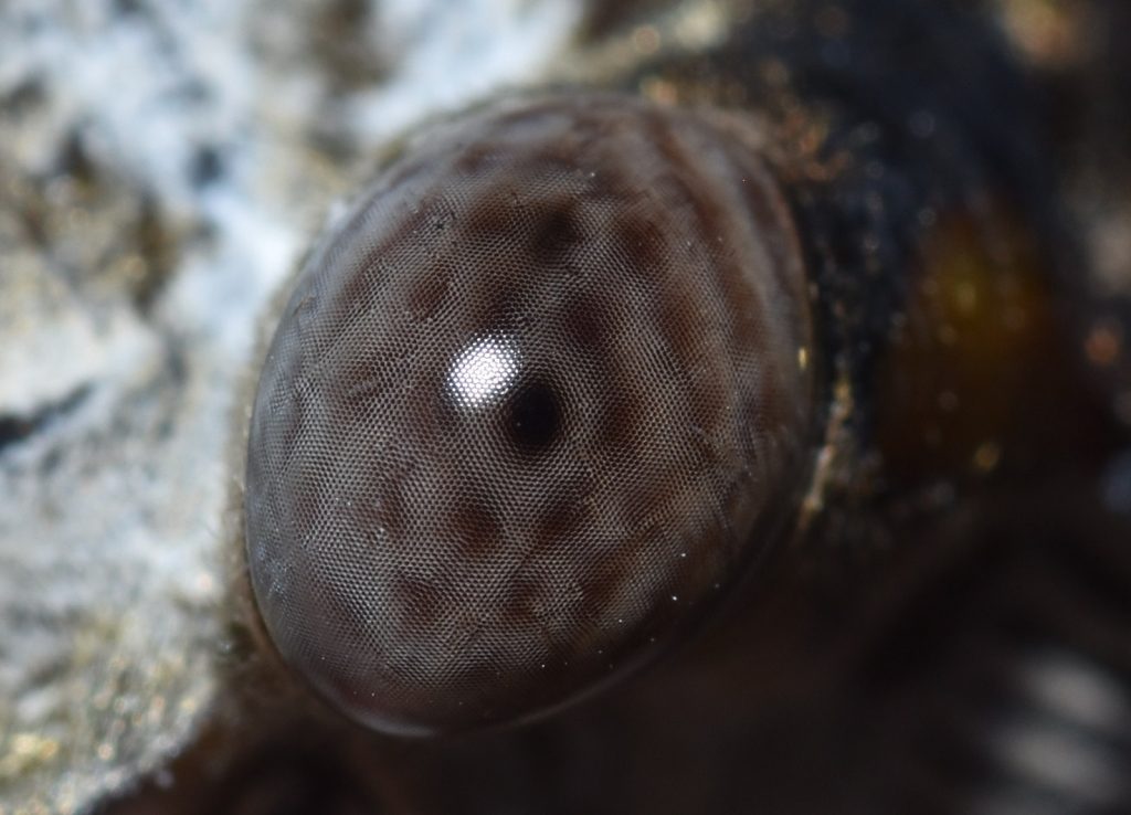
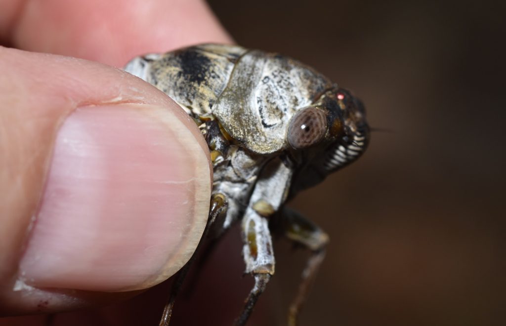
“An ant of velvet On a dusty hiking trail A wasp chirps softly” – Haiku1
Velvet ants are not really ants. They’re wasps. They’re called velvet ants because the females are wingless and look a bit like large, hairy ants. They’re also called cow killers. They earn this moniker by having excruciatingly painful stings.
You might be forgiven for thinking that the more painful a sting is, the more dangerous it is. In fact, the toxicity of a venom is unrelated to the level of pain it produces. The sting from a velvet ant is less toxic than that of a honey bee, but hurts way more. On the Schmidt sting pain index2, the velvet ant scores a 3. (The index only goes up to 4.) One of the few insects in Southern California with a more painful sting is the tarantula hawk wasp, which I covered in an earlier post. (See The Stuff of Nightmares.)
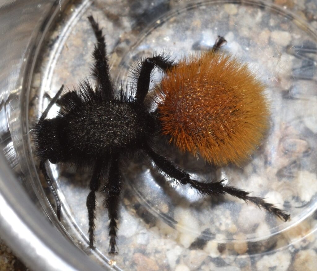
Dasymutilla magnifica – A beautiful desert species of velvet ant Anza Borrego State Park, San Diego County, CA 6/22/19
I’ve actually been stung by a velvet ant. While hiking near where I live, I saw a Sacken’s velvet ant, a species whose long white fur makes it look kind of like the seed-head of a plant — if a seed-head had long black legs. I wanted to show it to my grandnephew, who’s a huge fan of bugs (can’t imagine where he gets that from), so I caught it in a small plastic container. The young woman I was hiking with was worried that it wouldn’t have enough air, so, to make her happy, I decided to transfer it to a larger container that I happened to have with me. When I got home, I discovered that the container had gotten squeezed in my pack and the lid had popped loose …and it was empty. It seemed unlikely that the velvet ant would be able to find its way out of the pack, so it stood to reason that it was still in there. Somewhere…
I started very gingerly pulling things out. Finally, I upended the pack and dumped out the remaining items. No velvet ant. I had just about concluded that it had escaped altogether, when I detected movement out of the corner of my eye. I jerked my hand back just as the wasp struck. I must have reacted just in time to prevent the insect from injecting more than a tiny amount of venom, because the sting was very mild — much less painful than a typical spider bite — and the pain faded completely in a few minutes. Obviously, I was very lucky.
Here’s a couple of photos of a velvet ant like the one that stung me (but not the same individual):
Sacken’s velvet ant (Dasymutilla sackenii) Cuyamaca Rancho State Park, San Diego County, CA 8/8/20
Wasps are in the order Hymenoptera, which also includes bees, ants, and sawflies. All these insects are in many ways closely related. Bees have been described as vegan wasps (see my earlier blog post Vegan Wasps and Flying Mice) and ants could be thought of as wingless, social wasps.
Within the Hymenoptera, wasps form the suborder Apocrita and, within that, velvet ants make up the family Mutillidae. As noted, female mutillids are wingless. The males do have wings and often differ markedly in size, form, and coloration from the females. This presents a problem for entomologists. Velvet ants are solitary insects. The only time males and females hang out together are when they are actually in the process of mating. So, given any two velvet ants — one male and one female — how can you tell if they are the same species? Often the only way to know for sure is if you manage to catch them in flagrante. One entomologist who specializes in the group writes that, although he has frequently seen males in the vicinity of females (and actively searching for them), he’s only once seen them actually doing the deed. It seems that they prefer to conduct their affairs in private.
Obviously, this makes it very difficult for scientists to sort out the different species. Many times males and females of the same species have been given different species names. When the mistake is finally caught, one name (the first one given) becomes the official species name while the other becomes a synonym.5 Currently, there are around 150 species of velvet ant listed for North America. Of these, about a third are known from both males and females; another third are known only from males; and the final third are known only from females.
A male velvet ant in the subfamily Sphaeropthalminae Mission Trails Regional Park, San Diego County, CA 8/13/18
A closeup Note the three large ocelli on the top of the head. (See The Story of Eyes for an explanation of ocelli.)
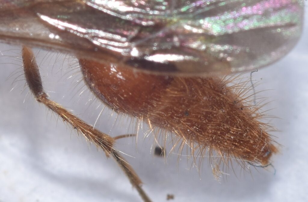
A closeup of the abdomen
Dietary Habits
Adult velvet ants feed on nectar. I mentioned earlier that I caught a female to show to my grandnephew. Since hummingbirds also feed on nectar, but will accept sugar water as a substitute, I figured mutillids might do the same. So, we mixed up some sugar water, added a little red food coloring (this was more for the kid’s benefit than the bug’s — he thoroughly enjoyed making “nectar”), and put a few drops into the container with the wasp. The insect didn’t immediately go for it — she was probably distracted by all the unwanted attention — but later I saw her grazing on the sticky, drying droplets.
What the larvae of velvet ants eat is a bit more gruesome. Mutillid larvae are parasitoids — parasites that kill their hosts. With a few exceptions, they prey on the young of solitary, ground-nesting bees or wasps. After mating, the female velvet ant goes in search of a host nest. When she finds one, she invades the nest and lays an egg next to each larva or pupa. When an egg hatches, the mutillid larva begins feeding on the host larva/pupa. It takes only a week or two for the wasp to consume its host, after which, it metamorphoses into an adult and leaves the nest.
Naturally, a bee or wasp whose nest is invaded by a velvet ant defends it vigorously. And since the hosts are usually solitary, the nests tend to be scattered far and wide, so the female velvet ant has to spend a lot of time wandering around in the open searching for them. This means exposing herself to an increased risk of predation. As a result of these selection pressures, velvet ants have evolved a remarkable array of defenses. One of these is their extremely painful sting, which we’ve already discussed. I’ll cover the other defenses in the following sections.
Velvet ant (Dasymutilla sp.) Mission Trails Regional Park, San Diego County 9/16/19
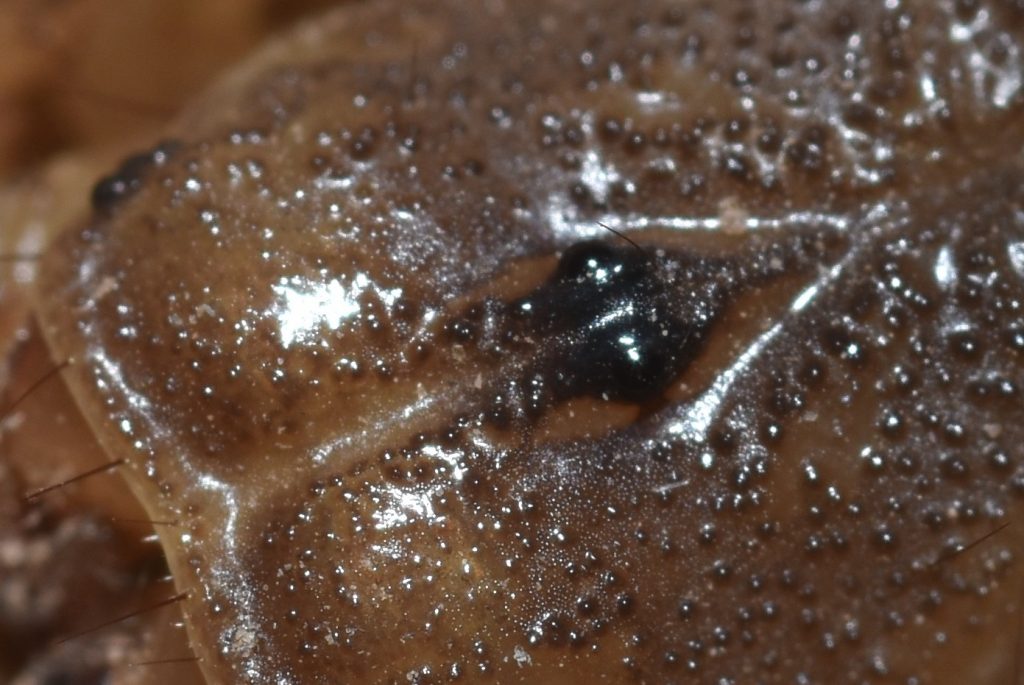
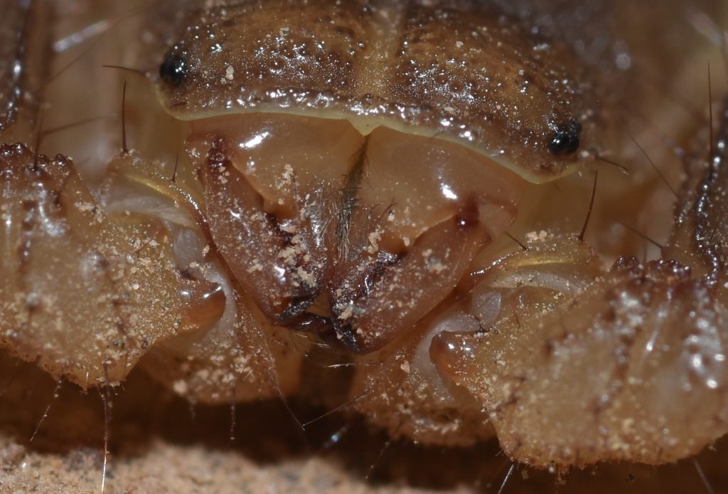
The velvet ant’s exoskeleton is heavily reinforced. So much so, that collectors often resort to using tiny drill bits to drill holes for the pins, instead of trying to force the pins through the bug’s carapace. If an entomologist does try to push a pin through by brute force, when it finally breaks through, it will often continue on through the bottom of the carapace and into the collector’s finger. So, a drill bit is probably the way to go.
The top and sides of a velvet ant’s exoskeleton are covered with pits and ridges. This serves to strengthen the exoskeleton without adding a lot of weight. The ridges may also serve to redirect attacks. When a mandible or stinger catches on a ridge, it tends to slide along it towards an even thicker part of the armor.
An aposematic signal is a warning directed toward potential predators. It’s a way of saying, “you really don’t want to mess with me”. Such signals often take the form of colorful, eye-catching patterns. Most velvet ants are covered in brightly colored fur. If a predator tries to eat one, it’s probably going to get a bad sting and, while the wasp in question may or may not survive the attack, the predator will quickly learn to avoid any similar-looking insects in the future. So, aposematic coloration helps to reduce predation on a species as a whole.
A defensive strategy often allied with aposematic coloration is Müllerian mimicry. This is when two or more species that share the same predators evolve very similar warning signals. If a predator learns to avoid members of one of the species, it will likely avoid members of all of the species, so sharing the same aposematic signals is mutually beneficial. There are many cases of velvet ants forming ‘Müllerian mimicry rings’ consisting of dozens of species. A study in “Current Biology” found that North American velvet ants form one of the largest known Müllerian mimicry complexes on the planet, consisting of eight extensive and distinct mimicry rings.9
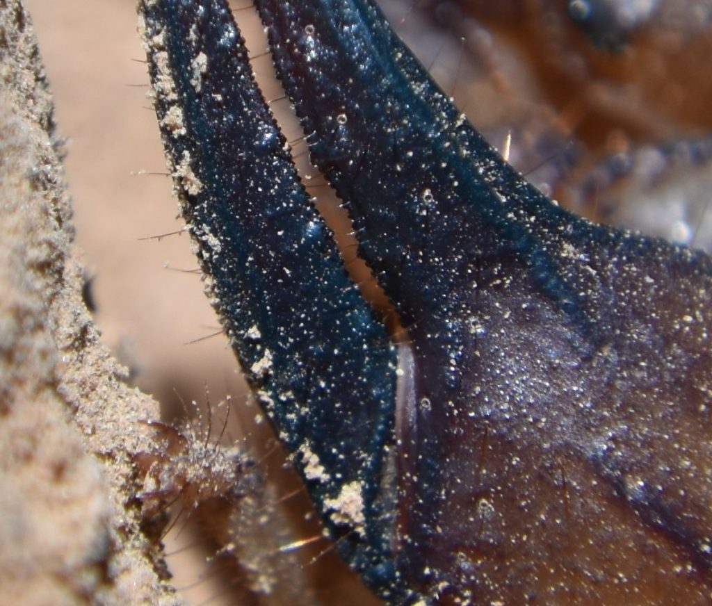
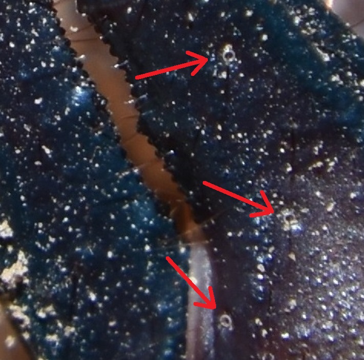
Aposematic coloration is a pretty good warning signal, but what if your attacker is color blind? Perhaps to fill that gap in its defenses, the velvet ant has evolved the ability to secrete an alarm chemical from a gland in its mandible. This chemical serves as an olfactory alarm signal. A predator quickly learns to avoid any insects that smell like it.
Ok, but what if your attacker is color blind and has a head cold? Better to give him an auditory warning, too, just to be safe. Insects don’t have vocal cords, but many of them are able to produce sounds by rubbing together various body parts. This is called stridulation. Crickets, for example, are famous for their chirping, which they make by rubbing together their wings.13 When disturbed, a velvet ant emits a sound that has also been described as “chirping” or sometimes “squeaking”. It does this by rubbing together two of the segments of its abdomen.
When I showed that velvet ant to my grandnephew, we found that we could get it to stridulate by gently stroking its back with the cap from an ink pen. The sound was soft and difficult to hear, but then, it’s probably tuned to non-human ears. The vibration of the abdomen may also serve as a deterrent by provoking a startle response. In one experiment, a shrew was observed to drop a velvet ant as soon as it started stridulating, even though it hadn’t been stung.
I should note that stridulation may not be used exclusively for defense. According to one article, the female also uses it to attract males.14
In the following video, I’ve increased the volume to make the stridulation easier to hear. You can also clearly see the movement of the abdomen. Note: the velvet ant in the video looks a bit bedraggled. This is probably because of the time it spent loose in my backpack. It was not otherwise harmed and I later released it near where I found it. (It even got a nice meal of sugar water for its trouble. That’s the red splotches in the video.)
Scientists have actually studied the susceptibility of female mutillids to predation. In one study, entitled “The indestructible insect: Velvet ants from across the United States avoid predation by representatives from all major tetrapod clades”15, velvet ants from both the Eastern and Western United States were exposed to various potential predators that they would be likely to encounter in their native habitats. These included birds, shrews, lizards, toads, and one mole. (Apparently, moles were in short supply.) The results of the study can be summed up by the following excerpt from the paper: “…out of over 100 interactions between potential predators and various species of velvet ant, there were only 16 occasions where feeding strikes occurred. Only four velvet ants were eaten and three of the four were immediately regurgitated. Only one velvet ant was consumed and retained by a predator.”
Female mutillids probably lost their wings as an adaptation to entering the burrows of their hosts. Wings would be easily damaged by attacking insects, so better to just do away with them. This presents a problem, though — it limits species dispersal — and this puts them at an evolutionary disadvantage, because if a species is only found in a small area, it will tend to adapt itself to the specific environment found in that area. If that environment changes drastically, the species could be wiped out.
Some mutillids have overcome this handicap by evolving phoretic copulation. Phoresy is a type of behaviour in which one animal hitches a ride on another animal. Some types of mites, for instance, will hitch rides on beetles or other larger animals. This way, they get more widely dispersed than would be possible if they just walked. In phoretic copulation, the male of a species carries a female to a new location either before or during mating. In the case of velvet ants, the male transports the female by flying. He actually carries her attached to his genitalia, so mating is done in mid-air. To facilitate this behavior, males of these species have evolved to be significantly larger than the females.
I began this article with a mission: to figure out how a woodpecker can spend its entire adult life battering its head against tree trunks without beating itself into a coma. Scientists estimate that the human brain can tolerate decelerations of up to about 100 g. In other words, you can (theoretically) beat your head against a wall with 100 times the force of gravity without experiencing brain injury.11 On the other hand, when researchers measured the force generated when a woodpecker pecks, they found that it experiences a deceleration of from 600 g to 1500 g — up to fifteen times the amount of g-force that a human being can withstand! And it doesn’t do this just now and then. Some types of woodpeckers peck up to 20 times a second and may make 8,000 to 12,000 pecks in a day!12 How did evolution engineer a bird that can take that kind of punishment day in and day out? How do you evolve a jackhammer for a head?
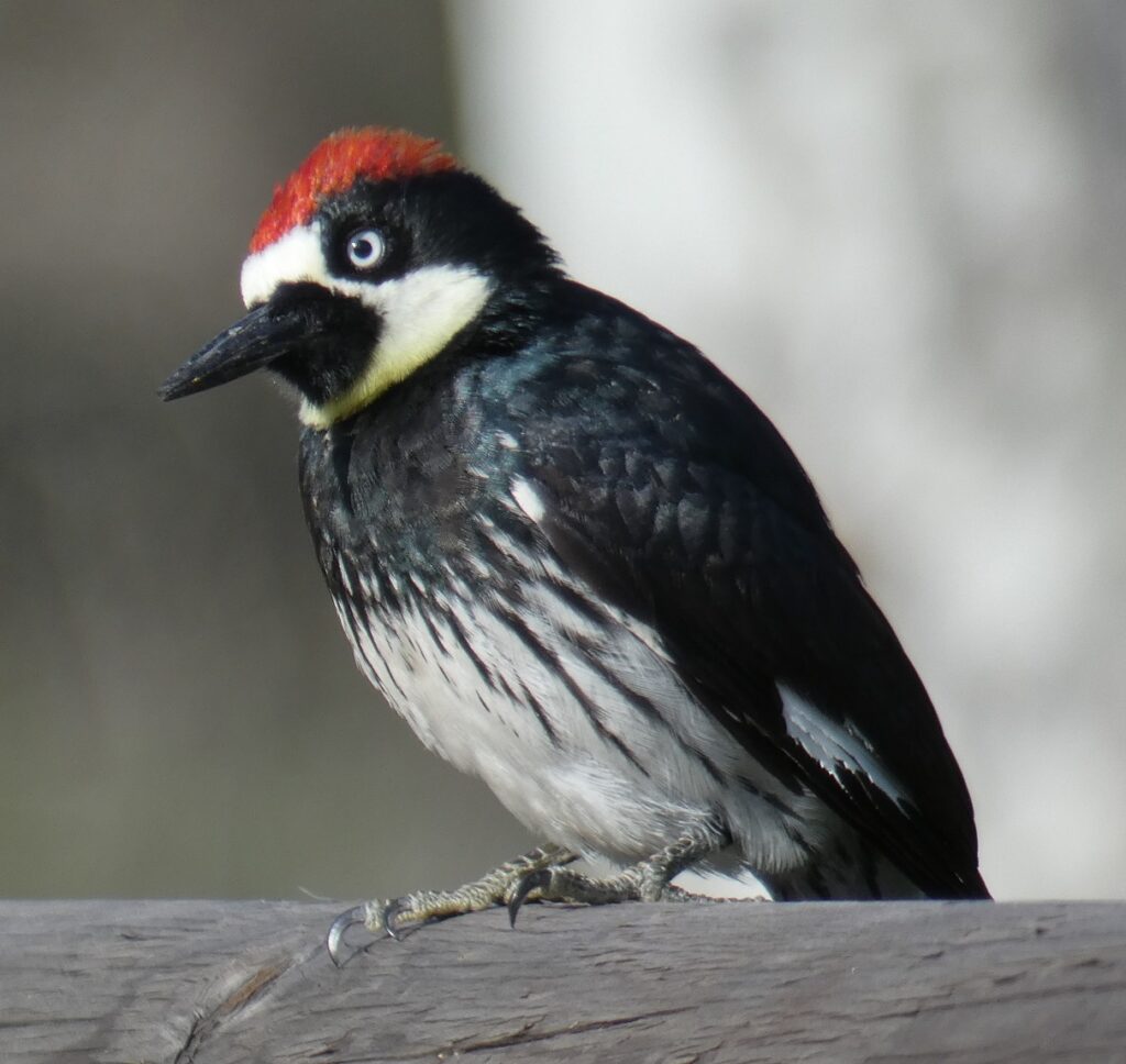
Before we begin our investigation, let’s get to know our test subject. The study I mentioned above, in which researchers measured the pecking force of a woodpecker, was done on Melanerpes formicivorus — the acorn woodpecker. This is fortuitous, since M. formicivorus is one of the most common woodpeckers here in Southern California. I often see them when I hike in wooded areas. Despite this, I’ve never managed to get any good photos of the bird. Fortunately, Christopher MacPherson kindly agreed to let me use his beautiful photo of one:
Acorn woodpecker (Melanerpes formicivorus) San Clemente Canyon, San Diego, CA 3/15/2019 Author: Christopher MacPherson Used with permission
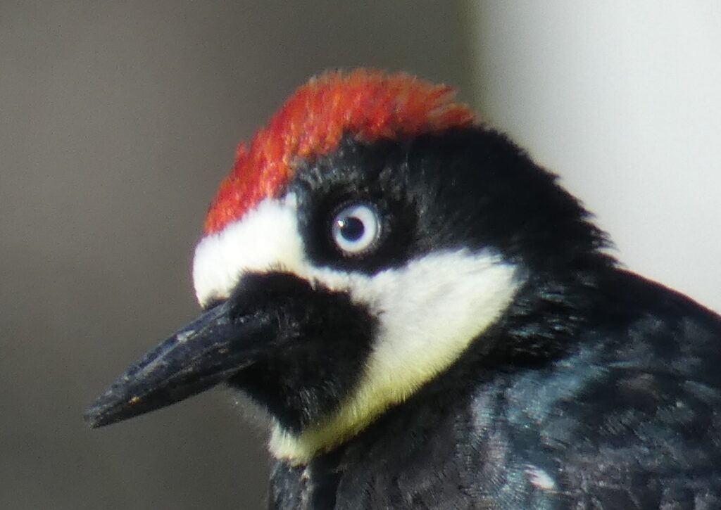
Here’s a closeup of the head from the same photo:
Closeup of the previous photo Author: Christopher MacPherson Used with permission
As you can see, the acorn woodpecker is a very distinctive bird with a clown-like face and wide white eyes. Their wings are black with a single broad white stripe down the middle. This makes them easy to identify as they flit from tree to tree.15
As woodpeckers go, M. formicivorus is pretty average in most respects. It’s medium-sized — about 21 cm or 8.3 in long. The force of its peck is nothing out of the ordinary for woodpeckers. It’s probably fairly representative of the group.16
There is one thing about acorn woodpeckers that is not average, though. As you may have guessed from the name, they eat acorns. That’s not all they eat. Insects are much higher in protein than acorns and make up a large part of their diet. They also consume fruit, tree sap, and even the occasional lizard.19 Nevertheless, acorn woodpeckers cannot survive without acorns because they rely on them to make it through the winter.
Even here in Southern California, where it doesn’t get very cold, insects are rather hard to come by in winter. (I know — they’re my favorite subjects for macro shots.) You can find some by turning over rocks and rotten logs, but birds tend to be bad at that sort of thing. A lot of beetles bore into wood or underneath bark. Woodpeckers locate them by listening for the sounds they make and peck a hole to extract them. But in the winter, such insects tend to go into a dormant state and don’t make much noise, which means the woodpeckers are out of luck.
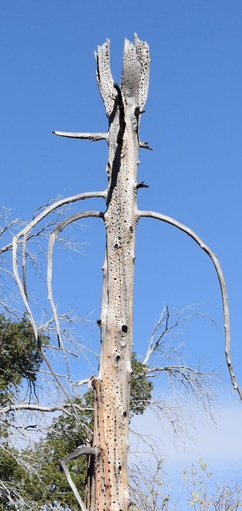
So, like squirrels, acorn woodpeckers turn to hoarding acorns to get them through the hard times. Unlike squirrels, however, they don’t bury the acorns. Instead, they pick out a tree with nice, soft bark — dead pines seem to be a favorite — and they peck thousands and thousands of holes in the trunk. They stick an acorn in each hole and hammer it in with their beak. This kind of tree is called a granary. A granary can have as many as 50,000 acorns stored in it! 20
As acorns dry, they shrink and become loose in their holes, so the woodpeckers have to move the acorns to progressively smaller holes. They also have to defend their granary from other hungry birds. As you can imagine, it’s a big job. Acorn woodpeckers spend a significant amount of their time stocking, maintaining, and defending their granaries.
I should note that not all acorn woodpeckers use trees for granaries. They sometimes use telephone poles, wooden fences, or even the wooden walls of buildings (which does not endear them to the buildings’ owners). In Arizona, some woodpeckers pecked holes through the walls of a wooden water tank and sank 220 kg (485 lbs) of acorns in it! Occasionally, they don’t bother excavating holes at all and, instead, just wedge acorns into cracks in rocks or in the bark of trees.21 But in my area, all the granaries I’ve seen have been of the more traditional variety. Here’s a picture of one:
An acorn woodpecker granary Airplane Monument Trail, Descanso, CA 11/11/2017
And here’s a closer view:
Acorn woodpeckers are unusual in one other way — they often engage in cooperative breeding. This is when three or more birds take care of the nestlings in a single nest. This type of behavior is estimated to occur in only nine percent of bird species. Sometimes two acorn woodpeckers will form a traditional monogamous pair. In other cases, they may form a coalition of as many as seven breeding males and three nesting females. Three males and two females are more common, though. Adult offspring may also stick around to help raise the next generation. A group may contain as many as ten such non-breeding helpers.
Such a coalition or ‘breeding collective’ will stake out a territory around its granary. Breeding females all share the same nest cavity, which is generally a deep chamber excavated in the trunk of a dead tree with the entrance hole at the top of the chamber. Surprisingly, a female will usually destroy any eggs in the nest before she starts to lay. This stops once all the females have begun to lay, so eggs from more than one bird are able to hatch. Such behavior is obviously a disadvantage to the individual birds whose eggs are destroyed, however, it helps to ensure the survival of the group as a whole, since it’s easier to feed and care for fewer chicks at a time. Breeding males will mate indiscriminately with all the nesting females in a group and all members of a group take part in caring for the young.22
Some Chinese Studies
When I started looking into the “pecking problem”, the first thing I came across online was a paper published in 2012 entitled “Numerical study of the impact response of woodpecker’s head”. It’s based on a study conducted in China. The paper is written in such broken English that it would be difficult to understand even without all the scientific terminology. One sentence summed up the researchers’ conclusions pretty well, though: “The woodpecker’s head is a high [sic] effective shock absorbing system, due to the special structure and viscoelasticity of the biomaterials.” 23
The next paper I found was also about a study that took place in China. It was published in “PLOS ONE” in 2011.24 Thankfully, this one was easier to understand. In this study, the researchers used two synchronous, high-speed video systems to record pecking behavior and a force sensor was used to measure the impact of the pecks. Micro-CT scanning and a mechanical testing system were used to study the structure and mechanical properties of the woodpecker head. Finally, a 3D mathematical model of a woodpecker’s head was developed to calculate the theoretical stresses to the bird’s skull.
The woodpecker involved in the study was the great spotted woodpecker (Dendrocopus major). For comparison, the researchers also looked at the Eurasian hoopoe (Upupa epops), which is not a woodpecker. One difference the study found between the two birds was in the bones of their skulls. The woodpecker had more “plate-like spongy bones” in its cranium, while the hoopoe had more rod-like bones. The distribution was different, too. In the woodpecker, the spongy bone was concentrated in the front and back of the skull.
This makes sense if this spongy bone is acting as armor for the brain. When a human suffers a blow to the head, his or her brain first slams against the inside of the cranium on the side where the impact occurred. It then rebounds, slamming into the opposite side. Since the impact of a woodpecker’s peck is felt mostly on the front of the bird’s skull, its brain is going to press up against the inside of the forehead first and then rebound against the back of the skull — the very areas that are most heavily reinforced with spongy bone.
There was a lot more in this paper, but I’m going to move on from scientific papers to an article about a scientific paper, just because the article in question does a nice job of explaining things. The article is “Why Woodpeckers Don’t Get Concussions” by Stephanie Pappas25 and the subject is yet another Chinese study — apparently a separate one from the other two.
The author points out that scientists had previously determined that woodpeckers use their thick neck muscles to diffuse the impact of a peck. If you watch a woodpecker pecking, you’ll notice that it keeps its neck rigid and uses its whole body to peck. (In essence, it’s the bird’s whole body that’s a jackhammer, not just the head.) This means that much of the force of the peck is redirected from the head to the body.
As noted earlier, a woodpecker’s skull is reinforced with thick, plate-like, spongy bone. The mesh that makes up this bone is formed by large numbers of trabeculae, which are microscopic, beam-like projections of bone. Other birds have these trabeculae, but in the woodpecker they’re closer together.
The woodpecker’s beak is also adapted for high impact. It contains many thinner trabeculae, as well as microscopic rod structures. The upper and lower beaks of a woodpecker each consist of a bone covered in a sheath of softer material. In the study mentioned earlier, researchers found that the bone in the lower beak was slightly longer than the bone in the upper beak. On the other hand, the sheath covering the upper beak extends farther than the sheath covering the lower beak. This configuration causes the beak to deform on impact in a way that channels much of the shock into the lower beak and, from there, into the neck and body, bypassing the brain altogether.26
It’s estimated that 99.7 percent of the strain energy produced by a peck ends up being channeled into the body where it’s dissipated as heat. Only 0.3 percent is left to be converted into heat in the head. Even so, temperatures inside the skull rise so quickly that the woodpecker has to take frequent breaks just to keep its brain from overheating.27
In addition to all these adaptations, woodpeckers have evolved a thick inner eyelid (in addition to the two regular eyelids) which closes just before the beak strikes the wood. While this doesn’t do anything to protect the brain, it does keep flying wood chips from damaging the eye. It also keeps the bird’s eyeballs from popping out.28 (Seriously.)
When I first considered writing a blog post about woodpeckers, it was the mystery of how they avoided head injury that inspired me. When I read about their tongues, there was no longer any question — I had to write about these bizarre birds! There are a lot of weird tongues in the animal kingdom, but when it comes to woodpeckers… Well, click here to watch a video and see for yourself.
I hope you took the time to watch that video. It’s pretty remarkable. That woodpecker’s tongue was longer than its head. So where does it put its tongue when it’s not in use? I’ll explain that in some detail in a minute, but it will help you to understand it better if you take a couple minutes to watch one more video. Please click here for that.
All vertebrates have a complex of bone and cartilage called a hyoid apparatus. In humans, the hyoid bone is located at the top of the throat and connects by muscles to the tongue. In birds, the small hyoid bones and cartilage extend to the tip of the tongue, making it stiffer and more spear-like than our tongues.30
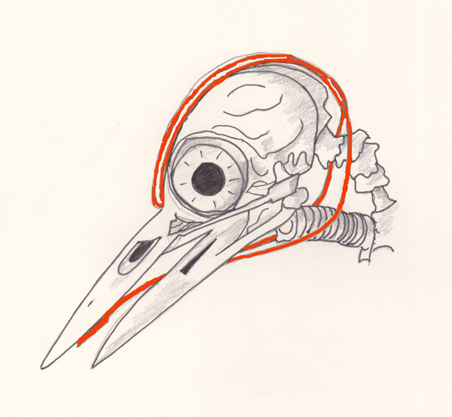
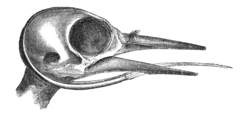
Behind the tongue, the hyoid bone divides in two like a long, thin wishbone with the two horns of the wishbone projecting toward the back of the head. In most birds, these horns are fairly short, terminating on each side of the trachea, but in woodpeckers they’re long — really long. Take a look at figure 1 below in which the tongue and hyoid bone are drawn in red. The horns of the hyoid bone snake around the back of the skull, just underneath the skin, and continue over the top of skull.
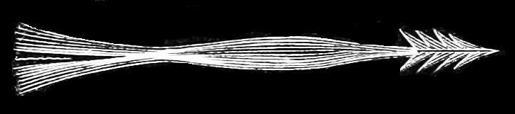
Muscles attached to the hyoid apparatus move it forwards and backwards. When it moves forward, the tongue is extended. The length of the horns determines how far the tongue can be extended. In some species, the tongue can be several times longer than the bill. Again, When the woodpecker retracts its tongue, the hyoid is pushed farther into the skull. When it extends its tongue, the hyoid is pulled partially out. It’s a bit creepy to imagine that thin, flexible bone slithering back and forth underneath the woodpecker’s scalp, but of course, it must feel completely natural to the woodpecker.
In some species, the ends of the horns join together and extend farther, sometimes even inserting into the right nostril (just in case you didn’t think things were weird enough), where they are anchored in place. It’s not entirely clear from the reading I’ve done, but I think that if the ends of the hyoid are not joined together, then the horns move back and forth within relatively snugly fitting sheaths. If the ends are joined together and anchored in place, this is obviously not going to work. In this case, when the tongue is retracted, the hyoid apparatus has a great deal of slack in it. When the tongue is extended, the slack is taken up.
Here’s one other drawing of a woodpecker skull with the tongue and hyoid bone. It’s probably not as informative as the last one, but it’s a nice drawing:
I should mention that woodpecker tongues do vary quite a bit. Some are very long; some not so much. Flickers have the longest tongues, but sapsuckers have possibly the strangest. Their tongues are short and end in bristles, which suck up sap through capillary action.
Believe it or not, I haven’t quite finished listing all the freaky things about woodpecker tongues. As noted, in at least some woodpeckers, the hyoid wraps around the entire head. So, why not let it double as a seat belt? I’m not sure if this has actually been confirmed or is just speculation, but a number of sources suggest that just before a peck, a woodpecker may tighten muscles attached to the hyoid, drawing it tight against the skull and, thus, bracing the head against the impact.31
There are a few more things that contribute to protecting the woodpecker brain. In humans, the brain is loosely packed inside the skull and the space between is filled with cerebrospinal fluid (CSF). In woodpeckers, however, the brain is tightly packed inside the skull, leaving almost no space for CSF. As a result, the woodpecker brain doesn’t “slosh around” as it pecks.
Our higher order mental processes, which were the last to evolve, take place mostly in the outer layer of the brain. We’ve all seen in photos and illustrations that the human brain is covered with folds and ridges. This folding increases the area covered by that outer layer. In other words, it increases our brain power. A woodpecker’s brain, by comparison, is smooth. While this may limit the woodpecker’s mental prowess, it does have the benefit of making its brain more compact and, as a result, less susceptible to damage.3233
In the course of this article, we’ve explored quite a few adaptations that go into making a woodpecker the pecking dynamo that it is. But maybe you’re not quite convinced. Maybe you still find it hard to believe that all these adaptations are enough to explain how a bird can spend its entire life blissfully beating its head against trees. If so, you’re absolutely right. The truth is, I haven’t yet mentioned the most important “adaptation” of all and it’s not really even an adaptation — woodpeckers are small.
The amount of deceleration that an animal’s brain can withstand without injury depends, more than anything else, on its size. If a human being were to jump off the roof of a two-story building and land on the ground, he would almost certainly be killed. If a mouse were to jump off the same building, it would walk away unscathed. And this is the main reason the woodpecker is able to handle all that hard pecking — it’s so much smaller than us.
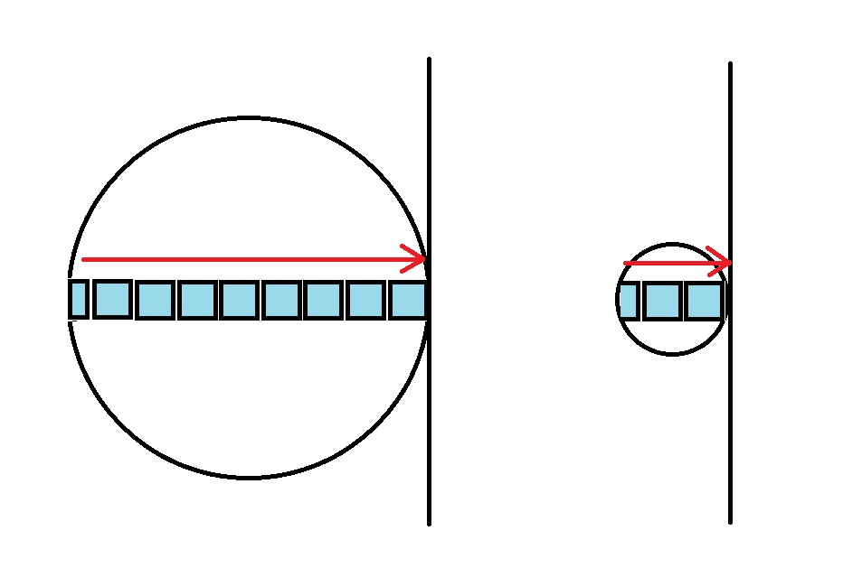
But why exactly is this the case? Why should an animal be able to survive an impact that would kill a human just because it’s smaller? Here’s how I like to think about it. For simplicity, let’s imagine the human head as a large sphere and the woodpecker head as a much smaller sphere. I’ve represented these spheres as circles in figure 3. Now imagine stacking as many equal-size cubes in each sphere as will fit. These are represented by the cyan squares in the figure. (I just drew the ones through the center of each sphere. You’ll have to imagine the rest.)
Figure 3. The human head is represented by the circle on the left. The woodpecker head is the circle on the right. The squares represent units of brain volume. Only the ones through the center are shown. The vertical lines represent walls.
Obviously, the large sphere will hold many more cubes than the smaller one. The cubes represent chunks of brain. Basically, I’m asking you to imagine each brain as divided up into equal-size chunks. Now imagine both spheres slamming into walls (the vertical lines in the figure) at the same speed. Each cube is going to slam into the cube in front of it. So, the cube nearest the point of impact will have all the cubes behind it pushing against it.
Hopefully, you see where I’m going with this. The cubes nearest the point of impact for the human head (the left sphere) have a lot more force acting on them than the corresponding cubes in the woodpecker head. This is indicated by the red arrows in the figure. So, these cubes (chunks of brain) would be much more susceptible to damage in the human brain.
For me, this is the explanation that makes the most sense. If you prefer a more mathematical treatment, check out this short series of Cornell Lab videos: Built to Peck: How Woodpeckers Avoid Brain Injury. The sixth video in the series goes into the effects of scale, so you can skip to that one if you want. The entire series is worth watching, though.
In either 1940 or 1941 (my sources are a bit confused about the exact year), cartoonist Walter Lantz was honeymooning with his wife, Grace, at a cabin in June Lake, California. An acorn woodpecker made such a racket with its pecking that it kept them awake at night. Later, when it started raining heavily, they learned the hard way that the woodpecker had drilled holes in the roof of the cabin. Supposedly, Lantz wanted to shoot the bird, but Grace suggested that he should make a cartoon about it instead. And thus, Woody Woodpecker was born.
While classic cartoon characters like Bugs Bunny and Daffy Duck are still well known among young people today, Woody seems to have mostly faded into obscurity. It’s a pity. His cartoons were very popular when I was a kid and his raucous laugh was unforgettable. Although it was the acorn woodpecker that originally inspired the character, the designers seem to have modeled his appearance more on the pileated woodpecker.
As for the famous laugh, it’s not clear. According to Wikipedia, Lantz modeled it on the acorn woodpecker’s call, but at least one other source argues that it sounds more like the call of the pileated woodpecker. If you click here and scroll down a bit, you’ll see a link to audio recordings of all three woodpeckers (acorn, pileated, and fictional), so you can decide for yourself which one was the real inspiration. Voice actor extraordinaire Mel Blanc voiced the original laugh, so, who knows, he could have just made it up out of thin air.
Update 9/15/20: The Purpose of That Long Tongue
One of my readers asked an excellent question — why does a woodpecker need such a long tongue? Wood-boring beetle larvae form tunnels just underneath the outer layer of wood. These tunnels stretch up and down the trunk or along the length of a branch and can extend for several inches or even feet. The woodpecker listens for the sounds of a beetle grub chewing on wood. It will also tap on dead wood and listen for the sounds of the larva scurrying away. When it locates a potential meal, it pecks its way into the tunnel and uses its long tongue to probe the contents. The tip of the tongue is stiff and barbed, so it can spear the grub and haul it out.38
As a boy, I first encountered wild raccoons while on a camping trip with the Boy Scouts in the mountains of Virginia. After we’d gone to bed, a group of raccoons invaded the campsite looking for food. Their rummaging around woke some of us up and we went outside to take a look. I still remember the glow of their eyes when we shined our flashlights on them. They weren’t large animals, but they weren’t especially small either, and their seeming lack of fear was a little unnerving. We eventually went back to bed and left them to their explorations.
My first (and only) encounter with raccoons in Southern California also took place at night — not surprising, given that they are mostly nocturnal. It took place next to a stream, which is also not surprising, since a lot of a raccoon’s diet consists of aquatic prey. I heard a rustling in the bushes and, when I trained my headlamp in that direction, I spotted a large raccoon climbing in a small tree. Here’s the video:
It’s not a great video, but, unfortunately, it’s the best I’ve got. For the rest of this article, I’ll be relying on photos that are in the public domain.
The Common Raccoon
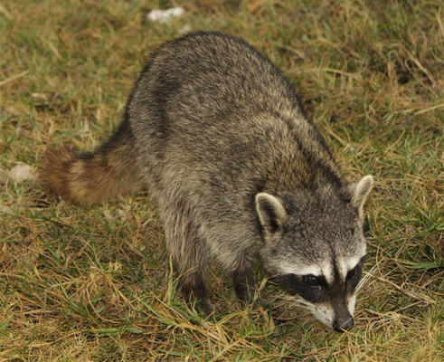
The common raccoon, Procyon lotor in science speak, is native to North America. I shouldn’t need to describe it here. Even in the unlikely event that you’ve never seen a live raccoon, you’ve certainly seen pictures. It’s one of those animals that almost any child can recognize by the age of three. Not only is it very distinctive, there’s pretty much nothing else in the United States that looks very much like it. For your reference, though, here’s a great photo of one that I found on Wiki Commons:
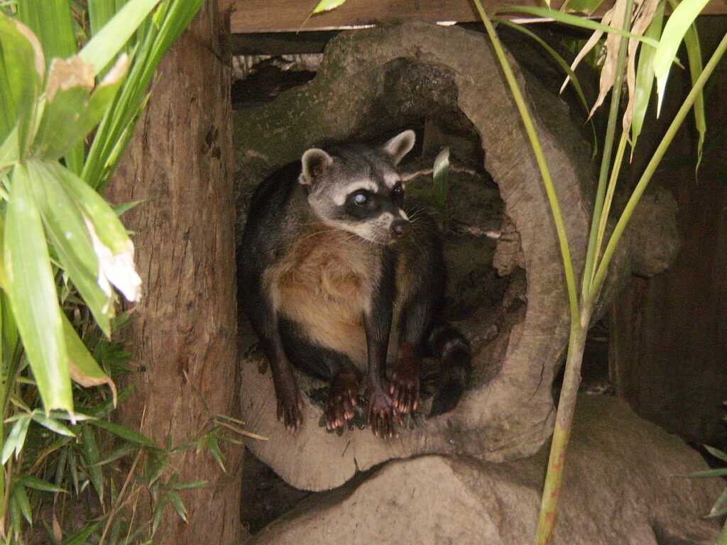
In addition to the common or North American raccoon, P. lotor, there are two other species in the genus Procyon. The crab-eating raccoon (P. cancrivorus) is found in Costa Rica and much of South America east of the Andes. As you can see from the photo, it’s quite a bit sleeker than the common raccoon and has shorter fur. P. cancrivorus and P. lotor are believed to have branched off from a common ancestor around 4.2 million years ago.
Then there is the Cozumel or pygmy raccoon (Procyon pygmaeus). It’s found only on the island of Cozumel, which is located off the eastern coast of Mexico’s Yucatán Peninsula. P. pygmaeus has a smaller head and body than the common raccoon and is probably an example of ‘island dwarfism’. This is the tendency for some animals that are confined to islands to evolve into smaller versions of themselves. There are several possible explanations for island dwarfism. The most likely is that a smaller animal has a better chance of surviving on an island where the food supply may be more limited than on the mainland.
There are many more extreme examples of island dwarfism. For instance, on the California Channel Islands, just a few hours from where I live, there used to live a pygmy mammoth, which was probably a dwarf version of the Columbian mammoth. These creatures ranged from 4.5 to 7 feet high at the shoulders, as compared to 14 feet for their relatives on the mainland. Weight-wise, the difference would have been even more startling, as the pygmy and Columbian mammoths are estimated to have weighed around 2000 pounds and 20,000 pounds, respectively.
Humans are not exempt from the phenomenon of island dwarfism. In 2003, the remains of an individual in the genus Homo were uncovered on the island of Flores in Indonesia. This individual would have stood about 1.06 m or 3 ft 6 in. Remains were found of a second individual that was just slightly taller at around 1.1 m or 3 ft 7 in. Stone implements of an appropriate size were also found. Initially, there was some debate over whether the remains belonged to a new species of human or were from modern humans with congenital defects, however, the current consensus is that they do represent a new species of very small humans. The species has been named Homo floresiensis, after the island on which it was discovered. The common name is ‘Flores Man’, but the original discoverers also gave them the nickname “hobbits”, which quickly caught on in the media.
H. floresiensis is believed to have lived on Flores as recently as 50,000 years ago, which is probably around the time modern humans reached the island. So it’s likely that the two species existed side-by-side for a short time before modern humans drove them to extinction.
There are lots of subspecies of P. lotor. I won’t go over them all here, since they’re mostly just slight variations of each other. I do think the Torch Key raccoon (P. l. incautus) is worth a mention, though, just for its general cuteness, if nothing else. This is a small raccoon with an unusually pale coat. It lives in the Florida Keys.
The word ‘raccoon’ is a corruption of a Native American word. It comes from the Algonquian word arakun (variously spelled aroughcun, arocoun, arakunem, and rathkone) and it means something like “he scratches with his hands.”
Of course, there were plenty of Native American tribes that were not part of the Algonquian peoples and they had their own names for the animal. The Sioux, for instance, called it wica, weé-cha, or way-atch-a, which translates to “little man”.
Although native to North America, raccoons have been introduced to many other parts of the world, including several European and Asian countries. In some cases, raccoons were introduced for hunting. In other cases, they managed to establish local populations after escaping from captivity. Naturally, non-English speakers have come up with their own names for the creatures. As you probably know, raccoons are famous for their habit of dousing their food in water and, apparently, many people think they look a bit like little bears. As a result, a surprising number of languages have combined these two characteristics in their word for the animal. In German, for instance, the word for raccoon is Waschbär (‘wash-bear’). In Italian, it’s orsetto lavatore (‘little washer bear’). And in both Chinese and Japanese it’s ‘washing-bear’ — dvivón róchetz and araiguma, respectively.
As far as the scientific name is concerned, Procyon can be translated as “before the dog” or “doglike” and lotor means “washer”. It’s not clear why Gottlieb Storr, who named the genus, chose Procyon. Maybe he just thought the animal looked doglike. It’s also possible that he was thinking of the animal’s nocturnal lifestyle and named the genus after the star Procyon, the brightest star in the constellation of Canis Minor (lesser dog). I suppose it could be both.
Probably the most notable characteristic of the common raccoon is the black fur around its eyes, which makes the animal look as if it’s wearing a mask. This probably makes it easier for raccoons to identify each other and to recognize each others’ expressions. It may also enhance night vision by reducing glare. Similarly, the dark rings on the tail may help in recognizing individuals and in determining body posture (which would presumably aid in identifying another raccoon’s intentions.)
The raccoon’s front paws have five fingers and are shaped much like tiny human hands. Although they lack opposable thumbs, raccoons are remarkably skillful with their hands. They use them to examine their food before eating it and they are very adept at using them to open windows, doors, and trashcans. According to a post by DiVon80 on the “Backyard Chickens” website, some raccoons got into a kitchen at a campsite and “…they opened jars, turned the water on by all the sinks, opened and dug in the fridge, even [opened] pop bottles…” 42
In captivity, a raccoon can live for quite a long time, although exactly how long varies, depending on the source. Wikipedia claims they can live for over 20 years, but another source 43 gives them a maximum lifespan of only 10 to 12 years. A raccoon’s chances of reaching an advanced old age are considerably reduced in the wild, where the average lifespan is often less than 1.8 years. You might think the reason for this would be their many predators — bobcats, bears, great horned owls, mountain lions, eagles, and coyotes, to name a few. The young are especially vulnerable. However, predation is actually not a significant cause of death, since many large predators have been exterminated or reduced in population in the areas raccoons inhabit. Human hunting, traffic, and severe weather are more important factors. Disease also takes its toll. In fact, the most significant cause of raccoon death in North America is distemper.
And then there’s rabies. It’s well-known that raccoons are a reservoir for this disease. In fact, they’re the most important reservoir in the United States.44 Before you start killing raccoons on sight, though, you should know that, according to the CDC, only one person in the U.S. has died from the raccoon strain of rabies.45 This is probably because rabid raccoons do not exhibit the aggressive behavior typical of rabid dogs. In fact, they become less aggressive when infected and tend to avoid people even more than usual. 46
With such a high mortality rate, you might expect raccoons to be endangered. In fact, nothing could be further from the truth. The International Union for Conservation of Nature (IUCN) lists their status as of “least concern”. So how have raccoons managed to beat the odds? I’d have to say it’s their adaptability. The common raccoon is able to live in a wide range of habitats from as far north as Canada to as far south as Panama. Raccoons are also highly omnivorous. It’s estimated that the average raccoon’s diet consist of around 27% vertebrates, 40% invertebrates, and 33% plant material. Fish, crayfish, frogs, bird eggs (and occasionally birds), insects, worms, fruits, and nuts are some of the many foods they consume. Sometimes they even eat carrion. They are also quite happy to raid our garbage cans.
Not really. Raccoons often use their front paws to dabble for food in the water near the shoreline. When they find something potentially edible, they pick it up, examine it, and rub it with their paws, probably to remove inedible or unappealing parts. They usually hold the food under water while doing this because the water softens the tough layer that covers their paws, increasing their tactile sensitivity. With dry foods like nuts, a raccoon will also examine the food with its paws before eating it, but it probably won’t bother dousing it in water. According to one author,49 both dabbling and dry feeding are done “while the raccoon gazes away into space and uses its acute sense of touch to investigate its meal”. (I just reproduced that quote because I enjoy the mental image of a raccoon meditatively “gazing away into space”.)
In captivity, raccoons will often douse even dry food in water before eating it. Scientists don’t believe they do this in order to wash the food, though. Instead, it’s probably an example of a ‘fixed action pattern’ — an instinctual behavior that is characteristic of a particular species. Its water dish is right there, after all — it’s not like it has to hike over to a stream — so it just does what feels natural to it.
Raccoons prefer to make their dens in the hollows of trees, but they will make do with crevices and caves, abandoned buildings, or the abandoned burrows of other animals. In a pinch, they will even sleep in bushes or on the ground under cedars. (There’s that adaptability again.)
During the winter months in cold regions, raccoons greatly reduce their food intake and spend most of their time sleeping. They don’t technically hibernate, though, and may sometimes be found outside their dens even in the depths of winter.
Mating generally takes place between January and March, but this varies from region to region. Males take no part in caring for the young and, in fact, they will move on to try and impregnate other females. Litter size varies from 1 to 8 cubs, but 3 to 5 is the normal range. Cubs are born deaf and blind. Their ear canals open after around 18 to 23 days and their eyes open a few days later. Cubs are usually weened by around 16 weeks. In the Fall, the mother raccoon will show them dens and feeding grounds. After that, they usually go their separate ways, although in colder climates, they may share a den with their mother for the first winter.5051
The first mammals were mostly tiny and mouse-like. Although they evolved toward the end of the Triassic, fairly early in the reign of the dinosaurs, they would remain small and furtive until the non-avian dinosaurs went extinct 65 million years ago (mya). Finding themselves no longer under the heels of their reptilian overlords, mammals rapidly diversified to fill the niches left vacant by them.
These ancient mammals split into five major groups, one of which, Creodonta, included the ancestors of all living carnivores. Creodonts took a wide variety of forms — some cat-like, some bear-like, and some hyena-like. During the Eocene, 55 – 37 mya, the Miacidae family of mammals appeared on the scene. The miacids were slender, five-toed, tree climbers. Changes in their teeth allowed them to exploit new kinds of food and new environments. All modern carnivores are believed to have evolved from this group.
Eventually, the order Carnivora split into two suborders, Caniformia and Feliformia, the “dog-like” and “cat-like” carnivores, respectively. Caniformia includes dogs, wolves, foxes, bears, badgers , and seals, among others. One of those “others” is raccoons. The raccoon family, Procyonidae, includes raccoons and their close relatives. The dog family (Canidae) and racoon family split off from the miacids during the Miocene. It’s not known whether the procyonids split off directly from the miacid line or were an early offshoot of the canids. Either way, the translation of ‘Procyon‘ as “before the dog” makes some sense in this context.
The Strange, Convoluted Tale of Rocket Raccoon555657
Many of you are probably familiar with the Beatles’ song “Rocky Raccoon”. The song was written mostly by Paul McCartney, although John Lennon may have contributed. Sadly, the song does not appear to be about an actual raccoon. The title character’s name was originally supposed to be “Rocky Sassoon”, but McCartney changed it to “Rocky Raccoon” because he thought it had more of a ‘cowboy’ ring to it. (The story is supposed to have taken place in the Old West.)
An interesting, quirky song, but hardly worth mentioning in an article about raccoons. But then, Marvel Comics came into the picture. Writer Bill Mantlo and artist Keith Giffen were inspired by the Beatles’ song to create a character named Rocky Raccoon for the comic book Marvel Preview #7. In this story, Rocky was an actual raccoon, albeit an anthropomorphized, talking version of one (with an English accent, no less). Rocky apparently only plays a bit part in the comic book. You can see an excerpt from it in a CBR.com article. Click here for that.
Rocky would next appear in 1982 in The Incredible Hulk #271. The story for this issue was entitled “Now Somewhere In the Black Holes of Sirius Major There Lived a Young Boy Named Rocket Raccoon” — an obvious play on on a line from the Beatles song. In this story, we learn that “Rocky” is actually short for “Rocket”. (He also loses the British accent.) You will find an excerpt in the same CBR.com article linked to previously. In 1985, the character was given his own four-issue limited series. In succeeding years, Rocket appeared in Quasar #15 and in three issues of Sensational She-Hulk, but in ten years time, he had amassed a grand total of only 10 comic book appearances. In other words, he wasn’t exactly taking the world by storm.
From 2006 on, however, Rocket appeared in a dizzying number of different comic books, including, significantly, the Guardians of the Galaxy Series. I won’t go into all the tedious details. You can read the Rocket Raccoon Wikipedia article if you’re interested.
As for Rocket’s biography, he started his career as the chief law officer on the planet Halfworld, an abandoned colony for the mentally ill. He was one of a group of animals that had been genetically modified to give them intelligence and bipedal-ism, so that they could act as caretakers for the inmates. At some point, the inmates get cured of their illness (apparently, they all had the same thing) and Rocky and friends take off into space for their own adventures. What followed was a long and, yes, convoluted, series of stories, which I also won’t go into. (Again, see the Wikipedia article.)
Rocket in these comics is a starship pilot, an expert marksman, a brilliant technician, a master military tactician and a born leader. (Well, genetically modified leader, I guess.) He also has all the abilities and attributes of a regular earth-type raccoon — sharp claws, acute senses of smell, hearing, sight, and touch, and the ability to climb trees and scale buildings with ease. (Somebody was really impressed with raccoons.) He has a sidekick named Wal Rus, who is — you guessed it — an anthropomorphic walrus; an enemy named Blackjack O’Hare, who is an anthropomorphic rabbit (get it?); and a love interest named Lylla, who is an anthropomorphic otter.
Somewhere in all this weirdness, Rocket finds out that a lot of the things he thought had happened on planet Halfworld hadn’t really happened — they were “mostly half-truths and deliberately crafted fake memories”. 58 Yeah, it gets complicated. Anyway, eventually he teams up with Star-Lord and the other Guardians of the Galaxy for a whole new series of adventures.
And then the Guardians of the Galaxy movie came out. If you haven’t seen this film and it’s two sequels, I highly recommend them. They are some of the funniest movies I’ve ever seen. One of my favorite scenes is when Peter Quill (Star-Lord) calls Rocket a trash panda. I won’t ruin it for you, though.
In the movies, Rocket’s biography is, thankfully, greatly stripped down. (He doesn’t even know he’s a raccoon or what a raccoon is.) He’s a great character and a great example of a raccoon in popular culture. And that’s how I’m justifying writing about him here. (Hey, nobody forced you to read it.)
There are three animals that will forever be associated with the American Southwest in my mind: rattlesnakes, scorpions, and tarantulas. My home state, Virginia, doesn’t have scorpions or tarantulas. There are a few timber rattlers, but they live way up in the Blue Ridge Mountains and I’ve never run across one. In fact, until I moved to California, I’d never encountered any of the three in the wild. Because of this — and their dangerous reputations — these three creatures have always had an exotic appeal to me.
So, when I started this blog, I knew I’d have to write about all of them sooner or later. It didn’t take long for me to get around to posting about rattlesnakes (see Rattlesnake!) and I covered scorpions in my last post, Scorpions, Scorplings, and Scorpionism. In this article, we’ll take a look at the last of the “big three”: tarantulas.
Even if you’ve never seen a tarantula in real life, you’ve probably seen them in magazines or movies. So you should have some idea what a tarantula is supposed to look like — big and hairy. Sort of like this:
Tarantula (Aphonopelma sp.) Anza Borrego Desert, San Diego County 10/23/16
The tarantula in the above photo is the first one I ever saw in the wild. I encountered it while camping in Anza Borrego State Park in the Sonoran Desert of California. It was about the size of my palm — far larger than any of our Virginia spiders and not at all bad for my first tarantula sighting.
Tarantulas actually come in a great variety of sizes. The largest is the Goliath birdeater (Theraphosa blondi), which is found in northern South America. It’s about the size of a dinner plate. True to its name, it has been known to eat small birds and even rodents, but it more commonly feasts on large bugs, worms, and amphibians.41
On the other end of the spectrum is the spruce-fir moss spider (Microhexura montivaga), which is about the size of a BB. It eats mites and springtails — tiny invertebrates that are barely visible to the naked eye. (See my earlier post, “Of Mites and Men… and Ticks” for a discussion of mites.) M. montivaga lives on the east coast of the United States in small, isolated pockets of the southern Appalachian Mountains — not the kind of place you would normally think of when you think of tarantulas. Unfortunately, its habitat is rapidly shrinking due to the warming climate and also due to an invasive insect that has killed off most of the Fraser firs in those mountains.4346
Despite the terror they often instill, tarantulas are not really very dangerous. They all have venom, but it’s not lethal to humans and, in fact, there has never been a documented case of a person dying from a tarantula bite. This makes them far less dangerous than even wasps and bees.
Before you go picking one up, though, I should mention that tarantulas — at least the ones in the Americas — have another defense mechanism that they can deploy against large, genuinely dangerous animals like us. On their abdomens, they have barbed, urticating hairs or bristles. “Urticating” basically means irritating and, if you get a tarantula irritated, it may return the favor by using its hind legs to flick a cloud of these bristles in your direction. These can embed themselves in your skin or, worse, in the corneas of your eyes, possibly causing permanent eye damage. So use caution if you plan on handling one of these spiders.
When a tarantula flicks off a cloud of urticating hairs, it often leaves a bald spot on its abdomen. The hairs will not grow back until its next molt.
. . .
Before we go any further, maybe I should take some time to explain just what makes a tarantula a tarantula. Obviously, a tarantula is a type of spider…
Spiders are members of the order Araneae. Everything that is a spider is in the order Araneae and everything in the order Araneae is a spider. Tarantulas are in this order. So far, so good.
Araneae is broken down into suborders and infraorders as follows:
Figure 1. How Araneae breaks down into suborders (Mesothelae and Opsthothelae) and infraorders (Arachnomorphae and Mygalomorphae) Adapted from “Are Tarantulas Spiders? (Or Are They Different?)“
The vast majority of spiders are in the infraorder Arachnomorphae. These are referred to as the “true” spiders. Tarantulas are not in this group — they are in Mygalomorphae. So, if tarantulas are not true spiders, does that mean they are they false spiders? No, tarantulas are still considered spiders. You see, biologists have this annoying habit of deciding that, given a group of animals X, a certain subset of X will often be designated as true X, even though the general public knows the entire group as X. For instance, there are true bugs, true frogs, true toads, true salamanders, and so on, ad nauseum. So it is with spiders. So, tarantulas truly are spiders; they just aren’t true spiders.
Let’s see if we can figure out how a tarantula differs from your “typical” spider. As mentioned in the last section, the vast majority of spiders are in the infraorder Arachnomorphae. Arachnomorphs have two main characteristics that differentiate them from the rest of the order. First, they have only one pair of book lungs. All other spiders have two pairs. The book lungs are usually visible on the underside of the abdomen as light-colored patches, so this is a good distinguishing feature. Second, their chelicerae — the appendages that end in the fangs — open and close from side to side. Other spiders have chelicerae that raise up and stab down. This means that an arachnomorph spider can rush up to a prey animal and immediately bite it. A tarantula, on the other hand, has to lift its chelicerae, unfold its fangs, and then stab down. This gives arachnomorphs an evolutionary advantage and helps to explain why there’s so many more of them.59
Within the Mygalomorphae, tarantulas form the family Theraphosidae. So another, slightly more technical, name for a tarantula is ‘theraphosid’. All tarantulas have either two or four spinnerets on the rear of their abdomens. Arachnomorphs usually have six. Like most spiders, tarantulas have eight eyes. Unlike most arachnomorphs, these eyes are crowded together on an ocular tubercle. (See the section on anatomy.)
So it’s pretty easy to distinguish tarantulas from arachnomorphs. Distinguishing them from other mygalomorphs is more of a challenge. The website The Natural History of Tarantulas lists a few characteristics that can be used. Check that site out if you’re interested. I usually just post photos of a specimen on BugGuide or iNaturalist and hope that someone more knowledgeable than me will tell me what it is.
As far as California goes, most of the tarantulas found here are in the genus Aphonopelma. All the sightings I’ve made have been in that genus. Unfortunately, it’s hard to identify them down to the species level.
Tarantula (Aphonopelma sp.) Mission Trails Regional Park, San Diego County 9/28/18
Here’s a closeup of the spider in the above photo. Note the long spinnerets:
Tarantulas are found on all continents except Antarctica. Most species are tropical, but lots are found in arid environments, too. Here in Southern California, they are found in both chaparral and desert.
Habit-wise, tarantulas can be broadly divided into three types: terrestrial tarantulas that have permanent burrows, terrestrial tarantulas that don’t have permanent burrows, and tree-living or arboreal tarantulas. The vast majority of tarantulas are of the first type — they live on the ground and have burrows. They may dig these burrows themselves or they may commandeer the abandoned dens of mammals or reptiles. They often tend to hang out near their burrows. Here’s a video of one doing just that. You can see it quickly scramble inside when I get too close:
Tarantulas line their dens with silk. This provides structural support and also makes it easier for the spider to climb up and down. Many species will extend the bottom part of this silken tube outside the den as a kind of “welcome mat”. The spider senses the vibrations of a prey animal walking on the mat and rushes out to invite it in for dinner.
The second type — terrestrial tarantulas that don’t have permanent burrows — may not actually exist, as this lifestyle has not been verified by researchers in the field. Be that as it may, there are a few species, such as Cyriocosmus ritae and Lasiodora parahyban, that are suspected of falling into this category. This type wanders around at night and hides during the day in whatever shelter it can find.
Arboreal tarantulas inhabit trees. Some species construct silken tubes to live in. Others live in rot holes or hide behind loose bark or in epiphytic plants.
Tarantulas don’t spin what we usually think of as spider webs. They use their silk for other purposes, such as the aforementioned silken tubes.
One notable difference between New World and Old World tarantulas is that the Old World varieties don’t have urticating hairs. They compensate by having more potent venom and by being much quicker to bite people. For that reason, Old World species are less popular as pets.
Tarantulas tend to be nocturnal, although they do sometimes come out during the day. If you want to find one, your best bet is to look for them at night in the middle of summer. (That’s also the best time to find scorpions, so you could get a two-for-one.)
Here’s a couple of videos of a tarantula just walking around in the desert. (This one was, in fact, out in broad daylight.)
Tarantula Pinyon Canyon, Anza Borrego State Park, San Diego County 11/20/16
Tarantula Pinyon Canyon, Anza Borrego State Park, San Diego County 11/20/16
How Does a Tarantula Dig?
One thing that mystified me for a long time was how tarantulas are able to dig burrows. I mean, they don’t exactly look like they’re made for digging. I finally got around to googling it and it turned out to be pretty simple. The spider sprays some sticky silk on the ground and uses its legs to roll up the silk, along with the soil that’s stuck to it, into a ball, which it then pushes out of the way. Rest and repeat. A lot. In this way, a tarantula can slowly excavate quite an extensive burrow. It just takes time and lots of patience, of which a tarantula, with its tiny brain, has plenty. Click here to watch a video of someone’s pet spider digging a den.
Tarantulas eat mostly large arthropods such as insects, centipedes, millipedes, and other spiders. The biggest species will sometimes kill larger prey such as lizards, birds, bats, mice, and small snakes. Mostly, they use ambush to capture prey — just sitting around and waiting for something tasty to wander by. As already mentioned, some burrowing species will extend the bottoms of the silken tubes that line their dens to form mats outside the entrance. Vibrations caused by a prey animal walking on this mat will alert the spider to its presence.
Lots of things, actually. Lizards, frogs, birds, scorpions, giant centipedes, and a few mammals (including even some humans) routinely dine on tarantulas. Some mammals, such as opossums, mongooses, and honey badgers, have evolved immunity to the venom.
The most interesting (and horrifying) predators of tarantulas are large members of the wasp family Pompilidae, collectively known as “tarantula hawks”. The females of these wasps hunt down tarantulas by smell, sting them, drag them back to a previously prepared burrow or sometimes leave them in their own burrows, lay a single egg on their abdomens, and seal the burrow. When the wasp larva hatches out, it begins eating the spider, which is still alive, but paralyzed. It starts with the non-vital organs in order to keep the tarantula alive and fresh for as long as possible.
I covered tarantula hawks in much more detail in an earlier blog post, The Stuff of Nighmares. I encourage you to read it or at least check out the photos and video. Tarantula hawks are surprisingly beautiful wasps and their relationship with tarantulas is as fascinating as it is gruesome.
‘Crispy Tarantulas Served With Lime and Pepper Sauce’63
Yes, people do eat tarantulas. Not so much in North America, but in some countries, like Venezuela and Cambodia, they are considered delicacies. The bristles are removed by roasting them over an open fire. Click here for an amusing article by a guy who tried them in Cambodia.
Unlike insects, whose bodies are divided into three main parts (head, thorax, and abdomen), a spider’s body is divided into just two parts: the prosoma and opisthosoma. The prosoma, which is also called the cephalothorax, is basically a fusion of what was ancestrally the head and thorax. The opisthosoma is the abdomen. A narrow, flexible waste connects the two. This waste allows the spider to move the opisthosoma independently of the prosoma, which is important when weaving silken structures.
Tarantula (Aphonopelma sp.) Anza Borrego State Park, San Diego County 1/12/17
The section of exoskeleton that forms the top or dorsal surface of the prosoma is called the carapace. Near the center of the carapace is a dimple called the fovea. This is where the exoskeleton has “enfolded” to form a reinforced area inside the exoskeleton and it is to this area that many of the spider’s muscles are attached.
Dead Tarantula (Aphonopelma sp.) Canyon Sin Nombre, Anza Borrego State Park, San Diego County 12/2/18
All spiders have eight legs. At least, they should have. It’s actually remarkably common for a spider to lose one, two or even more of its legs. Often, this happens during a molt. One or more of the legs may get tangled up in the old exoskeleton and the spider will end up just biting them off. A spider may also lose a leg to a predator. Either way, it’s not a usually a big deal. A spider can get along just fine with seven or even six legs and the missing legs will grow back the next time it molts. (Assuming it has any molts left. If not, then it will have to get by with its remaining legs for the remainder of its life.) So the legs are kind of made to break off easily.
In one particularly amazing case, a pet tarantula named Bob escaped from its cage and crawled onto a sticky pad that was supposed to catch ants. Unfortunately, it was pretty good at catching tarantulas, too. Its owner rescued it, but it lost all but one of its legs in the ordeal. Remarkably, Bob survived and eventually all his legs grew back!65 If you’re on Facebook, you can check out his page: Bob the Former Disabled Tarantula.
This unfortunate ground spider (not a tarantula) only has 5 legs left. La Mesa, CA 5/5/20
A closeup of the same spider. You can see that the missing leg broke off cleanly after the first segment (the coxa).
At the front of a spider’s prosoma is an additional pair of appendages, which look similar to legs, but are usually shorter. These are the pedipalps. They carry sense organs for taste and feel. On sexually mature males, the last segment of each pedipalp has a swelling called an embolus (pl. emboli) or palpal bulb. This is used to transfer sperm to the female. (See the section on reproduction.)
Between the pedipalps are the chelicerae or jaws, which end in an impressive pair of black fangs. As noted earlier, in tarantulas and other mygalomorphs, the chelicerae move vertically up and down. To envenomate prey, the spider lifts its chelicerae, unfolds its fangs, and stabs down.
Here’s a view of the ventral (bottom) surface of a tarantula:
Tarantula (Aphonopelma sp.) Anza Borrego State Park, San Diego County 9/4/17
… and let’s have a closer look at those fangs:
Tarantula (Aphonopelma sp.) Anza Borrego State Park, San Diego County 9/4/17
Note the pair of tiny claws on each leg:
Tarantula (Aphonopelma sp.) Anza Borrego State Park, San Diego County 9/4/17
Near the end of its abdomen, a tarantula has two or four spinnerets. Often it has one long pair and one shorter pair. In this, they differ from most araneomorphs, which usually have six spinnerets.
I should probably mention that the bristles on a tarantula (urticating or otherwise) are not really hairs. Only mammals have true hairs. A tarantula’s bristles are actually made of a fine cuticle. The technical name for these bristles is “setae” (singular seta).
A male tarantula’s genitals are located on the underside of its abdomen between its book lungs. The sperm are secreted through an opening at this location. How the spider gets the sperm from this opening to the corresponding opening on the female’s abdomen is a bit complicated.
The male begins by spinning a “sperm web”. This is a mat of silk that is elevated just enough for the spider to fit underneath. The tarantula spins the web right-side up. When he’s finished, he turns upside-down and crawls underneath it. He then rubs his abdomen on the mat, releasing sperm onto it. When the web is loaded up with semen, he inserts the emboli on the ends of his pedipalps into the pool. (See the section on anatomy.) The emboli absorb the semen and keep it fresh until the spider locates a mate. By the way, tarantulas aren’t the only spiders that spin sperm webs. Similar webs are used by all spiders to charge their emboli.
Now that the male is “loaded for bear”, so to speak, he goes out hunting, not for bear, but for a female tarantula. He does this by following the scent of her pheromones. When he finds a prospective female, he taps his pedipalps on the ground or other surface in a behavior known as “courtship drumming”. If the female is interested, she will move closer and allow him to go through a series of courtship gestures that help to determine whether or not the two spiders are of the same species.
If all goes well, the male approaches the female and uses his front legs to lift her fangs safely out of the way. This also serves to expose her belly, where the genital opening is located. (In many species, the male has “tibial hooks” on the front pair of legs that help him keep a grip on those nasty fangs.) He then inserts the ends of his pedipalps into the female’s epigynum (her external genital structure) and begins transferring sperm.
Once the transfer is complete, the male does not hang around to smoke a cigarette. Instead, he prudently gets the heck out of Dodge before the female decides to make a meal of him. (This actually doesn’t happen very often with tarantulas, although how much this is due to the male’s quick retreat, I don’t know.)
After a male tarantula reaches sexual maturity, he doesn’t usually molt again. He also doesn’t have much longer to live — generally just one to one and a half years. Female tarantulas, on the other hand, go on to live quite a long time after reaching sexual maturity and continue to molt periodically. Females have been known to live for 30 to 40 years. (The record seems to be in some dispute. I’ve seen various figures. One or two sources even claimed over 50 years!) Males have much shorter lifespans and, for that reason, females are more popular as pets.
When the female is ready to lay her eggs, she spins a silken bowl, lays the eggs in it, and wraps them up, forming a sac. A tarantula will lay anywhere from 50 to 2000 eggs, depending on the species. Usually, the female will turn the egg sac periodically to keep the eggs from deforming. It generally takes around 6 weeks before the eggs begin to hatch. At that point, the baby spiders, which are called spiderlings or slings, exit the egg sac.
When a sling first hatches, it is still attached to the embryo and looks kind of like an egg with eight legs sprouting from it. As a result, newly-hatched spiders are sometimes referred to as “eggs with legs”. After their first molt, the slings begin to look a bit more like proper spiders. A spiderling is considered a juvenile when it begins to look like an adult, i.e. when it begins to assume the adult coloration and patterning. This takes a number of molts, depending on the species. A tarantula’s growth rate also depends on the species, but environmental factors, such as temperature and availability of food, play a part, as well.
Like all spiders, tarantulas shed their exoskeletons periodically as they grow. You probably won’t be surprised to hear that the shed exoskeleton includes, not only the outer layers of the main body, but also the outer layers of the legs. What may surprise you is that it also includes the lining of the mouth, stomach, and lungs and even the corneas of the eyes! That’s right, a spider’s corneas are part of its exoskeleton! The outer layers of the fangs come off, as well, leaving them an unexpected milky white. (They’ll turn black again when the new exoskeleton hardens.)
The molting process for a tarantula is an awkward, stressful process lasting usually one to a few hours. Some older spiders may take as long as 24 hours. The spider begins by spinning a molting mat. This is, for all intents and purposes, a bed that the spider will lay on during its molt. When the tarantula is ready to begin the process, it will lay on its side and flip over onto its back on the mat. Tarantulas do sometimes molt right side up, but it’s rare.
The old exoskeleton will split open and the spider will begin to wiggle out. As you can imagine, getting all those parts out of their old coverings is not an easy process. As mentioned earlier, a spider may even have to abandon a leg that gets stuck and won’t come free. Once the molting process is finished, it takes several days for the new exoskeleton to harden. During that time, the tarantula is very vulnerable and will try to remain hidden. Some tarantulas will go through the entire molting process inside their dens.
The following series of photos shows a tarantula’s shed exoskeleton (called an exuvia). The oval piece is the old carapace. With that detached from the exoskeleton, you can see the leg holes on the inside.
Tarantulas do not have the ability to make vocalizations. Nevertheless, some species are able to “hiss”. The technical word for this is stridulation and it’s usually done by rubbing some of the setae (bristles) against each other. Typically, a tarantula stridulates as part of a threat display, but it may also be used in mating displays or territorial behavior. Not all species stridulate, but there are examples of both New and Old World species that do so. Click here for an excellent video of a tarantula stridulating.
How to Handle a Tarantula
Tarantulas don’t like to be handled. Ideally, you should just watch them, take a few photos, and let them go about their business. Ideally. Of course, some of us just can’t resist. So, let it be understood that I’m not advising you to handle a tarantula, but if you choose to take the risk (both to yourself and to the spider), here’s a few tips…
First, if you have the spider in the palm of your hand, don’t stand up with it. Keep your hand close to the ground, so that if the tarantula tries to run, it won’t have more than a few inches to fall. Tarantulas, as scary as they may look, are actually quite delicate creatures. They aren’t designed to survive falls.
Second, don’t get your eyes too close to it. As noted before, a frightened tarantula may kick urticating hairs from its abdomen and you really don’t want to get those in your eyes.
So how should you go about picking up a tarantula? There are two ways to do it. The first way is to grasp the spider around the waste using your thumb and index finger. Obviously, you should only try this if the tarantula is large enough for you to easily get your fingers between its legs and around its waste. The video below shows a spider that was the perfect size for this technique. The advantage to this method is that the spider shouldn’t be able to bite you from this position and it would probably be difficult for it to kick hairs at you, as well.
The second method is probably less stressful for the spider. Place your hand palm up on the ground directly in front of the tarantula. Give it a gentle push with your other hand to get it to crawl onto your open palm. It’s probably okay to lift your hand up a few inches to observe the spider, but don’t stand up with it.
Here’s a video of me demonstrating the wrong way to do this. (At the time, I didn’t realize how easily they could be injured.)
Sadly, information about tarantula evolution is very sparse. Only two fossil specimens have been found that have been convincingly identified as belonging to the tarantula family. These were both found in amber and both date back about 16 million years. It’s likely that tarantulas go back much farther than that, however.
Fossils of other mygalomorph spiders have been found that date back to the Triassic, which ended about 203.1 mya, so it’s quite possible that tarantulas go back to that period, as well. We don’t know when they split off from the other mygalomorphs, though, so we can’t be certain.
Despite the shortage of fossils, scientists have been able to get some understanding of the evolutionary tree of tarantulas through phylogenetic studies — i.e. studying the genes of living tarantulas. This gives us some idea of the relationships of the different groups and the evolution of various features. For instance, one study concentrated just on the evolution of the urticating setae.75 I’ve only read a brief abstract of the study, so I don’t know the details, but it’s fascinating to think that researchers are able to use genetic analysis to follow the development of a specific feature.
In another interesting study,7677 scientists found that tarantulas have evolved a blue coloration on at least eight separate occasions. Not all tarantulas exhibit the rather drab colors of the ones here in California. A number of species sport a bright, cobalt blue on much of their body.
In this study, scientists used previously published phylogenetic trees to create a “supertree” of 53 tarantula species. Then, with the help of digital images from Flikr and Google Images, the researchers mapped out where genera with one or more blue species appeared on the tree. From this, they were able to determine the lowest possible number of times the blue color could have evolved independently. The result was eight.
This is particularly surprising, given that all the different species exhibit almost exactly the same shade of blue. The wavelength of the blue color in the photos only varied within a 20 nm range around a central wavelength of 450 nm.
The researchers also used an electron microscope to examine blue hairs from eight different tarantula species. It turns out the color is not due to pigment, but to the nanostructure of the hairs. In other words, these super-fine structures produce color as a result of physical processes such as diffraction, interference, and coherent scattering. The researchers found that the shapes of these nanostructures varies between species, indicating that they are producing the blue color in different ways — more evidence that the coloration evolved independently in the different groups.
So what could cause different species of tarantula to all evolve almost the exact same shade of blue? We don’t know. In many animals, bright colors evolve as a result of sexual selection — they increase the animal’s chances of attracting a mate. This is unlikely to be the case with tarantulas, however, because they have very poor eyesight. They probably can’t even see the colors they produce. It’s possible that the blue color helps to conceal the spider when it hunts at night. Or perhaps it serves as a warning to predators that might want to eat it. It’s all just speculation at this point. The answer will have to wait on future studies by other clever scientists.
Most people know that spiders have spinnerets on their butts. What most people don’t know is that male spiders have additional spinnerets called epiandrous fusillae on the bottoms of their abdomens. They use these to spin their sperm webs. (See the section on mating.)
Even more weirdly, observations have suggested that the zebra tarantula (Aphonopelma seemanni) may have silk-producing organs on its feet! This has led to speculation that silk production in spiders may have evolved to aid in climbing and only later evolved to be used for web making.
Spiders, of course, aren’t the only arthropods that have the ability to produce silk. The larvae of many insects spin silken cocoons and some caterpillars spin entire shelters where they congregate en masse. Some adult insects also have spinnerets. Webspinners or footspinners (order Embioptera) have silk glands in their forelegs. Colonies of these insects live in silken galleries spun from these glands.
My one lucky observation of a webspinner, specifically a black webspinner (Oligotoma nigra). The swellings on the forelimbs contain the silk glands. Mission Trails Regional Park, San Diego County 7/31/18
One thing that biologists have learned is the importance of control genes. The genes that are responsible for the development of a particular structure are present in the nucleus of all the cells. Control genes are responsible for turning these genes on or off in response to the cell’s environment (what other types of cells are around it, for example). By manipulating these control genes, a fruit fly, for instance, can be made to grow legs in place of antennae. Apparently, mutations in some such control genes can cause an arthropod to grow silk glands in almost any part of its body. If having the ability to produce silk in that part turns out to give the animal an evolutionary advantage, the animal’s descendants may retain the mutation and refine it.
Sorry, I’m getting off topic here. Please consider this a hint of a possible future post about silk.
Parting Shots
Here’s a few photos of a tarantula (Aphonopelma sp.) that I saw just last weekend during a night hike. Enjoy!
Tarantula (Aphonopelma sp.) Mission Trails Regional Park, San Diego County 7/12/20
A closeup of the abdomen. I suspect those hairs are of the urticating variety.
A closeup of the ocular tubercle
Not the biggest tarantula I’ve ever seen, but a goodly size for a spider
Alongside rattlesnakes and tarantulas, scorpions are one of the most iconic animals of the American Southwest. That’s not to say they’re limited to that area — far from it — scorpions are found in tropical and temperate regions all over the globe and in just about every sort of terrestrial environment from deserts and high mountains to inter-tidal zones. They’ve also been around for a very long time. Scorpions date back at least as far as the Silurian period, around 435 million years ago.
California Common Scorpion (Paruroctonus silvestrii) Mission Trails Regional Park, San Diego County, CA 7/1/17
Despite their long evolutionary history and wide distribution, their basic form hasn’t changed much. Like spiders, they have eight legs. At the front of the body are another pair of appendages called pedipalps, which end in large claws or pincers.
Perhaps their most distinctive feature is their long “tail” — really just a narrowing of the abdomen — ending in a large, bulbous stinger. The fact that this tail is really an extension of the abdomen can be seen by the location of the anus, which is on the last segment, just before the stinger. A scorpion will often hold its tail curved over its body with the stinger pointing forwards. It may forgo using its stinger on small prey, but when it needs to subdue a larger animal, the stinger stabs forward with blinding speed.
Although all scorpions are venomous, most are not dangerous to people. In some cases, the sting is said to be less painful than a bee sting. (This is not the case with the two most common scorpions in my area. More on that later.) Of the approximately 1750 described species of extant (living) scorpions, only around 25 have venom that is dangerous to humans. Most of these are in the family Buthidae. This includes the deadliest species in the United States, the Arizona bark scorpion (Centruroides sculpturatus). As you might expect, C. sculpturatus is found mostly in Arizona, but there’s also a small population in the southeastern corner of California.
The venom of the Arizona bark scorpion causes severe pain, usually lasting between 24 and 72 hours. If the sting is in a hand or arm, the victim may experience temporary loss of use of the appendage. In especially bad cases, the victim may experience numbness, paralysis, convulsions in the affected limb, and frothing at the mouth.
Despite the severity of these symptoms, death is extremely rare. Every year, thousands of people in Arizona are stung by C. sculpturatus, yet only 2 fatalities have been recorded in the state since 1968. This is not to say that stings should be treated lightly. Part of the reason for the low fatality rate has been the development of antivenin. If you think you’ve been stung by C. sculturatus, you should seek medical attention as soon as possible. Children, the elderly, and immuno-compromised people are most at risk.
While we’re on the subject of stings, I know you’ll want to see a scorpion in action, so here’s a video of a California common scorpion trying to sting my hiking pole. (No, I don’t regularly harass scorpions. I just wanted to see one in action, too.) 6667
The general term for envenomation by scorpion, by the way, is ‘scorpionism’. Why we need a general term for this, I don’t know.
No, scorpions do not produce milk. They are ‘milked’ for their venom. Why, you ask? Scorpion venom is a mixture of many different biochemical substances that have all sorts of physiological effects. It’s very possible that some of these substances will find a use in medicine. Researchers are actively exploring the use of scorpion venom for such things as treating multiple sclerosis and cancer and to help with heart transplants. It may also be useful in pain management.
One thing researchers are concerned about, though, is the possibility of addiction to scorpion venom. Yes, you read that right. It’s no secret that many of the medications currently used for pain management are extremely addictive. There’s no guarantee that medications derived from scorpion venom will be any different.
And there’s cause for concern. There is currently a craze in India of people deliberately letting themselves be stung by scorpions. Apparently, the pain is followed by a feeling of euphoria. There are even dealers on the side of the road between Delhi and Bombay that you can go to if you want to pay to be stung. Many clients seem to get addicted to the stings.
So, danger of addiction aside, how do you go about milking a scorpion? It’s actually not that hard. You cover the mouth of a container with some sort of membrane, position the membrane in front of the stinger, and give the scorpion a tap on the back. When the scorpion strikes, the stinger penetrates the membrane and leaves a tiny drop of venom clinging to the inside.
Of course, you’re not going to collect massive amounts of venom this way. Neither is it particularly safe. People who collect venom professionally (for sale or research) usually use electrical stimulation of the venom bulb to get it to release venom. Click here to watch a video of someone using this method. A robot has even been developed to speed up the process of collecting venom for cancer research.82
Despite the advent of scorpion-milking robots, scorpion venom is produced in such tiny quantities that it supposedly holds the record for being the most expensive liquid in world. A single liter will set you back over ten million dollars!83
Scorpions are nocturnal predators. They usually hide in burrows during the day and come out at night to hunt. Their prey typically includes insects, spiders, and other arthropods. Some species also feed on grubs and earthworms. Really large scorpions have even been known to eat lizards and small rodents. Most species are generalists, feeding on many different things, but some specialize in one type of prey, such as burrowing spiders.
They will also eat each other. Because of this, it’s perhaps not surprising that the vast majority of scorpions are nonsocial. There are a few species that overwinter in groups, however. Presumably, those types are less inclined to cannibalism.
Scorpions are not active hunters. Mostly, they sit and wait. Their eyesight is poor, but the sensory hairs on their legs and pedipalps allow them to sense the vibrations of approaching prey.
Scorpions tend to be very abundant in arid regions — more so than is possible for most predators. They’re able to pull this off because they have unusually low metabolic rates. According to a study published in the “Journal of Experimental Biology”,88 the metabolic rates of scorpions from two genera were measured to be less than 24% that of typical terrestrial arthropods such as spiders and insects (measured at 25° C). This means they don’t require nearly as much energy and don’t have to eat as often. According to one website,89 scorpions can go up to a year without food and require less than a tenth as much oxygen as most insects.90
Another fascinating thing about scorpion metabolism is that they seem to have the ability to slow it down and quickly speed it back up when the situation demands. Click here to watch a “National Geographic” video that discusses this. While many animals slow their metabolism when hibernating or estivating, it typically takes a while for them to raise it back up to normal speed. A scorpion, however, can be resting in a lowered metabolic state and still be ready to strike at full speed if a tasty morsel wanders by.
This slow metabolism undoubtedly plays a part in their ability to survive in a wide range of environments. Although they usually live in hot, arid places, researchers have frozen scorpions overnight and then watched them thaw out and walk away!91
While most scorpions reproduce sexually, there are a few species that have decided to skip all the mess and fuss of exchanging genetic material. These species reproduce through a process called parthenogenesis in which embryos develop from unfertilized eggs. All the scorpions in these species are female and all the babies are clones of their mother.
In species that do reproduce sexually, the courtship starts with a dance. The male grasps the female’s claws in his own and starts leading her around. Depending on the species, the dance may involve other behaviors such as the male grasping the female’s chelicerae (mouth-parts) with his own — almost like a kiss. In fact, the behavior is sometimes called a cheliceral kiss. There may also be some juddering and, in some species, the male even injects a small amount of venom into the female to keep her calm.
The dance, which is called a “promenade à deux” (French for a “walk in twos”), is not particularly romantic. Its main purpose is probably to keep the male from being eaten while he searches for a nice, smooth place to deposit his spermatophore. This is just a capsule that contains his sperm. Once he finds a good place, he extrudes a glandular secretion that forms a stalk to which the spermatophore is attached. He then positions the female over it. Physical contact with the spermatophore causes the sperm to be ejected into her genital opening. (See the section on anatomy.) The sperm, of course, then make their way to the eggs and fertilize them.
Unusually for non-mammalian animals, scorpions give birth to live young. The eggs are retained inside the mother and the embryos are nourished in utero for a period of several months to a year, according to species. Litter sizes range from 1 to 100, but average about 25. Immediately after birth, the young scorpions (or scorplings) climb up onto their mother’s back, where they will remain for 1 to 50 days. During this time, they absorb water through their mother’s cuticle. At birth, the scorpling’s cuticle is soft. It won’t harden until after the first molt, at which time the scorpling will leave its mother and assume its mostly solitary life.
Scorpions I Have Known
Here in Southern California, we have a number of species, but, so far, I’ve only encountered three of them. The most common one seems to be the California common scorpion (Paruroctonus silvestrii).94 The adults tend to be around 7 cm (2.5 inches) long. There’s a photo of one near the beginning of the article. Here’s another one:
California Common Scorpion (Paruroctonus silvestrii) Mission Trails Regional Park, San Diego County, CA 8/13/18
The body of this individual appears to be a bit thicker than the one in the earlier photo. This may indicate that it’s a female, as they’re supposed to have thicker bodies. I’m not an expert, though, so I don’t know for sure. The scorpion in the next two photos, however, is almost certainly female. In fact, I wouldn’t be surprised if it was pregnant. (But again, I’m not an expert.)
California Common Scorpion (Paruroctonus silvestrii) San Diego County, CA 7/18/17
California Common Scorpion (Paruroctonus silvestrii) San Diego County, CA 7/18/17
P. silvestrii is found throughout California and in a great variety of habitats. I usually see them in chaparral country, but I’ve seen them in the desert, as well. They can even be found on sandy coastlines and forested mountains.95
As noted earlier, the newborn or young of scorpions are called scorplings. The next few photos show a very young scorpling of P. silvestrii. I don’t know how young, but it was tiny compared to the adults — maybe a quarter of an inch or around one centimeter.
Young California common scorpion (Paruroctonus silvestrii) Mission Trails Regional Park, San Diego County, CA 8/21/17
Young California common scorpion (Paruroctonus silvestrii) Mission Trails Regional Park, San Diego County, CA 8/21/17
Young California common scorpion (Paruroctonus silvestrii) Mission Trails Regional Park, San Diego County, CA 8/21/17
The sting of P. silvestrii, by the way, is reportedly quite painful. According to enonumus, writing on the website “BugGuide” 96, when he got stung on the bottom of the foot by one of them, “It just felt like someone stuck an ice pick straight through my foot!!!”
The second most common scorpion I’ve seen in Southern California is the California swollen stinger scorpion (Anuroctonus pococki).97 Here’s a few pics.
California swollen stinger scorpion (Anuroctonus pococki) San Diego County 8/11/17
California swollen stinger scorpion (Anuroctonus pococki) San Diego County 8/11/17
This scorpion is about the same length as P. silvestrii, but its more robust body and much larger claws make it look a bit more fearsome. This may be misleading. As a general rule, the larger the claws, the less potent the venom. This is because having large claws is an indication that the scorpion relies more on its claws to subdue prey than on its sting.98 So, despite its appearance, A. pococki could have a less painful sting than P. silvestrii. This is not a hard and fast rule, however, and anecdotal evidence says that it’s sting is still plenty painful.99
The next photo shows a closeup of A. pococki’s stinger. Note the swelling just before the business end. This structure is what inspired the scorpion’s common name. The scorpion mating process can be hazardous to males, since females sometimes have difficulty distinguishing between a potential suitor and a potential meal. Males of some species get around this problem by stinging the female to calm her down. Of course, it wouldn’t do to inject a full load of venom into the female as killing her would tend to defeat the whole purpose of mating. Supposedly, the swelling is there to keep the male from overdoing things — maybe by preventing him from stabbing too deeply? I’m not quite sure how it’s supposed to work.100
Closeup of the stinger of a California swollen stinger scorpion (Anuroctonus pococki) San Diego County 8/11/17
The third and final scorpion species that I’ve encountered in Southern California is Stahnkeus subtilimanus.101102 If it has a common name, I haven’t been able to find it. I’ve only seen this scorpion once. I was hiking in the desert in winter and I turned over a rock, hoping to find something interesting to photograph. I was successful. It’s around the same length as the other two.
Stahnkeus subtilimanus Jojoba Wash in Anza Borrego State Park, San Diego County, CA 1/19/20
The easiest way to find a scorpion is: (a) look for them when they’re most likely to be out and about — namely, at night in the middle of summer — and: (b) use a black light. Black light or UV radiation makes scorpions glow a blue-green color.
Just in case you’re too young to remember all the black light paraphernalia of the 60’s and 70’s, a black light is just a light bulb (or, more likely these days, a grid of LEDs) that emits light in the ultraviolet or UV range of wavelengths, which is invisible to the human eye. When certain materials are exposed to UV light, they absorb some of it and re-emit it at lower wavelengths that are visible to us. Various creatures have bits that glow when exposed to UV light, but scorpions are unusual in that their entire bodies glow.
Black light flashlights are relatively cheap and can easily be ordered online. It’s worth the small investment if want to experience the thrill of seeing a scorpion in the wild. And they’re quite effective. It’s possible that you may find yourself standing around at night under the light of an ordinary flashlight with nary a scorpion in sight. You turn off your regular flashlight and turn on your black light and, suddenly, they’re all over the place! Scorpions are not uncommon in the American Southwest and they’re not hard to find if you know where (and when) to look for them. A black light certainly helps with that endeavor.
That said, it’s perfectly possible to spot a scorpion without using a UV flashlight. In fact, to be honest, I’ve never found one that way. I spotted my first scorpion while hiking at night with a regular headlight for illumination. After I saw my first one, I had no problem spotting more. Your eyes just learn what to look for. Note: Unless you’re willing to look under rocks for them, you will need to hike at night to find them. I have seen one or two in daylight, but it’s rare. And it has to be pretty warm. Scorpions, at least the ones in Southern California, only come out when it’s toasty.
California common scorpion (Paruroctonus silvestrii) glowing under a black light Mission Trails Regional Park, San Diego County, CA 7/1/17
The same scorpion glowing a lot more brightly. (Either I replaced the batteries in my black light or I adjusted the aperture setting of my camera. Probably the latter.)
You may be wondering what causes a scorpion to glow under a black light. The exoskeleton or cuticle of a scorpion contains several chemicals that fluoresce when exposed to UV radiation. These chemicals are present in the hard, thin out layer of the cuticle called the hyalin layer. Interestingly, a scorpion is not able to fluoresce immediately after molting. It starts to fluoresce again only after the new cuticle hardens. This could mean that the substance that causes the glow is produced as a byproduct of the hardening or it could mean the substance is not secreted until shortly after the molt.
So what are these chemicals you ask? Well, if you really want to know, they are β-carboline, 7-hydroxy-4-methylcoumarin, and some kind of phthalate ester that was only recently isolated from scorpion cuticle. You probably know about as much about these chemicals as I do, but I can tell you that at least one of them must be powerful and long-lasting. When a scorpion is preserved in alcohol, the alcohol itself will eventually begin to fluoresce. More remarkably, cuticle preserved in fossil scorpions hundreds of millions of years old still fluoresces under UV light!
Perhaps more interesting than how a scorpion fluoresces is why do they bother? I mean, what’s the point of all that glowing? The answer to that is still up in the air. There are a number of theories, but the one that seems to be backed up by the most experimental evidence is that it helps them hide from the moon.
Perhaps I should explain. It turns out that some UV light is present in moonlight and scorpions glow dimly when exposed to it. The theory is that converting the UV light to visible light allows the scorpion to detect it. In other words, it’s able to tell when it’s being illuminated by the light of the moon. Since scorpions have many predators, they prefer to avoid this. So a scorpion may remain in its burrow or keep to the shadows when the moon is visible.
One really odd thing about scorpions is that they have photosensitive elements in their tails. These elements are most sensitive to green light. The theory seems to be that the tail is used to detect the blue-green glow emitted by the cuticle. The brighter the glow, the brighter the moon. So the hyalin layer in combination with the photosensitive elements in the tail form a whole-body UV detector that helps the scorpion hide more effectively.
The Various Parts and How They Work (Scorpion Anatomy)107108109
The body of a scorpion is divided into three main parts — the prosoma, the mesosoma, and the metasoma. The prosoma (colored green in figure 1) is also called the cephalothorax. This term may be inappropriate, however, since in other arthropods it’s generally used for a body part that forms from a fusion of the head segment (the cephalon) with the following segment (the thorax) and there is no evidence that scorpions or their ancestors ever had a body part corresponding to a thorax. So prosoma may be the more correct term. The mesosoma and metasoma together are called the opisthosoma.
Figure 1. Unidentified Scorpion This work has been released into the public domain by its author, Pearson Scott Foresman. This applies worldwide. In some countries this may not be legally possible; if so: Pearson Scott Foresman grants anyone the right to use this work for any purpose, without any conditions, unless such conditions are required by law.
Legend for Figure 1:
Cephalothorax or prosoma – The area in green
1 to 7 – Segments of mesosoma (abdomen) in tan
M1 to M5 – Segments of metasoma (tail) in pink
Opisthosoma – The abdomen and tail together. Includes the mesosoma (in tan) and metasoma (in pink).
GO – Genetal opercula (covers the gonopore or genital opening)
PT – Pectins or pectines
The prosoma usually has one central pair of eyes on top. Often, there will also be two or three pairs of lateral eyes near the front edge. None of these eyes form sharp images, but the central pair is among the most light-sensitive of any animal. Scorpions are believed to use the shadows cast by starlight to navigate at night! The lateral eyes or ocelli are only used to detect changes in light intensity. They help the scorpion maintain its daily cycles.
In the photo below, you get a good view of the pair of central eyes. There is a cluster of lateral eyes visible, as well.
California common scorpion (Paruroctonus silvestrii) San Diego County 8/12/18
Here’s a better view of the lateral eyes. It looks like this species has three pairs grouped very closely together.
California common scorpion (Paruroctonus silvestrii) San Diego County 8/12/18
Four pairs of walking legs are attached to the underside (or ventral surface) of the prosoma. Extending from the front of the prosoma are a pair of appendages called pedipalps, which end in large claws. And between the pedipalps are a pair of pincer-like appendages called chelicerae that function as mouthparts, tearing up food. Prey is caught with the claws, passed to the chelicerae, and, from there, the shredded parts are passed into the preoral cavity. Digestive juices secreted into the preoral cavity partially digest and liquefy the food. Only liquefied food is passed on to the rest of the digestive tract. Parts that cannot be liquefied are filtered out and expelled from the preoral cavity.
The next photo shows the chelicerae of a California common scorpion. Its lateral eyes are also visible.
California common scorpion (Paruroctonus silvestrii) San Diego County 8/12/18
In the photo below, we can see the rather vicious-looking mouth-parts ofa California swollen stinger scorpion. The central and lateral eyes are visible on the prosoma.
California swollen stinger scorpion (Anuroctonus pococki) San Diego County 8/11/17
Behind the legs on the bottom of the prosoma are the genital opercula (GO in figure 2), which cover the openings to the genitals. Behind those are a pair of pectens (or pectines). These are comb-like appendages that serve as sense organs. Some species use them to locate prey. Males also use them to detect pheromones released by females. One way to tell males from females is that male pectines are larger and have more and larger teeth.
As noted earlier, the tail or metasoma is really an extension of the abdomen. The anus is located on the last segment of the metasoma, right before the telson. The telson is a venom bulb with a wicked stinger attached. This bulb contains two venom sacs which inject venom through a pair of openings on each side of the stinger, just before the tip.
On the ventral (bottom) side of mesosomal segments three through six are three pairs of book lungs. The ancestors of scorpions were aquatic and book lungs are evolved from gills. If you think of them as gills that have been enclosed inside the body to keep them from drying out, you probably won’t be far wrong.
And finally, the following photo shows a claw of a California swollen stinger scorpion. You can see that there are a great many “hairs” or setae sprouting from it. Setae are sensory hairs. Most of them are probably used to feel things that come in contact with the claw, but some of them are a special type of setae called trichobothria. These sprout from the centers of tiny circular openings. The second photo below shows a blowup of a section of the claw with several trichobothria indicated with red arrows. The trichobothria are used to detect airborne vibrations and currents.110
Claw of a California swollen stinger scorpion (Anuroctonus pococki) San Diego County 8/11/17
A closeup showing several tricobothria
The Grasshopper Mouse: A Mouse That Eats Scorpions 111112113
Most mice are vegetarians. Grasshopper mice in the genus Onychomys are an exception. They feed on a wide range of animals including insects (such as the eponymous grasshopper), spiders, snakes, centipedes, and even other species of mice. They also feed on scorpions. The southern grasshopper mouse (Onycomys torridus), in particular, routinely kills and eats Arizona bark scorpions (Centruroides sculpturatus). This is especially impressive given that, as already mentioned, this is the most venomous scorpion in the United States.
It looks so normal: Southern grasshopper mouse (Onychomys torridus) Author: National Park Service Source: Rodents – Tonto National Monument
So how does this rather unassuming-looking mouse manage this? A scorpion’s venom is a cocktail of chemicals that have various usually not very good effects on the animals it stings. One of these toxins has the job of causing intense pain. Researchers have found that the southern grasshopper mouse has a single mutation that changes the way this toxin interacts with pain neurons in such a way that, instead of exciting the neurons, it shuts them down, essentially anesthetizing the mouse. When it’s first stung, the mouse feels a quick stab of pain, but then it goes numb and is able to continue to feed with impunity.
Of course, this doesn’t explain how the mouse is able to handle all the other toxins in the venom. (There seems to be around 6 in total). Apparently, the grasshopper mouse has evolved resistance to those, as well, but researchers haven’t figured out the details yet.
There’s lots more I could write about this fascinating rodent, but this is a post about scorpions, so that will have to wait for a future article.
First, a quick bit of taxonomy. Scorpions are arthropods (phylum Arthropoda), which includes all animals with a segmented body, jointed legs, and an exoskeleton. Within Arthropoda, they are included in the subphylum Chelicerata, which groups together all animals that have chelicerae. Within Chelicerata, they are usually included in the class Arachnida, along with spiders, mites, ticks, and the like. (There is some debate about whether scorpions are really arachnids. We’ll get into that later.) Finally, scorpions themselves form the order Scorpiones.
To sum up, here’s the last paragraph in heirarchical form:
Arthropods were among the earliest animals and were part of the famous Cambrian explosion around 541 million years ago, which marks the beginning of the Cambrian period. All the major animal forms appear in the fossil record during this period. The last common ancestor of arthropods has not been found, but it’s been reconstructed as a modular organism in which each module or segment had a single pair of legs and an armor plate. It would have evolved during the period immediately preceding the Cambrian, the Ediacaran.
The Chelicerata evolved around 445 mya from marine arthropods. (All animal life was aquatic at this time.) Scorpions were among the earliest Chelicerata to evolve. Fossil scorpions as old as 430 million years have been found.
Scorpions are closely related to the extinct eurypterids (order Eurypterida) or sea scorpions, but the exact relationship is not known. Some scientists believe that scorpions evolved from eurypterids. Other scientists believe they both evolved from a common ancestor. Either way, the eurypterids are a fascinating group of animals and deserve a section of their own…
The Eurypterids
Sea scorpions go back at least to the Ordovician period around 467.3 mya. They may have evolved as early as the late Cambrian. The photos below attest to their obvious similarities to the true scorpions.
The earliest eurypterids lived in marine environments, but many later species inhabited brackish or fresh water. It’s possible they may have even invaded the land, although this is not known for certain. Some studies suggest that a dual respiratory system was present in some species, which would have allowed them to breathe both in water and air. But this, too, is not certain.
Eurypterids were very successful in their time and diversified to fill many niches. The smallest were less than an inch long. The largest known eurypterid, Jaekelopterus, may have reached 2.3–2.6 metres (7.5–8.5 ft)! It would have been the apex predator of the Early Devonian (roughly 419 to 393 mya).
Size of two species of Jaekelopterus Author: User:Slate Weasel This image is in the public domain.
Despite their similarities, it’s important to re-emphasize that scorpions may not have evolved from eurypterids. It’s quite possible that the two groups split from a common, unknown ancestor. There just isn’t enough fossil evidence to settle the matter one way or the other.
The first scorpions were either marine or amphibious. The earliest fossils are associated with other marine organisms, so it’s likely they were entirely marine. They appear to have had gills and their legs appear to have been adapted to a bottom-dwelling lifestyle. The first fossils of scorpions that were definitely terrestrial are from the Upper Devonian or Lower Carboniferous between 370 and 323 mya. The transition from external book gills to internal book lungs is what made their colonization of land possible.
Early scorpions (as well as eurypterids) had compound eyes. These evolved into the simple eyes and ocelli of modern scorpions. (See my earlier blog post The Story of Eyes: Part 1.)
Ancient scorpions included some impressively large specimens. The current record holder, Brontoscorpio anglicus, apparently grew to around a meter long! (The only fossil we have of B. anglicus is an “incomplete single free finger of a pedipalp, almost 10 centimetres (3.9 in) long”,120 so this is necessarily an estimate.)
I mentioned earlier that there is some debate over whether or not scorpions should be classified as arachnids. The fossil record of scorpions is not terribly good and embryological and morphological studies have not been able to establish a clear relationship between the two groups. Some scientists believe that scorpions do belong in Arachnida and may even be the ancestors of other members of the group. Others believe that they don’t belong in Arachnida and instead are members of the class Merestomata. In this view, their closest living relatives would be the horseshoe crabs.
Living Fossils? Not So Much
It used to be very common for certain plants and animals to be described as “living fossils” — generally because their outward appearance hasn’t changed much from their distant ancestors. Although you’ll still hear the term used occasionally, modern scientists prefer to avoid it. The fact is, all organisms change over time, even if the changes aren’t immediately obvious.
Scorpions are a good example. A paper in “Nature” 121 describes a study of the genome of the scorpion Mesobuthus martensii. According to this paper, “Although M. martensii appears to evolve conservatively, it has a greater gene family turnover than the insects that have undergone diverse morphological and physiological changes, suggesting the decoupling of the molecular and morphological evolution in scorpions.” In other words, while the scorpion’s overall form has been conserved, physiological features, such as basic metabolic pathways, signalling pathways, and venom makeup, have evolved at a remarkable pace. So much for scorpions being living fossils!
It’s interesting to note that the concept of “decoupling” is also used in modern computer programming, especially object oriented programming. I won’t go into a lot of detail, but the idea is that programmers try to design the various “entities” — usually classes — in a program in such a way that any given entity can be redesigned without having to modify other entities that use it or depend on it. More and more, biologists are finding that evolution also uses decoupling to improve the “evolvability” of organisms. The decoupling of morphology and physiology in scorpions is a nice example. Evolvability is an important concept in modern evolutionary theory, but that’s a topic for another time.
My introduction to the genus Euphorbia was a sandmat like the one pictured below. This one was growing in Southern California in chaparral country. It’s called smallseed sandmat (Euphorbia polycarpa). You can see why a macro photographer such as myself would be attracted to it. During the height of the blooming season, a single plant can form a fairly large mat, but it’s the tiny, delicate “flowers” that give this plant a such a striking appearance.
Smallseed Sandmat (Euphorbia polycarpa) San Diego County, CA 3/9/20
Smallseed Sandmat (Euphorbia polycarpa) San Diego County, CA 3/9/20
If you’re wondering why I put “flowers” in quotes, it’s because they technically aren’t. They’re inflorescences — clusters of flowers arranged around a main stem. The plant does have real flowers and they’re visible in the above photos, but just barely. Here’s a closeup, taken with a microscope. It’s of a similar species, Arizona spurge (Euphorbia arizonica), which grows in the desert:
Arizona spurge (Euphorbia arizonica) Anza Borego Desert State Park, San Diego County, CA 12/22/18
What looks like white petals are actually bracts — a type of modified leaf often associated with inflorescences. Although they’re not true petals, they frequently serve the same purpose — attracting pollinators by being bright and showy. The dark red, oblong structures at the base of each bract are nectar glands.
The real flowers are in the center of the inflorescence. They don’t look much like flowers because they lack petals and sepals. In the very center is a single female flower, consisting of a bare pistil (the female organ). The three red, forked structures together make up the stigma of the pistil (the top part above the style and ovary). Arranged around the pistil are multiple male flowers. These consist of bare stamens (the male organs). Each stamen consists of a short red filament topped by a yellow anther. Only two stamens are visible in the photo, but there should be four — one for each bract. The other two may be hidden by the pistil.8991122
Here’s a couple more photos:
Arizona spurge (Euphorbia arizonica)
Arizona spurge (Euphorbia arizonica)
Note: Recent research suggests that this ‘inflorescence’ may not actually be an inflorescence after all. See the section Evolution of the Cyathium later in the article for details.
Euphorbia is a large genus in the family Euphorbiaceae. The terms “Euphorbia” and “Euphorb” are sometimes used to refer to any member of the Euphorbiaceae family, but in this article I’ll be using it in its more restrictive sense for just the plants in the genus Euphorbia. There are over 2000 species, including many very popular ornamentals.
A common name for plants in this genus is “spurge”. The word comes from the Middle English/Old French espurge, meaning “to purge”, and reflects the use of the sap as a purgative in folk medicine. Indeed, all spurges contain a milky sap called latex which is toxic in varying degrees, depending on the species.
Euphorbia is a hugely diverse group, ranging from small herbs like sandmat all the way to trees. A great many species are succulents. Many also have thorns. If that sounds to you like a cactus, you’re not the only one who’s thought so. Euphorbias are often mistaken for cacti. The two groups are not closely related, though.
Cacti evolved in North America as an adaptation to arid environments. On the rare occasion of a rain, the shallow roots of a cactus quickly suck up any water they come into contact with and send it to the thick, fleshy stems for storage. The spines are actually modified leaves. In addition to its obvious defensive purpose, a thick mesh of spines also helps to shade the stems.
It’s not clear where euphorbs originated, but the succulent varieties, at least, appear to have evolved in Africa. In fact, they are Africa’s answer to cacti. Many of the spurges in Africa evolved in similarly arid environments and experienced similar evolutionary pressures. Sometimes, when two unrelated organisms experience very similar challenges, they come up with very similar solutions. This is called convergent evolution and it’s what happened with cacti and succulent euphorbs.
Despite their extraordinary variety, all spurges have some things in common. I already mentioned their toxic latex. Another thing is those weird inflorescences…
‘Inflorescence’ is a general term used with all flowering plants. The inflorescences of plants in the Euphorbia genus, however, are so unique that they’ve been given a special name: cyathia (singular cyathium). Every species of Euphorbia has cyathia, but no other type of plant has them. In most plant genera, the species differ in the details of the flowers themselves. In spurges, it’s mostly the cyathium that differs.
I already described a cyathium in the last section, but this is such an important feature of Euphorbia that it’s worth going through it again with maybe a few more details. According to Wikipedia126, a cyathium has the following characteristics:
Four or five bracteoles (small bracts)
One to ten nectar glands at the base of the bracteoles. These are sometimes fused.
Usually, there’s a single female flower in the center, consisting of a single pistil.
The female flower is surrounded by multiple male flowers — one for each bracteole. Each male flower consists of a single stamen.
The photo below shows a flower of an unidentified species of Euphorbia. A legend follows the photo. Even without referring to the legend, you can probably spot the female flower in the center and the bright yellow nectar glands surrounding it. The male flowers are a bit more difficult to identify, since they haven’t finished developing. “jmf” in the photo stands for “juvenile male flower”.
Researchers Gerhard Prenner and Paula Rudall at the Royal Botanic Gardens, Kew (England) have been studying the origin of cyathia. Euphorbia evolved from plants in the Old World — they have relatives in places like Australia, New Caledonia, Africa, and Madagascar. These relatives have a more open type of inflorescence called a thyrse. A thyrse is defined as a main axis that grows indeterminately (i.e. it doesn’t have a terminal bud), surrounded by cymes (floral arrangements where there is a terminal bud surrounded by younger buds). Figure 1 illustrates this.
In the particular kind of thyrse that these researchers believe evolved into the cyathium, there is a terminal female flower surrounded by cymes of male flowers. (This seems to contradict the requirement that the main axis be indeterminate, so I’m a bit confused on that point. At any rate, that’s the type of inflorescence they’re calling a thyrse.) It’s not too hard to imagine how such a cluster of flowers might have gradually condensed into the form of a cyathium.
Prenner and Rudall’s research has also led to the fascinating suggestion that the cyathium may actually be neither an inflorescence nor a flower, but some kind of hybrid of the two. It appears that some of the regulatory genes that would normally determine the development of individual flowers have been recruited to help control the development of the cyathium. This could help to explain why the cyathium looks so much like a flower, even though it evolved from an inflorescence.
The Fruit
In the cyathium in the center of the following photo, the fertilized female flower has developed into a fruit. As you can see, it’s grown so large that the pedicel can no longer support it. The male flowers that used to surround it have all withered away.
Smallseed Sandmat (Euphorbia polycarpa) San Diego County, CA 3/9/20
Another fruit is visible in the next photo. It has three compartments, which is typical of most euphorbs, although a few species have fruits with only two.
Smallseed Sandmat (Euphorbia polycarpa) San Diego County, CA 3/9/20
Now that we’ve covered the general characteristics of the genus, let’s take a look a few more species…
Note: This should not be confused with purple spurge (Euphorbia peplis).
Petty spurge is also called radium weed, cancer weed, and milkweed. (Milkweed is also used for plants in the unrelated genus Asclepius.) Petty spurge is native to Europe, northern Africa, and western Asia, but it’s been naturalized in many other parts of the world, including here in Southern California. It’s a small, annual plant that tends to grow in disturbed areas.
Petty spurge (Euphorbia peplus) Ruffin Canyon, San Diego County, CA 4/13/19
The following photo shows one of the fruits. As with E. polycarpa, it has three compartments.
Petty spurge (Euphorbia peplus) Ruffin Canyon, San Diego County, CA 4/13/19
The next several photos show the cyathium in various stages of development.
Petty spurge (Euphorbia peplus) Ruffin Canyon, San Diego County, CA 4/13/19
Petty spurge (Euphorbia peplus) Ruffin Canyon, San Diego County, CA 4/13/19 Petty spurge (Euphorbia peplus) Ruffin Canyon, San Diego County, CA 4/13/19
In European folk medicine, the latex from petty spurge is sometimes used for removing warts, corns, sun spots and non-melanoma skin cancers. The sap is applied to the spot once a day for two or three days. The latex “burns” the spot off, forming a scab that drops off after about a week.
Medical research has been done on the use of this sap in cancer treatment. Laboratory experiments have shown that the active ingredient in the sap, ingenol mebutate, is selective in targeting leukemia cells and that the cells are sensitive to small doses. Clinical trials with the latex have shown significant clearance of human non-melanoma skin cancers. (Note: Please don’t try to self-medicate with petty spurge. Always consult with your doctor for treatment options.)
Caper spurge is native to southern Europe, northwest Africa, and parts of Asia. Unfortunately, it’s something of an invasive here in Southern California. All parts of the plant are toxic. It’s sold by some nurseries because it’s believed to repel moles. For this reason, it’s sometimes called mole plant. Another common name is gopher spurge or gopher plant.
E. lathyris has been used in folk medicine as a purgative and for removing corns, warts, and non-lymphoma cancers. (The plant is highly poisonous, so don’t try this yourself. There are safer, modern treatments for all those things.) Beggars have even been known to apply the leaves to their skin in order to raise sores and blisters in an attempt to elicit pity and, thereby, increase their revenue.
Caper spurge (Euphorbia lathyris) Rancho San Diego, CA 9/27/17
Caper spurge (Euphorbia lathyris) Rancho San Diego, Jamul, CA 9/27/17
A Few Ornamentals
The species of Euphorbia that I’ve been discussing up to this point were all found growing wild. However, spurges are such popular ornamentals and take such strange and varied forms that I felt I should include a few of the cultivars that I see growing in my neighborhood. So the rest of this article will be devoted to a few of the more common examples.
Legend has it that the crown of thorns worn by Jesus at the Crucifixion was made from the stems of Euphorbia milii. Hence the common names: crown of thorns, Christ plant, and Christ thorn. Not surprisingly, there’s no hard evidence to back up this legend. It’s not entirely out of the realm of possibility, though. The stems are flexible and, as you can see from the following photo, they would certainly do the job. E. millii is native to Madagascar, but according to a number of websites, there exists “substantial” evidence that it was brought to the Middle East before the time of Christ. (Substantial though it may be, I haven’t been able to track down any of this evidence. Admittedly, I haven’t spent a lot of time trying.)
Crown of thorns (Euphorbia milii), cultivar 3/26/20 La Mesa, CA
Crown of thorns (Euphorbia milii), cultivar 3/26/20 La Mesa, CA
Crown of thorns (Euphorbia milii), cultivar 3/26/20 La Mesa, CA
A cyathium with the remnants of stamens and pistil
A cyathium with the remnants of stamens and pistil
Pencil cactus or pencil tree is, of course, not a real cactus. It’s a euphorb that’s native to Africa and it’s an example of a succulent euphorbia. It can grow into a small tree up to 7 meters tall.
Pencil cactus is a very popular ornamental in many parts of the world. It should be handled with care, however. Its latex can cause extreme irritation and a burning sensation if you get it on your skin. More alarmingly, it can cause blindness if you get some in your eyes. (I believe the blindness is temporary, but it’s not something I’d want to chance.) So it’s probably a good idea to wear gloves and eye protection when handling it, especially if you’re going to be pruning or cutting it.
Pencil Cactus (Euphorbia tirucalli) La Mesa, CA 3/24/20
The leaves, such as they are, are tiny and easily fall off. You have to wonder why the plant bothers.
Yes, the famous poinsettia is a euphorb. The red “petals” are actually bracts. In this case, it’s pretty obvious that they are modified leaves — they look almost exactly like the other leaves, just a different color.
Pointsettia (Euphorbia pulcherimma) Source: Max Wahrhaftig Permission is granted to copy, distribute and/or modify this document under the terms of the GNU Free Documentation License, Version 1.2 or any later version published by the Free Software Foundation; with no Invariant Sections, no Front-Cover Texts, and no Back-Cover Texts. A copy of the license is included in the section entitled GNU Free Documentation License.
Poinsettias are native to Mexico and Central America, ranging as far south as Guatemala. Most of the cultivated varieties are thought to be derived from a single population in the Mexican state of Guerrero.
Poinsettia gets its common name from Joel Roberts Poinsett, the man who was first responsible for popularizing the plant outside of its native range. Poinsett is an interesting character in his own right. Born in 1779 in Charleston, South Carolina, he had a long and storied career as a physician and diplomat. He was also an amateur botanist and maintained hothouses on plantations that he owned in Greenville, South Carolina.
From 1825 to 1829, Poinsett served as the first United States Ambassador to Mexico. He encountered poinsettias while visiting the Taxco area in 1828 and sent some back to his hothouses in South Carolina. He propagated the plants there and began sending them to friends and to botanical gardens.
Although not native to California, poinsettias nevertheless have a close connection to the state, thanks to the Ecke family. (Ecke is pronounced EH-kee.) Albert Ecke emigrated to Los Angeles from Germany in 1900, where he opened a dairy and orchard. Albert became interested in poinsettias and began selling them from street stands. Albert’s son, Paul, developed a grafting technique that made the plants significantly more attractive. It was Albert’s grandson, Paul Ecke Jr., however, that really got the ball rolling.
Paul Jr. seems to have been something of a marketing genius. He sent free plants to television stations where they would be displayed from Thanksgiving to Christmas. He even appeared on television shows like The Tonight Show and Bob Hope’s Christmas specials. The plants got screen time with Johnny Carson and Dinah Shore. By good fortune, poinsettias happen to flower during the Christmas season. (Remember, the plant is native to hot climates, so it’s not that too surprising that they would flower during the winter.) So the family began promoting poinsettia as “the Christmas flower”.
In 1923, the Ecke family moved their operation from Los Angeles to Encinitas, CA in San Diego County (only 30 miles from where I live, incidentally). The Paul Ecke Poinsettia Ranch is still located there.
The Eckes made pointsettias the best-selling potted plants in America. At its height, the family supplied 90 percent of the American market. Eventually, a grad student was able to figure out the secret grafting technique that was used to improve the appearance of the plants and, as a result, the company lost some of its market share. Nevertheless, the Eckes still retain a 70 percent share of the market.
Although it’s widely believed that poinsettias are highly toxic to humans and pets, this is an urban legend. Poinsettia sap is toxic, but much less so than many other members of the genus. It may cause diarrhea or vomiting if swallowed, but the bad taste makes this unlikely and simple contact with the plant by children or pets often has no effect. Researchers trying to determine a poisonous dose by feeding the plant to rats were unable to find one. They fed rats the equivalent of 500 leaves and the rats still survived! So you shouldn’t have to worry too much about keeping poinsettias around the house.
Many other types of flowering plants have inflorescences that look like flowers. The general term for this type of inflorescence is pseudanthium (pl. pseudanthia). Other terms you’ll hear are flower head, composite flower, and capitulum.
The most well-known plants with composite flowers are members of the aster or sunflower family, Asteraceae. The pseudanthium of a sunflower consists of a large number of disk flowers surrounded by ray flowers. The ray flowers look for all the world like petals, but they’re not. Each one is an individual flower. Likewise with the disk flowers.
The flowers of some Asteraceae, like the dandelions, have only ray flowers. Others, like the thistles, have only disk flowers, but in all cases, what looks like a single flower is actually many flowers.
Here’s a photo of the flower head of a California sunflower:
Another good example of flowering plants that have pseudanthia is the arum family, Araceae. Members of this family have a type of composite flower called a spadix, which is a tall column or spike, usually with female flowers arranged toward the bottom of the column and male flowers toward the top. The spadix is almost always surrounded by a modified leaf called a spathe.
As discussed earlier in the article, researchers have found that the cyathium of a spurge may actually be a hybrid of a flower and an inflorescence. Some of the regulatory genes that determine the development of a flower have been recruited to help control the development of the cyathium. It would be interesting to know if this is the case with other types of pseudanthia. I don’t know if anyone has studied this.
Snails in the group Pulmonata have eyes on the ends of retractable tentacles. These are true eyes with lenses and corneas. The snails have no muscles for focusing the lenses, though, so their vision is probably blurry, at best.
In additional to its eyes, the snail has photoreceptor cells distributed all over its body. These cells enable the snail to sense when a shadow falls over it. Since this usually means a predator, the snail reacts by quickly withdrawing into its shell. This is called the shadow reflex.
An unidentified snail in the taxon Pulmonata
If you watch the following video all the way to the end, you’ll be treated to the weird experience of seeing a snail’s eye disappear into its flesh without a trace!
Insects are believed to have evolved from crustaceans and, like crustaceans, many insects have compound eyes. Many insects also have several very simple, single-lens eyes called ocelli (singular ocellus). These are used to detect motion and do not form images. The photo below shows the head of a tarantula hawk wasp. (See my earlier post The Stuff of Nightmares.) In the center of the head, between the two compound eyes, you can see three small ocelli.
Tarantula Hawk Wasp (Pepsis mildei) Sweetwater River Trail 9/8/17
Tarantula Hawk Wasp (Pepsis mildei) Sweetwater River Trail 9/8/17
The compound eyes of the tarantula hawk wasp are too dark for you to see the individual facets. Not so with the large compound eyes of the cicada pictured in the following photos. As you can see, this is a very advanced eye with thousands of facets.
Eye of an unidentified cicada
Closer view
Closer still
Unidentified cicada Emporia, VA 7/3/18
You may be wondering about the large, dark spot in the middle of the cicada eye. This is a ‘pseudopupil’. As the name implies, it’s not a true pupil. In a sense, it’s not even really there. Each of the tiny facets is the top of a tall columnar structure called an ommatidium. (See figure 3 in part 1.) The ommatidia in the dark spot look black because we’re looking directly down into their dark interiors where the photoreceptors are absorbing the light. We’re viewing the rest of the ommatidia at an angle, so we see the light reflected from their sides. If we could move around the eye and look at it from a different angle, the pseudopupil would appear to move with us.140
Invertebrates have come up with many variations on the compound eye. These can be divided into two basic types: apposition eyes and superposition eyes.
All compound eyes consist of collections of ommatidia. The main parts of an ommatidium are the cornea, the lens, and the rhabdom — a rod-like structure containing photoreceptors. (See figure 3 in Part 1). In an apposition eye, each ommatidium is an independent unit. Light entering the unit is focused only on the rhabdom belonging to that unit. Pigments surrounding the rhabdom prevent light from leaking into neighboring ommatidia. This is the simplest type of compound eye and was almost certainly the first type to evolve. Visual acuity can be very good in an apposition eye, but sensitivity is limited, because each rhabdom receives light from only a small area of the outside world. So organisms with this kind of eye are mostly limited to environments with high light levels. Marine animals with apposition eyes probably live near the surface. Terrestrial ones are probably diurnal.
Figure 4 below shows a typical apposition eye.
Figure 4. Apposition compound eye Author: Bugboy52.40 Permission is granted to copy, distribute and/or modify this document under the terms of the GNU Free Documentation License, Version 1.2 or any later version published by the Free Software Foundation; with no Invariant Sections, no Front-Cover Texts, and no Back-Cover Texts.
A superposition eye solves the sensitivity problem by focusing light from multiple lenses onto a single receptor. This decreases the acuity, but this can be offset by increasing the size of the eye and adding more ommatidia. Figure 5 illustrates one type of superposition eye.
Each of these two major categories has several subcategories. Apposition eyes are subdivided into focal apposition, afocal apposition, and neural superposition eyes. (The latter is unrelated to true superposition eyes.) I won’t go into these in any detail other than to mention that only butterflies have afocal apposition eyes and neural superposition eyes are limited to true flies (order Diptera).
Superposition eyes are subdivided into refracting, reflecting, and parabolic superposition eyes. Again, I won’t get into the details, but I will mention that reflecting superposition eyes use mirrors to magnify the image, instead of lenses, much like a reflecting telescope! Nature, it seems, never misses a trick! (Actually, it does, but it’s still amazing to see the unexpected innovations it comes up with.)
Eye of the Spider
Meet Sassy. Sassy is a buttonhook leaf-beetle jumping spider (Sassacus vitis). She lives in my bathroom. I first encountered her when I was in the shower. Before you freak out, I should mention that Sassy is only a few millimeters long — much too small to be a threat to a human. I caught her in a small plastic container and, after her photo session, I returned her to the bathroom. I still see her now and then.
Buttonhook leaf-beetle jumping spider (Sassacus vitis) San Diego County 1/7/20
Most spiders have eight eyes. Spiders that build elaborate webs to catch their prey have very small eyes and poor vision. They detect prey by the vibrations they make when they get tangled in the web, so they don’t have much need for good eyesight.
Spiders that actively hunt their prey, like Sassy, are a different matter. Jumping spiders, in particular, have the best eyesight of any spider. The larger of the two pairs of eyes on the sides of the head are for detecting movement. The wide-set pair on the front of the head are for depth perception and the principal eyes — the large pair in the front center — provide a detailed image, probably in color.142143 As for the remaining pair of eyes — the smaller of the two pairs on the side of the head — their function isn’t clear. They may be vestigial in some species.144
The principle eyes are ‘simple’ eyes — as opposed to the compound eyes of most insects. Like our eyes, they use a single lens to form an image. The remaining pairs of eyes are thought to be derived from the compound eyes of the spider’s ancestors, but they no longer have multiple facets.145
Jumping spiders hunt during daylight hours. Wolf spiders, on the other hand, hunt at night or in twilight. Four of their eyes have a tapetum (plural tapeta) — a reflective layer behind the retina. This is actually a very common feature in many kinds of nocturnal animals. When light hits the retina, not all of it gets absorbed. The light that passes through hits the tapetum and gets reflected back, so the retina has a second chance to absorb it. In this way, the sensitivity of the eye is greatly increased. Oddly, the principal pair of eyes in wolf spiders do not have tapeta.146 This seems counter-intuitive to me — I would expect the principal pair to be the most sensitive — but I haven’t been able to find an explanation for why this is not the case. Maybe having a tapetum would decrease the eye’s acuity somehow, but that’s just a guess. If any of you readers know the answer, please enlighten me in the comments.
Even with a second pass not all of the light gets absorbed. Some of the reflected light escapes the eye entirely. As a result, the eyes of animals with a tapetum, like wolf spiders and cats, glow in the light of a flashlight. If you hike at night in areas with a lot of wolf spiders, you’ll soon get used to spotting the tiny glow of their eyes. Many people say it’s a greenish glow, but it just looks white to me.147
Wolf spider (Schizocosa mccooki) San Diego County. 10/02/19
Wolf spider (Schizocosa mccooki) San Diego County. 10/02/19
Wolf spider (Schizocosa mccooki) San Diego County. 10/02/19
Some animals can see patterns formed by polarized light. This, of course, begs the question: What is polarized light?
Quantum mechanics tells us that light can behave as either a wave or a particle, depending on the circumstances. I won’t go down that particular rabbit hole. For the purposes of this discussion, we can think of light as a wave. If two people stretch out a rope between them and one of them moves her end rapidly up and down, a vertical wave will travel down the rope. If she moves her end from side to side, a horizontal wave will be produced. She can even create a circular wave by moving it in a quick circle. Think of that rope as a light wave. If the wave is oscillating vertically, then the light wave is vertically polarized. If it’s oscillating horizontally, then it’s horizontally polarized. (It can also be polarized at any angle in between horizontal and vertical.) And, of course, if it’s oscillating in a circle, then it’s circularly polarized.
The rays of light coming from the sun are randomly polarized. (Imagine a whole bunch of ropes oscillating in every direction.) However, when non-polarized light is reflected or refracted, it may become polarized. For instance, light bouncing off the surface of a body of water is polarized.
The sky looks blue because light from the sun is scattered by particles in the atmosphere. Why it’s blue and not some other color isn’t important for our purposes. The important thing is that the scattered light gets at least partially polarized. When the sun is directly overhead, the light is polarized horizontally throughout the sky. At dusk, the light is polarized vertically north to south — that is, perpendicularly to the direction of the sun. If you’re having trouble visualizing this, click here to go to a website that has some good illustrations of what I’m talking about.
The take away from all this is that polarized light forms patterns in nature and some animals are able to see those patterns. How are they able to do this? It turns out that a photoreceptor cell is most sensitive to light that is oscillating in a particular direction, depending on how the cell is rotated. That is, it’s more likely to absorb light that is polarized in that direction. In the retina of most animals, the photoreceptors are rotated randomly, so no matter what direction a ray of light is polarized, some of the receptors are going to be rotated at the correct angle to detect it.
So how does an animal evolve to detect polarized light? It just evolves groups of photoreceptor cells that are all aligned at the same angle. For instance, honey bees use the polarization of the sky (together with an internal clock that tells them what time of day it is) to navigate. If all the ommatidia in a bee’s eye were sensitive to the polarization of light, it would wreak havoc with it’s normal vision — it wouldn’t be able to make out objects in the environment. Instead, only the ommatidia in the upper part of the eye — the ones that are pointing at the sky — are sensitive to polarization.
I’ll be mentioning one or two other animals that can sense polarization later in the article, but before we get to that, it might interest you to know that humans are also able to detect the polarization of light, although we aren’t very good at it…
Humans did not evolve to see polarization, but because of a quirk in the way the photoreceptors are arranged in our retina, it is possible for us to detect the pattern formed by polarized light in the sky. This pattern appears as kind of a faint cross with two blue or purple arms and two orange ones. Click here to see an illustration. The pattern is called Haidinger’s brush in honor of the scientist who discovered it. It takes some practice to see the pattern and not everyone can do it. If you want to try, click on that last link and follow the directions given there.
Most animals have at no more than five types of photoreceptors. Many have fewer than that. Humans, for instance, have only four. The mantis shrimp has, depending on the species, between 12 and 16!
Mantis shrimp are not really shrimp — they’re a kind of marine crustacean belonging to the order Stomatopoda. Mantis shrimp are unique in a lot of ways. For instance, some species have clubbed claws with which they are able to smash open shells. These claws move so fast during a strike that they create vapor-filled bubbles called cavitation bubbles in the water. Their prey is hit twice — once by the club and a second time by the shock wave produced by the collapsing cavitation bubble. This wave is powerful enough to stun or kill the prey, even if the actual strike misses.
Mantis shrimp have the most complex visual system known to science. If you look at the photo below, you’ll notice a band going across each eye like a belt. Some of the photoreceptors in this band are used to detect polarized light. Most animals, if they can see polarization at all, are limited to detecting linear polarization. Some species of mantis shrimp are unusual in being able to detect both linearly and circularly polarized light. The ability to see polarized light helps the mantis shrimp detect its prey. Animals that blend into the background under ordinary vision often stand out clearly to predators that can see polarization.
The mid-band also contains various photoreceptors for ultraviolet, blue, and a longer wavelength in the green or red range. Other photoreceptor cells contain colored droplets of oil that act like yellow sunglasses, filtering out much of the blue light. Presumably, these are used to see yellow. There are also photoreceptors containing rhodopsin for night vision.
Some of the ommatidia in the rest of the eye (outside the mid-band) are directed toward the same point in space as the ones in the mid-band. This apparently helps with depth perception, but it also means that the animal’s vision is mostly limited to a narrow strip. That is, if the mid-bands of both eyes are held horizontally, then it won’t be able to see what is above or below it very well . The mantis shrimp compensates for this by being able to rotate its stalked eyes in any direction. It can even hold the mid-band of one eye in a vertical position, while the other mid-band is held horizontally!
The description of the mantis shrimp visual system that I’ve given here just scratches the surface. Wikipedia has a much more in-depth treatment. Check it out, if you’re interested.
Amphibians represent the transition from an aquatic to a terrestrial environment in vertebrates. They evolved directly from lobe-finned fish and their eyes show it. Like fish, they have round, inflexible lenses and, like fish, they focus their eyes by moving the lens forward and backward. Your typical fish, however, doesn’t have to worry about keeping its eyes moist. Amphibians had to evolve a lacrimal system (tear glands and supporting structures) to keep their eyes from drying out when they’re not in water. They also needed lids to spread the tears. Probably, they evolved tear glands first, closely followed by lids.
Amphibians also evolved a transparent, nictitating membrane that can be drawn across the eye to protect it and to remove dirt. Reptiles, which evolved from amphibians, retained this membrane and improved on it. Mammals, which evolved from early reptiles, sometimes retained it and sometimes didn’t. (Humans, obviously, are in the didn’t category.)
A couple of fossils of lobe-finned fish show evidence of tear ducts that drained into the nasal cavity, so a lacrimal system was probably well on the way before true amphibians evolved. Lids don’t fossilize, though, so it’s not known if these fish had them.
Like most fish, many amphibians are tetrachromats — that is, they have four types of cone cells for detecting color. So some of them, at least, can see more colors than we can.
It sounds crazy, but frogs use their eyes to help them swallow. Frogs, salamanders, and newts have a muscle called the retractor bulbi that connects to the back of the eyeball. This muscle is used to pull the eye down and into the roof of the mouth. When a frog swallows a bug, it pulls its eyes in to help push the food down its throat. This is necessary, because the frog’s tongue attaches near the front of the mouth, instead of at the back, so it can’t be used for this purpose. There is evidence that this mechanism goes way back — the last common ancestor of the placoderms and the ray-finned fish probably had a retractor bulbi muscle.
American bullfrog (Lithobates catesbeianus) San Diego County, CA 8/19/17
American bullfrog (Lithobates catesbeianus) San Diego County, CA 8/19/17
As mentioned in the section on the lens and cornea in part 1, most land vertebrates use the refractive properties of the cornea to supplement the eye’s internal lens. This doesn’t work in water, however. Because a cornea is itself mostly water, it has essentially the same refractive index as water. This means that light passing from the water to the cornea is not bent, so the cornea in fish can’t be used as a lens. Another consequence of this is that any bumps on the cornea will not significantly affect the fish’s vision, so fish corneas tend to be rather roughly textured.
When the lobe-finned fish and, later, the amphibians, started using their eyes in air, a rough cornea became a problem. At the same time, the greater clarity of air opened up the possibility of improved acuity. Amphibians responded by evolving a smoother, more compact, and more transparent cornea. It also became much steeper, so that it could act as a lens. In the frog eye, the cornea has taken over most of the refractive load from the internal lens.
Amphibian vision is specialized for detecting motion. In fact, the only way for an amphibian to see an object is if it is moving across its visual field. An article on the Evergreen website put it as follows:
“…if a frog possessed the mental ability to read, it could only do so if the words were continually passed before it’s eyes. Even then, his visual acuity might not be sufficient to distinguish letters; but if he tried to read a stationary page, it would just appear a gray blur. When you see something in peripheral vision, attention is drawn. You may know that something has moved, but not exactly what. Imagine that your whole visual field is like the peripheral, and that you automatically lunge toward and attempt to eat any moving object that seems a lot smaller than you and that moves erratically enough to seem lively. Imagine that anything static (not moving) in your visual field blurs and fades to obscurity. This is what it would be like if you were an amphibian.”155
Most amphibians have four types of photoreceptors — two types of cones and two types of rods, including the usual ‘red rod’ that most vertebrates have and also a ‘green rod’ (which is actually attuned to blue rather than green). The green rods are different in form from normal rods and certain of their features suggest that they originated as cones that transformed into rod-like cells as frogs and salamanders transitioned to a nocturnal lifestyle. In some salamanders, these rods have reverted back into cones.
Reptiles evolved from amphibians. The reptile eye began as a diurnal eye. It tends to have a rounded globe shape, especially compared to birds, which evolved from reptiles. The cornea is clear and steep, more so than in amphibians. The uvea or vascular tunic is the eye’s middle coat. It contains the blood vessels that nourish the retina. In reptiles, terrestrial vision is still in the early stages of development and their uveas are quite thin compared to birds. As vision improved, the retina would thicken and the uvea would have to thicken to support it.
A major innovation in reptile eyes is the method of accommodation. Fish and amphibians focus by moving the lens backward and forward. Reptiles, birds, and mammals focus by deforming the lens. The structure that is responsible for this is called the ciliary body. It’s part of the uvea and is located just behind the iris.
This mechanism of accommodation, combined with the use of the cornea to help focus, allows the lens to be much thinner. This, in turn, allows the lens to be enlarged without filling the entire eye. A wider lens captures more light, so the sensitivity of the eye can be increased without having to enlarge the entire eye.
Skink (Plestiodon sp.) Sheephead Mountain, San Diego County, CA 9/21/19Skink (Plestiodon sp.) Sheephead Mountain, San Diego County, CA 9/21/19
The theropod lineage of dinosaurs evolved into the birds. In fact, birds are now classified as avian dinosaurs. (See my earlier post, The Coolest Thing About Birds.) The theropods were the top predators of their time and, not surprisingly, they developed good vision. Their bird descendants continued the tradition, eventually evolving the best eyesight of any animals.
Burrowing owl
Flight requires keeping your weight to a minimum, so birds tend to have small heads. There’s no way to push visual acuity to a maximum without enlarging the eyes, however, so avian eyes evolved to fill their sockets. In most birds, the eyes are bigger than the brain. Bird eyes have grown so large, that they’ve crowded out most of the extraocular muscle. Because of this, birds can generally only move their eyes a small amount in their sockets.
My pet dinosaur, Elvis. A cockatiel.
Most birds have roughly spherical, globular eyes like their therapod ancestors, but a couple of other eye shapes have evolved. Aquatic birds that dive for their food, developed flattened globes. As with fish, the cornea cannot help the lens refract light when a bird is underwater, so aquatic birds have evolved flattened corneas, as well. But unlike fish, they still have the soft, relatively thin lenses of their reptile ancestors. To compensate, some diving birds have come up with a novel solution. Muscular contractions of the ciliary body force the lens to bulge out through the pupil. This forces the lens into more of a spherical shape, increasing its refractive power.
The other eye shape that has evolved in birds is the tubular eye, which is seen only in owls and some goatsuckers (Caprimulgiformes), both of which are nocturnal. Having a long tube for an eye allows it to act like a telescope, magnifying the image and, thus, increasing the sensitivity of the eye. This is obviously important for nocturnal animals. The tubular eye of an owl is so large that it has even less room for extraocular muscles than is the case in other birds. They’ve compensated by developing the ability to turn their heads at great speeds through almost 270 degrees.
Goats have horizontal pupils. This looks pretty weird if you’re not used to it. Actually, many large herbivores have this kind of pupil. Ambush predators like cats and rattlesnakes, on the other hand, usually have vertical pupils. A study published in 2015 by Martin S. Banks et al. explores the relationship between pupil shape and an animal’s ecological role. The researchers looked at 214 different species of land animals and analyzed how the shape of their pupils affected their vision.
Obviously, prey animals need to be able to spot predators a long way off. A medium-sized herbivore like a goat, which tends to live in open country, probably doesn’t need to worry too much about predators swooping down from the air or jumping from trees. What it needs is wide, panoramic vision. And that’s what horizontal pupils give it. It also helps that its eyes are on the side of its head. This combination of features allows the animal to see a wide swath of its surroundings. (Ambush predators, on the other hand, usually have eyes on the front of the head so that they can focus directly on their prey.)
As important as it is for a prey animal to have a panoramic view of its surroundings, it still needs to be able to see in a forward direction well enough to avoid obstacles when it runs. Bank’s study found that a long, horizontal pupil enhanced the quality of the image both ahead and behind the animal.
Another fascinating thing the study found is that the extraocular muscles of a prey animal automatically rotate the eyes in such a way that the pupils remain in a horizontal position when the animal raises and lowers its head.
Unidentified goatNotice how the eye protrudes from the socket, giving the goat a view of what’s in front of it.
Eye of the Rattlesnake
In the previous section, I discussed a study that examined why different animals have differently shaped pupils. This study found that many ambush predators have vertical pupils because it improves their ability to judge the distance to prey.159 One such animal is the rattlesnake. You can clearly see the vertical pupils in the images below.
I’ve already covered rattlesnakes in a previous post (see Rattlesnake!), so I won’t spend a lot of time on them here, but I do want to mention the pit organs. These are also visible in the photos. The snake uses these to detect infrared radiation (a.k.a. heat) given off by prey. The pit organs contain infrared receptors in the form of heat-sensitive proteins. The mouth of the pit is smaller than its diameter, so the pit functions as a crude, infrared version of a pinhole camera. In other words, it actually forms an image! The pit organs don’t contain photoreceptors, so they are not usually thought of as eyes, but maybe they should be — a rattlesnake can see well enough with its pit organs to hunt in complete darkness!
If you haven’t already read my post on rattlesnakes, I urge you to at least read the section on pit organs, where I cover these amazing structures in more detail.
Probably a southern Pacific rattler (Crotalus oreganus) The silvery oval is the pit membrane. Mission Trails Regional Park, San Diego County 8/14/2017Red diamondback (Cotalus ruber) Mission Trails Regional Park, San Diego County 8/7/2018
Most mammals have dichromatic vision. That is, they have two different types of cone cells, containing two different photoreceptor proteins. This allows them to see violet, blue, green and yellow. This is a much smaller color range than that of many reptiles and birds, which are often tetrachromatic — having four different types of cone cells.
Why did mammals get short-changed? Back when the dinosaurs ruled, mammals were small, nocturnal, probably burrowing animals. They didn’t have as much need for color vision and, in evolution, if you don’t use it, you tend lose it. I don’t know why mammals didn’t lose color vision completely. Perhaps some color vision was helpful — they just didn’t need the wider range of color vision of birds and reptiles.
Some marsupials have better color vision than placental mammals (the group that most modern mammals belong to). Marsupials evolved before placentals and it’s speculated that some of them never lost the more extended color vision of their ancestors. They still don’t have the range of color sensitivity of birds and reptiles, though.
Most Old World monkeys and primates (including humans), are trichromatic. That is, they have three types of cone cell. Their ancestors, however, were dichromatic, which means that they somehow managed to re-evolve a third cone type. They did this by gene duplication.
When DNA gets copied, occasionally an error occurs that causes a gene to be duplicated. If this happens in a germ cell, it gets passed down to the offspring as a mutation. Depending on the gene, having an extra copy may cause the animal serious problems or it may have no noticeable effect at all. In the latter case, the animal ends up with an extra gene just “lying around” that evolution is free to play with.
So, in our case, the extra gene eventually accumulated mutations that caused it to produce cone cells that were sensitive to a different range of wavelengths and this led to a lineage of mammals with a third kind of color receptors. Specifically, the new receptors allow primates to see red and orange hues.
It’s important to note that this new type of cone cell is not a reappearance of the one that was lost when mammals became nocturnal and burrowing. It’s an independently evolved photoreceptor that extends the color vision of primates and Old World monkeys.
Homo sapiensHomo sapiens
This third cone cell would not have been maintained in the lineage if it didn’t give its owners some advantage. What might that advantage have been?
A howler monkey is a type of Old World monkey that eats mostly leaves. Young leaves tend to be the most nutritious. They also tend to be more reddish or orange, because they haven’t developed enough chlorophyll to mask the colors of the red and orange pigments. (These are the same pigments that make leaves change color in the Fall as they lose their chlorophyll.) In 1999, a team of scientists working in a national park in Uganda spent ten months studying what the various monkeys in the park ate. The researchers found that the howlers were using their extended color vision to selectively eat the most nutritious leaves.
So having trichromatic vision appears to have given howler monkeys an evolutionary advantage. But most monkeys today eat fruit, not leaves. Why would the ancestors of Old World monkeys and primates have evolved an adaptation to eating leaves in the first place?
Molecular studies of the photoreceptor proteins in primates have allowed scientists to estimate when the gene duplication that led to trichromatic vision occurred. It turns out that the lineage of mammals with three color genes goes back to between 40 and 30 million years ago.
It was around this time that the Earth was starting to get significantly cooler. Vast ice sheets pressed down from the poles, pushing back the tropical forests, full of palms and figs, and replacing them with grasslands and deciduous forests with more leafy trees. Gone were the large, abundant fruits, so now our ancestors had to rely much more on eating leaves.
The ice sheets have advanced and receded multiple times in the last 40 million years, but the Earth has never gone back to a climate where tropical forests covered most of the planet. It appears that we have that cooler climate to thank for our ability to experience such a colorful world.
A while back, I wrote a blog post about the parietal eye of lizards. (See Mysteries of the Third Eye.) In the course of doing research for that article, I read a bit about the evolution of eyes in general and I realized that this would make a fascinating topic for a future post — fascinating and challenging. The first eyes probably evolved around 600 million years ago and natural selection has been diversifying and refining them ever since. The variety of eyes and eye-like structures in modern organisms is truly mind-boggling. In this article and the one that follows, I’m going to cover a broad sampling of visual organs and try to give you some idea how they came to be.
Let’s start with a quick overview. Most eyes consist of soft tissue, which rarely fossilizes, so their fossil record is very sparse. Nevertheless, scientists have a pretty good idea of the major stages in eye evolution. These are shown in the illustration below. An explanation of the various stages follows the figure.
Figure 1. Steps in the evolution of the eye. Author: Matticus78 at English Wikipedia Permission is granted to copy, distribute and/or modify this document under the terms of the GNU Free Documentation License, Version 1.2 or any later version published by the Free Software Foundation; with no Invariant Sections, no Front-Cover Texts, and no Back-Cover Texts.
Stage 1: Sensing the presence of light. (Figure 1-a) One of the earliest ‘proto-eyes’ to evolve was the eyespot. An eyespot consists of a patch of photoreceptive cells. This relatively simple organ is enough to let an organism know whether it’s in light or darkness. This may allow the organism to regulate it’s circadian rhythms. It may also allow it to move around at random until it finds the level of light or darkness that suits it.
Stage 2: Sensing the direction of light. (Figure 1-b) A very small organism, especially a microscopic one, is often transparent, so light can reach its eyespot either by shining directly on it or by first passing through the body. Eventually, some organisms evolved a layer of opaque pigment underneath the eyespot. Now, only light coming from outside the body could reach it. This enabled organisms to get some sense of the direction from which the light was coming.
Eventually, in some organisms, the eyespot became depressed, forming an eyecup. If light shone on the cup at a sharp angle, the rim of the cup would block part of it, so that only some of the photoreceptors in the eyecup were exposed. This allowed the animal to get a better sense of the light’s direction. Over many generations, the depression deepened into a pit and the organism’s ability to sense the direction of light steadily improved.
Stage 3: The pinhole camera eye. (Figure 1-c) Eventually, a limit was reached beyond which increasing the depth of the pit no longer helped. Now, however, evolution was in a position to take a very simple, but incredibly important step — the opening of the pit started to narrow, forming a pinhole camera.
Maybe you played around with one of these in science class back in the day. Take a shoe box and punch a small hole in one side. Light entering the box through this hole will form an image on the opposite wall of the box. (You can cut a larger hole in the top to peek at the image.)
So, as the opening got smaller, the pit started to act as a pinhole camera and a crude image began to form on the layer of photoreceptors lining the pit. The image got sharper as the opening got smaller, but, unfortunately, it also got dimmer. This is a serious limitation of pinhole cameras. Nevertheless, we now have our first true eye.
Step 4: Lenses and other refinements. (Figure 1 d-f) Covering the opening with a transparent layer of cells, displacing the water with a transparent ‘humor’, and adding a lens to collect more light (and thus improve sensitivity) led to the first ‘lens eyes’. Many other enhancements followed, some of which we’ll be covering later.
How Long Did All This Take?
So how long does it take to go from eyespots to eyes? Not as long as you might think, according to a paper by Dan-E. Nilsson and Susanne Pelger.159 These researchers estimated the time required to evolve an eye based on things like the rate of mutation and the relative advantage to the organism. They came up with a conservative estimate that the vertebrate eye could have evolved from a patch of photoreceptor cells in just 364,000 years — so, not even half a million years! This is a remarkably short period of time from an evolutionary standpoint and it helps to explain why there are so many varieties of eyes in modern animals.
Eyespot of the Planarian
Planeria are small, flat, worm-like animals, commonly called flatworms. What looks rather ridiculously like two crossed eyes, are actually eyespots. The dark areas are pigment cells that shade the photosensory neurons from one side, allowing the planarian to detect the direction of light.162 The eyespots are also slightly depressed, forming very shallow eyecups. This further aids the animal in sensing direction.163 I was able to find one site that shows the anatomy of a planarian eye. Click here if you want to check it out.
Phototaxis is the tendency of an organism to move either toward or away from light. Planaria exhibit negative phototaxis, meaning they tend to move away from it.164165
Planaria Kumeyaay Lake, San Diego County 7/17/17
A Quick Note
In the course of these two articles, I’ll be interspersing non-technical sections with slightly more technical ones. The latter will have a cyan background. Feel free to skip over the more technical parts, if you’re not feeling ambitious.
The First Photoreceptors and the Evolution of Multi-cellular Life166
Bacteria are prokaryotes — single-celled organisms that lack a cell nucleus. Cyanobacteria, as the name suggests, are a type of bacteria. They’ve been around for between 3.75 and 2.6 billion years. Cyanobacteria use the photoreceptive compound chlorophyll for photosynthesis. Chlorophyll may have been the first photoreceptive molecule used by living things, although there are other possibilities.
Another important group of prokaryotes is the Archaea. Archaea are descended from bacteria and look very similar. In fact, they were originally thought to be bacteria. When scientists finally got around to doing molecular studies on them, however, it was found that they are very different genetically. In fact, Archaea are more closely related to eukaryotes — organisms that do have nuclei in their cells. Eukaryotes include single-cell Eukaryoptes called protists (amoebae, paramecia, etc.) and all multi-cellular organisms, including us.
One type of Archaea, halobacteria, uses a photoreceptive molecule called halorhodopsin both as an energy source and to detect light. This could have been the first photoreceptor used for sensory purposes. Halorhodopsin is a member of an important family of photoreceptors called rhodopsins. Various rhodopsins are used in the eyes of all animals.
Eukaryotic cells are much larger and more complex than prokaryotic cells. In addition to the nucleus, they contain various other membrane-enclosed structures called organelles that serve specialized functions within the cell. All eukaryotic cells, for instance, contain mitochondria. These are the ‘powerhouses’ of the cell. They produce the energy that facilitates high-energy activities such as locomotion. Another organelle, the chloroplast, is found only in plant cells. Chloroplasts, like cyanobacteria, contain chlorophyll, so they are used to harvest energy from sunlight.
Molecular studies of mitochondria and chloroplasts have found compelling evidence that both of these organelles originated as free-living bacteria. Chloroplasts, in particular, were originally cyanobacteria. At some point in evolutionary time, a larger single-cell organism must have either engulfed a cyanobacterium in an attempt to eat it or a cyanobacterium tried to parasitize the larger cell. Instead of the cyanobacterium being digested or the larger cell being killed, they somehow formed a ‘truce’. The bacterium reproduced inside the larger organism and, when that organism reproduced by dividing, the descendants of the captured bacterium got distributed to the new cells. Eventually, the cyanobacteria evolved a mutually beneficial relationship with the enclosing cells — they became chloroplasts. This process — of a captured cell being incorporated into a larger cell as an organelle — is called endosymbiosis.
Chloroplasts (and also mitochondria) have their own DNA, separate from the DNA in the nucleus of the parent cell. (This DNA, by the way, is very similar to bacterial DNA, which is what first gave researchers the clue that they originated as bacteria.) They reproduce independently and are passed down to the daughter cells in the cytoplasm. Over time, many of the genes that were originally in the bacterial DNA have been incorporated into the DNA of the parent cell, leaving the chloroplasts and mitochondria with much reduced, but still recognizably bacterial, genomes.
Recent research suggests that it was an Archaean that formed the first endosymbiosis with a bacterium, thus leading to the first eukaryotes and, eventually, to the first multi-cell organisms.
Volvox is a genus of single-cell organisms that forms large colonies. A Volvox colony behaves much like a single organism. It was probably such a colony that evolved to form the first truly multi-cell organism. Volvox contain a photosynthetic rhodopsin compound that is very similar to the rhodopsins in animals. It’s likely that all the rhodopsins used by animals have a common origin.
Not every animal that uses eyespots is ‘primitive’. Threadsnakes (a.k.a. blindsnakes) spend most of their time underground, so they don’t have much use for full-blown eyes. Their ancestors had eyes, of course, but living underground eliminated the selection pressures on them. What does this mean? Suppose a mutation occurs in one of the genes that’s responsible for the development of part of the eye, resulting in poorer vision. Well, it doesn’t matter, because the snake isn’t using its eyes, anyway. Over thousands of generations, such mutations will tend to accumulate, because natural selection is no longer weeding them out. Eventually, the threadsnake is left with nothing but eyespots.
Does this mean that they will eventually lose the eyespots, too? Threadsnakes do occasionally come to the surface — usually at night or late in the day — so it’s helpful if the snake can at least tell night from day. Eyespots are good for that. So it’s possible that future generations of threadsnakes will keep the eyespots.
Southwestern Threadsnake (Rena humilis humilis)
Southwestern Threadsnake (Rena humilis humilis) and some kind of seed
Eye evolution has taken two major directions — simple eyes (an eye with one lens) and compound eyes. All vertebrates have simple eyes (if they have any at all). Invertebrates may have either or both. We’ve already seen how simple eyes evolved. Compound eyes evolved by duplication. That is, a simple eye got duplicated multiple times to form a cluster and nerve impulses from all the eyes in the cluster were integrated in the brain to form a single image.
A simple lens eye doesn’t have to be very good. In many animals, the main purpose of a lens is to improve sensitivity by concentrating light. The ability to form an image is secondary. (The first eyes evolved in the depths of the ocean, so just detecting light would have been a priority.) Another way to increase sensitivity is to evolve multiple eyes, so that even more light is collected.
In this way, some animals evolved clusters of simple eyes. Now, suppose that such a cluster happens to form a bulge. Light hitting the bulge at a sharp angle would be blocked by the bulge itself, so that the light couldn’t reach all of the eyes. In this way, the bulge would give the organism a sense of the direction of the light. Increasing the bulge would improve that sense of direction.
So now we have a cluster of lens eyes, with each one oriented in a slightly different direction. In other words, we have the beginnings of a compound eye. In a true compound eye, the individual simple eyes — now just sub-units of the compound eye — are called ommatidia. (See figure 3 for a simplified illustration of an ommatidium.)
As the simple eyes evolved into ommatidia, they stretched out, becoming thin and deep, so that only the rays of light that hit the top of an ommatidium more or less directly were able to penetrate into its depths to reach the photoreceptors. In this way, each ommatidium received light from only a small section of the visual field. Input from all the ommatidia was integrated in the brain to form an image.
Millipedes are arthropods, like crustaceans, spiders, and insects. Some are eyeless; some have compound eyes; others, like the one pictured here, have patches of simple eyes that are only loosely integrated and maybe don’t quite qualify as fully compound.
Millipede (Tylobolus sp.) Mission Trails Regional Park, San Diego County 12/03/19
(Note: Part of this section is taken from my earlier post Crayfish Journeys.)
Crayfish are crustaceans, like crabs, lobsters, and shrimp. Like most crustaceans, they have compound eyes. Each eye consists of thousands of thin, columnar structures called ommatidia. The main parts of an ommatidium are a cornea, a lens, and photoreceptor cells. (Figure 3 shows a generic drawing of an ommatidium.) Each of the tiny, individual facets visible in the following photo is the cornea of an ommatidium. When excited by incoming light, the photoreceptor cells produce electrical impulses representing the intensity and color of the light. These impulses are integrated by the nervous system and brain of the crayfish to form an single image.
Red swamp crayfish (Procambarus clarkii) Sweetwater River near Jamul, CA
Red swamp crayfish (Procambarus clarkii) Sweetwater River near Jamul, CA
Figure 3 – A generic ommatidium A and B represent the cornea and lens, respectively. E is the rhabdom, which contains the photosensitive proteins. This image is in the public domain. It was downloaded from Wiki Commons at this url.
The beginning of the Cambrian explosion is dated to about 541 million years ago and it lasted from around 13 to 25 million years. It was a remarkable period of diversification in which most of the major groups that we see in modern animals appeared. It marks the beginning of the Cambrian Period.
The Ediacaran Period, which immediately preceded the Cambrian, is sparcely represented in the fossil record, probably because most of the organisms at the time had soft bodies that rarely fossilized. In the Cambrian, by comparison, we see the emergence of animals with hard shells or exoskeletons. We also see the emergence of animals with fully developed eyes.
Trilobites, for instance, were a very large, diverse, and highly successful group of arthropods with a wide range of visual organs. Some were blind, but many others had compound eyes with rigid lenses made out of pure, transparent calcite. Each lens was an elongated prism. Some trilobites had only one such lens in each eye (in which case, I suppose it was really a simple eye, not a compound one). Others had thousands of lenses in each eye, rivaling some modern insects. Calcite is a mineral, by the way, so you could argue that trilobite eyes were made of rock (or, at least, the lenses were)!
Trilobites didn’t have corneas. The lenses were fixed to the exoskeleton of the animal. This means it was impossible to focus it, so trilobites were unable to accommodate for distance. Otherwise, though, some of them appear to have had surprisingly good vision. Studies have shown that some types of trilobite eyes had excellent depth of field and minimal spherical aberration.
Scientists are still debating what led to the proliferation of animal forms during the Cambrian. One likely explanation is the evolution of eyes. Having the ability to see other animals at a distance would have been a game changer. Suddenly, predators were able to see and stalk their prey. Their prey, in turn, evolved hard coverings to protect themselves. Of course, all of this took time. The changes that eventually led to the types of animals we see in the Cambrian explosion must have gotten their start in the Ediacaran. According to some estimates, true eyes may have evolved as early as 600 million years ago.
Nautilus is a genus of cephalopods — the group that also includes squids and octopuses. They are one of the few surviving members of an ancient lineage that evolved in the late Cambrian. The best known species is the chambered nautilus (Nautilus pompilius), pictured below.
N. pompilius is most famous for its shell, which is usually displayed split down the middle to reveal the beautiful geometry of its interior. What doesn’t get as much attention is its large, pinhole camera eyes. These eyes have no cornea or lens. The pupil takes the form of a circular hole near the center of the eye, with a narrow slit trailing down vertically from it. This slit will close completely as the pupil contracts. In life, the eye is filled with a gelatinous substance that is probably similar to the vitreous body of our eyes, but the pupil has no covering, so seawater can freely pass in and out.
The nautilus lives in the Pacific at depths ranging between 150 and 600 meters. It’s active mostly at night and in twilight, so it’s most important source of light is probably bioluminescence. A nautilus can only see a few meters and it’s vision must be rather blurry, but living as a scavenger and opportunistic predator, that’s apparently good enough. Also, its hard shell offers pretty good protection, so it doesn’t have much need to see predators coming a long way off.
The earliest fish, the ostracoderms, were jawless fish related to the lampreys. The first fish with jaws were the Acanthodii. They showed up around 410 million years ago during the Silurian. Not much is known about their eyes.
We know a bit more about the eyes of another group of jawed fish, the placoderms, which also appeared during the Silurian. These fish were very heavily armored with bony plates on their heads and bodies. They had more bony plates in their jaws that served as cutting teeth. One of the largest placoderms, Dunkleosteusterrelli, grew up to 6 meters (almost 20 feet) in length. When it opened its jaws, the suction would be strong enough to suck in even large prey.
Skull of a Dunkleosteus sp. at Sam Noble Museum This work has been released into the public domain by its author, Mitternacht90 at the Wikipedia project. This applies worldwide. In case this is not legally possible: Mitternacht90 grants anyone the right to use this work for any purpose, without any conditions, unless such conditions are required by law.
The eyes of some placoderms were encased in bony capsules. The molding of these capsules have allowed paleontologists to study their extraocular muscles (the muscles that attach to the outside of the eye and control its movement). It’s also allowed them to study the choroidal gland — a layer of connective tissue and blood vessels that supplies the outer layers of the retina with oxygen and nourishment. This layer allows an animal to develop a much thicker and more powerful retina than would be possible otherwise.
Although the eyes of modern ray-fin fish (Actinopterygii) are not encased in bone, they are enclosed in a ‘cartilaginous cup’. Many also have scleral ossicles. These are bones that are embedded in the sclera (the white of the eye) and form a ring around it, kind of like an iris of bone (but larger than the actual iris and unrelated in its function).
The scleral bones in fish likely serve as protection for the eye. In reptiles and birds, they serve the additional purpose of preventing distortion of the eyeball during focusing, which the animal does by changing the shape of the lens and cornea. (Fish focus by moving the lens back and forth and the cornea doesn’t change at all, so they don’t have this problem.)
Both the cartilaginous cup and the scleral ossicles are probably homologous to the bony casing of the placoderms. That is, they likely evolved from a similar structure that was present in the common ancestor of the two groups.177
Placoderms, by the way, provide the earliest evidence of a ‘third eye’ or parietal eye. (See Mysteries of the Third Eye.) So this organ must have evolved quite early in vertebrates. The lamprey, which probably evolved earlier than the placoderms (and is still around today), has a pineal gland, which is closely associated with the parietal eye. One source suggests that the parietal eye or something like it could even have evolved before the lateral eyes.178
The moray eel, as its name implies, is a type of eel. Despite the unusually elongated and flexible body, eels are ray-finned fish — the most common type of fish. The moray’s eyes are rather small and it probably doesn’t have very good vision. It looks wicked scary, though, so I had to include it. (Weird nostrils, too!)
Most vertebrates have six or seven ‘extraocular’ muscles that attach to the eye from the outside. Six of these muscles are used for the obvious purpose of changing the direction the eyes are looking. (The seventh, if present, is used to retract the eye. I’ll write more about this in the section on amphibians in part 2.) They serve another purpose that’s even more important, though. If a set of photoreceptors receives continuous stimulation, it becomes “fatiqued” — essentially, it runs out of the chemicals that are responsible for producing electrical impulses and stops working. To compensate for this, extraocular muscles move the eye in microsaccades — fine, fast movements that prevent the light from stimulating the same receptors over and over. This is true of humans, as well. Even when you stare fixedly at an object, your eyes are continually moving in microsaccades. Somehow your brain is wired to ignore these movements, so you’re totally unaware of them.
Barreleyes are small, deep-sea fish in the family Opisthoproctidae. The name comes from their bizarre, barrel-shaped eyes. These eyes can be directed straight up when the fish is resting on the bottom or rotated to point forward when it’s hunting.
Various barreleyes Source: https://www.flickr.com/photos/biodivlibrary/7413852664 Author: Chun, Carl This image is in the public domain because it is a mere mechanical scan or photocopy of a public domain original, or – from the available evidence – is so similar to such a scan or photocopy that no copyright protection can be expected to arise.
At least one species of barreleye, Micropinna microstoma, has its eyes totally enclosed inside a transparent skull! Supposedly, even its brain is visible (although, in the video that I link to below, they say something about two large scales behind the eyes, so maybe they block the view of the brain). I so wish I could show you a photo of this insane creature. Unfortunately, I haven’t been able to find one that’s in the public domain, so all I can do is provide a link to a video. Click here to watch it. I strongly urge you to check it out. You’d be hard put to find a stranger animal.
Most land vertebrates, such as us humans, use the cornea in conjunction with the lens to refract light and form an image on the retina. In fact, the cornea often does the bulk of the work. The lens serves mostly for accommodation — that is, changing the focus. The eye accomplishes this by using muscles attached to the lens to distort it — making it thicker or thinner as needed. The lens in land animals also tends to be less thick, since it doesn’t have to do all the imaging by itself.
In aquatic animals, this system doesn’t work, because the index of refraction of the cornea is almost the same as that of the surrounding water, so light is not bent as it passes through. The animal has to rely entirely on the lens to form the image. For this reason, the lens tends to be close to spherical in shape and the cornea is usually rather flat. The lens is also stiffer. Focusing is accomplished, not by distorting the lens, but by moving it forwards and backwards, like the lens in a telescope.
In cartilaginous fish (sharks, rays, etc.), the resting state of the lens is toward the front of the eye, which means that the fish are naturally focused on more distant objects. The fish focuses on near objects by pulling the lens backwards, closer to the retina. Bony fish focus in exactly the opposite way — the resting state of the lens is closer to the retina, meaning that the fish is naturally focused on nearby objects. To focus on distant objects, it has to move the lens away from the retina. This marked difference in focusing mechanisms suggests that the eyes of the common ancestor of cartilaginous and bony fish had little or no ability to accommodate. This ability must have evolved independently in the two lineages.
Eye of the Flatfish
Flatfish are members of the order Pleuronectiformes. They include such familiar fish as sole, halibut, and flounder. As far as I know, there’s nothing especially noteworthy about the anatomy of a flatfish’s eye or how it functions. What is noteworthy is the remarkable journey one of the eyes undergoes as the fish grows from a newborn fry to an adult.
In the adult, both eyes are on the same side of the head. The fish lies on the bottom, where it blends in almost perfectly with the seafloor. It may even cover itself with a thin layer of sediment, so that only its eyes are visible.
It doesn’t start out like this, however. A flatfish begins life as a very tiny and very ordinary looking fry (the standard term for a fish larva) with its eyes in the usual position, swimming around as part of the plankton. After a few weeks, though, a bizarre metamorphosis occurs. The bones of the fish’s skull distort as one of its eyes slowly migrates to the opposite side of the head. The fish’s sense of balance seems to undergo a transformation, as well, and it begins to tip over, swimming at an angle.
The side of the fish that ends up with both eyes develops a speckled coloration, matching the seafloor. The other side, now eyeless, loses pigmentation, turning pallid. Eventually, the fish settles to the bottom as the adult flounder we all know and love. Click here to check out Ikumi Kayama’s excellent illustration of the metamorphosis of a summer flounder (Aralichthys dentatus).
This is all very interesting, but how on Earth did such a weird fish evolve? Evolution requires intermediate forms and it’s difficult to imagine how any such intermediate form could have been useful. What would be the evolutionary benefit of an eye that was only part-way migrated towards the opposite side of the body?
In a paper published in Nature in 2008, 183 Matt Friedman re-examined two types of fossil fish, Heteronectes and Amphistium, both about 45 million years old, found at a site in Italy. Prior to this, no one had thought that these fossils were related to flatfish, but Friedman used CT scanning to examine the fossils in great detail and found evidence that, not only were they members of this group, but that they represented intermediate steps in flatfish evolution. In both cases, one of the fish’s eyes had migrated towards the top of the head, but had not yet passed over it to the other side. (Various characteristics indicated that the fossils were of adult fish, so these were not fry in an intermediate stage of development.)
Friedman points out that modern flatfish often use their dorsal and anal fins to prop up their bodies above the substrate. Flatfish are ambush predators and such behavior would have allowed their ancestors to “peek” at their surroundings with the bottom eye, increasing their field of vision. A partially migrated eye would have further improved the fish’s view. (I do wonder why this “propping up” behavior persists in modern fish now that the migration has been completed, but maybe it’s still useful for tilting their field of vision towards something that attracts their attention.)
One last thing about flatfish — in most vertebrates, the inner ear contains structures that give the animal a sense of balance. Most fish use their inner ears in combination with their eyes to maintain an upright position. Even in the dark, they can tell which way is up, just as we can. Flatfish seem to be an exception. Flatfish larvae that have been blinded swim on their sides, as if their eyes had already migrated. One researcher used infrared imaging to film fry in complete darkness. Without light, the larvae were incapable of remaining upright. Some bobbed around with their heads pointing down. Older larvae leaned over and drifted to the bottom, landing on the side that would eventually be eyeless.
So in flatfish, the eyes seem to overrule the inner ear. This neatly explains why a fry swims leaning over as one of its eyes is in the process of migrating.
Photoreceptor cells are basically nerve cells that are used to detect light. In most animals, they are located in the retina. Humans and other mammals have three types of photoreceptor cells: rods, cones, and intrinsically photosensitive retinal ganglion cells. (I’m guessing you didn’t cover that last one in science class. In fact, they were only discovered in the 1990s.)
Cone cells are so called because they are roughly cone-shaped. The three types of cone cells in humans are referred to as S-cones, M-cones, and L-cones because the photoreceptor proteins they contain respond more readily to short, medium, and long wavelength light, respectively. Any of the cone types can respond to any of the visible wavelengths of light, but they are much more likely to respond to light in their particular category. In other words, it’s a statistical thing.
As you’ve probably guessed, Rod cells are roughly rod-shaped. While cone cells require a relatively high level of light exposure before they fire, a rod cell can be triggered by a single photon of light. When I was a kid, the numerals of alarm clocks were often painted with a substance that contained radium-226. The radioactive decay of radium produced a faint glow that made the numerals visible in the dark. (So I was probably getting a tiny amount of radiation poisoning, but I guess it wasn’t enough to have any long-term consequences. Knock on wood.) Lying in bed at night, if I stared at the numbers on the clock, I could see the tiny flash of a photon produced by a single decaying atom! 185
The human retina contains around 120 million rods and 6 million cones. Rods and cones are intermingled on the retina, but the cones are concentrated more towards the center — the part directly opposite the lens. At the direct center of the retina is a small pit called the fovea, which contains only cone cells. This is the area of highest visual acuity. When we look directly at an object, we’re positioning it in our visual field so that light from that object falls directly on the fovea. Because cones are much less sensitive than rods, a very dim star may disappear completely when you look at it directly, but reappear when you look off to the side.
A ganglion cell is usually defined as any nerve cell that’s located outside of the brain and spinal cord.186 The impulses produced by the rods and cones feed into bipolar cells (which I won’t get into) and then into ganglion cells, which do some preprocessing before the signals are sent on their way to the brain. Around 1% of the ganglion cells in the retina are intrinsically photosensitive retinal ganglion cells (ipRGCs). The first evidence that there was a third type of photoreceptor cell in the eye was discovered in 1923 when it was found that the pupils of mutant mice born without rods and cones still responded to light by contracting. Recent research suggests that ipRGCs support both the pupillary reflex and circadian rhythms.
As far as I can tell from my reading, most animals possess rods and cones of some sort. The ratio of rods to cones can vary greatly, however. A nocturnal animal will probably have very few cones compared to rods. The cone types can vary greatly, as well. Many birds and reptiles have four types of cones, while most mammals have only two. Some mammals, including humans, have independently evolved a third type. (I’ll cover this in more detail in part 2.)
The four-eyed fish (genus Anableps) is a fish with… wait for it… two eyes. They only appear to have four because each eye contains two pupils, one above the other. Four-eyed fish live in Mexico and Central and South America. They inhabit mostly fresh and brackish water. The fish usually holds its eyes right at the surface of the water, so that one pupil is above the water line and the other is below it. Internally, different parts of the eye handle the two views separately. In this way, the fish can ‘keep an eye on’ things going on in both environments.
Among insects, whirligig beetles (family Gyrinidae) independently came up with the same trick. Their compound eyes are each divided into two parts — one for looking above the surface and one for looking below.188
In Guildford, England, woodlice are known as ‘cheesy bobs’. I don’t live in Guildford — or in England, for that matter — but I love that name. Maybe I’ll start calling them that and see if it catches on. Anyway, here in Southern California, most people call them roly-polies or pillbugs, if they call them anything. (These names usually apply only to members of the genus Armadillidium, which can roll up into balls.)
Woodlice have a ridiculous number of common names. Wikipedia lists 25 different names and this is just a small sampling. I clicked at random on one of the footnotes in the list and it brought up a reference to a paper (yes, an actual scientific paper) entitled “Vernacular Names of Woodlice with Particular Reference to Devonshire”, which went on to list many more names. This is kind of surprising, given that most people know jack-all about woodlice. Many people don’t even know what they are, which brings me to my next topic: 189
Since there are so many great common names, I thought it would be fun to sprinkle some of them throughout this article. Just to anchor us, I’ll also frequently refer to them as ‘woodlice’ (singular ‘woodlouse’). Whenever I use one of the other common names, I’ll put it in single quotes and note the source in a footnote. For instance:
‘Chucky pigs’ 192 are crustaceans (like crabs, lobsters, shrimp, etc.) They form the suborder Oniscidea within the order Isopoda. One interesting thing about woodlice is that they’re the only terrestrial crustaceans. That is, they’re the only ones that have managed to adapt completely to living on land. Not all species live on land and not all of them live on land all the time, but most do.
Actually, there is some ambiguity in the literature about whether or not woodlice really are the only terrestrial crustaceans. Some articles state this as fact. Others point to a few other types of crustaceans, such as some species of amphipods, that may also qualify as fully terrestrial. This discrepancy may be due to the difficulty of defining ‘fully terrestrial’.
Common Pill Bugs or Roly-polies (Armadillidium vulgare) San Diego County, CA 6/15/17
As I already mentioned, ‘hog-lice’ 193 are in the order Isopoda. Woodlice, and Isopods in general, have seven pairs of equal-sized legs. This is the reason for the name ‘Isopoda‘, which is Latin for ‘equal feet’. Isopods also have segmented exoskeletons, two pairs of antennae, and five pairs of appendages on the abdomen that are used for respiration. In woodlice, only one of the pairs of antennae is easily seen. The other pair are very small and may be vestigial (no longer used). (See figure 2.)
Common Pill Bugs (Armadillidium vulgare) San Diego County, CA 6/15/17 The smaller woodlouse is not riding on the older one for any particular reason. It just happened to crawl on top of it.
Female isopods carry their young in a pouch underneath their bodies. This pouch is called a ‘marsupium’. The marsupium starts off full of water. After the young have developed to the point where they are capable of breathing outside water, air is injected into the marsupium and the young continue to develop for a time before leaving the brood pouch. This is one of the nice adaptions the woodlouse has made to living on land.
Armadillidium nasatum Woodlice in the genus Armadillidium are famous for their ability to roll up into balls. They are often called pillbugs or roly-polies. Author: Ilona Loser This file is licensed under the Creative CommonsAttribution-Share Alike 4.0 International license.
More on Anatomy
The ‘ sow bug’ 194 body is divided into three major parts: the cephalothorax (which is technically the head fused with the first segment of the thorax), the pereon (the remaining segments of the thorax), and the pleon (the abdomen). (See Figure 1.)
Figure 2 shows a ventral (bottom) view of the head of a woodlouse. A number of important features are visible in this figure. The maxillipeds were originally legs, but they have evolved to serve as sort of a lower lip. You can see how they’ve fused together. Above these, you can see the mandibles and, above them, the upper lip. Note the large main antennae and the very tiny pair of antennae between them. The eyes are also visible.
Figure 2 Illustration from the book “The British Woodlice” by Webb, Wilfred Mark and Sillem, Charles (1906) This media file is in the public domain in the United States. This applies to U.S. works where the copyright has expired, often because its first publication occurred prior to January 1, 1925, and if not then due to lack of notice or renewal. See this page for further explanation.
The following photos show a woodlouse in the genus Porcellionides (according to bugguide.net).
Porcellionides sp. Ruffin Canyon, San Diego County 11/1/19Porcellionides sp. – ventral surface
In the next photo, you get a pretty good view of one of the eyes. Although Figure 2 describes them as compound eyes, they may not technically fit that definition if they haven’t been integrated into a single organ. (See my previous article on millipedes.) However, according to one article,195 the eyes can vary from small groups of ocelli (simple eyes) to compound eyes, according to the species. So maybe some of them do have true compound eyes.
Porcellionides sp.
Here’s some photos of another species of ‘butcher boy’ 196 It’s been identified as two completely different genus/species on two different forums, so I’m not sure what it really is, other than some kind of woodlouse.
Woodlouse San Diego County 12/14/19
See if you can match up the mouth-parts in the following photo with those in figure 2.
Closeup of the mouth-parts
At the beginning of this section, I referred to woodlice as ‘sow bugs’. This is actually one of the more common names. As a side note, someone on the Internet mentioned that she once met a kid who insisted on calling them ‘sour bugs’. When she tried to correct him, he said “Have you ever tasted them?”, which effectively ended the argument. (I wish I could give this person credit, but, unfortunately, I don’t remember where I read the story.)
Eating and Being Eaten
‘Boat-builders’ 197 eat mostly decomposing plant material (detritus). They love compost. As such, they are very beneficial bugs, helping to recycle organic material.198 Woodlice are not usually carnivorous, but they will occasionally eat dead and decaying animals. They are also cannibalistic — they will happily eat dead woodlice or even live woodlice that are vulnerable because they’re in the process of molting.199
Of course, in addition to eating, they often get eaten. Some of their many predators include toads, lizards, snails, newts, foxes, and small owls.200 ‘Chucky pigs’ 201 also have to contend with the woodlouse spider (Dysdera crocata). This spider has large chelicerae (fangs) that are able to pierce the woodlouse’s exoskeleton. Laboratory studies have shown that D. crocata doesn’t actually have any preference for ‘monkey-peas’.202 — they just hang out in the same habitats (in leaf litter, under logs and rocks, etc.) 203
Woodlice are sometimes eaten by humans, although it’s probably not a widespread practice. According to one article, they taste a like shrimp. This isn’t too surprising — they are closely related, after all.204 Other authors are less complementary. Wikipedia references an article from Discover that describes them as tasting like “strong urine”.205 I’m not sure how the author knows what strong urine tastes like, but, there you go.
Woodlouse genitalia are on their abdomens. Females have two genital openings, one on each side. When a male woodlouse finds a receptive female, he crawls onto her back and, according to the source referenced in the footnote for this section, “spends a few minutes licking her head and tapping on her back with his front legs”. (The source doesn’t bother explaining how he goes about licking her without the benefit of a tongue. Your guess is as good as mine.) The male then moves to one side, bends his body under the female, and inserts his genitalia into the genital opening on the opposite side. After transferring sperm, which takes about 5 minutes, he moves to the opposite side of the female and repeats the process with her other genital opening.
Gills or Lungs?
According to Wikipedia, ‘Grammersows’ 207 “breathe through trachea-like lungs in their paddle-shaped hind legs (pleopods), called pleopodal lungs.” 208 Other articles describe woodlice as breathing through gills. The confusion may be due to the difficulty of telling where gills leave off and lungs begin. Here’s a link to a very interesting video that refers to them as gills: Click here. The video goes on to explain that “folds in the pleopod gills developed into hollow, branched structures — almost like tiny lungs.”
Another source of confusion could be that different species of woodlouse vary in the extent to which the ancestral gills have been modified. In any case, woodlice and their relatives tell a fascinating story of how a group of aquatic animals evolved to conquer the land. As such, they’re of great interest to evolutionary biologists.
Wolbachia is a genus of bacteria that infects arthropods (including woodlice) and some nematodes. It’s remarkably widespread. One study estimated that 25 to 70 percent of all insect species were potential hosts for this microbe.211 In some arthropod species, the relationship has evolved to be mutualistic, so that the host actually benefits in some way from the infection. There are even examples of species that cannot reproduce, or even survive, without the bacteria.
The most remarkable thing, though, is what the bacteria does to its hosts. Wolbachia is passed from mother to offspring through the egg cells. It doesn’t pass from father to offspring, because the bacteria are too big for the sperm cells. What this means is that Wolbachia benefits if there are more female hosts than males. It also benefits if there are fewer uninfected hosts to compete with the infected ones. Various strains of Wolbachia have evolved different ways to take advantage of this:
1. The bacteria may kill the males during larval development, so that only infected females live to reproduce.
2. Feminization of the males. In some cases, Wolbachia actually overrules a host’s genes, causing genetic males to develop as functional females. In other cases, the males may develop as infertile ‘pseudofemales’, which, from the bacteria’s point of view, is just as good as killing them.
3. Parthenogenesis, a.k.a. asexual reproduction. The bacteria modifies the female host so that she is able to reproduce without the help of males. In other words, she lays unfertilized eggs that hatch into new females. Remarkably, some researchers have suggested that asexual reproduction is always due to Wolbachia.212
4. Cytoplasmic incompatibility. Wolbachia alters the chromosomes of the infected hosts in such a way that infected males can only successfully reproduce with infected females.
As far as I can tell, Wolbachia infection in woodlice hasn’t been widely studied. I did find one paper from 2001 that described a study done on the species Oniscus asellus.213 I won’t bore you with the details — you can check out the link in the footnote, if you’re interested — but, in general, Wolbachia seems to exhibit a wide variety of strategies in woodlice, just as it does in other arthropods.
Invertebrate iridescent virus 31 (IIV-31), a.k.a. ‘isopod iridescent virus’, is a virus that infects and eventually kills isopods. But before it kills them, it turns them blue. In the advanced stages of an infection, the crystalline arrangement of the virus particles that have built up in the bug’s exoskeleton cause it to turn a bright, iridescent blue or sometimes a bluish-purple. If you ever encounter a woodlouse of this color, you may be sure that it’s been infected with IIV-31 and, moreover, that it’s not long for this world.
A fossil isopod has been found preserved in Myanmar amber that actually displays iridescent blue patches, suggestive of an IIV-31 infection. While it’s not known for certain that this is a result of the virus, it seems likely. Here’s a photo:
A fossil isopod in early Cretaceous amber Author: George Poinar Jr. Source: George Poinar Jr., “Evolutionary History of Terrestrial Pathogens and Endoparasites as Revealed in Fossils and Subfossils,” Advances in Biology, vol. 2014, Article ID 181353, 29 pages, 2014. doi:10.1155/2014/181353 This file is licensed under the Creative CommonsAttribution 3.0 Unported license.
More Random Weirdness
Honestly, there are so many weird facts about ‘chucky pigs’ 216 that it’s hard to figure out how to discuss them in any kind of logical order. So here’s a few of them listed at random: 217
Like all arthropods, a woodlouse has to periodically shed its exoskeleton as it grows. Unlike most arthropods, it does this in two stages. First, it molts the back half of the exoskeleton, then it molts the front half a few days later. If you see a woodlouse whose front and back halves are different colors, it’s probably in the process of molting. Molting in two stages may make the woodlouse less vulnerable to predators. It probably also reduces water loss.
‘Granfer-grigs’ 218 can drink water through their anus. Woodlice have tube-like structures called uropods on their back ends. They press the uropods together and touch them against a moist surface. Capillary action sucks water up through the uropods and into the anus. They can also drink water the usual way through their mouths and they even seem to be able to absorb water vapor directly through their exoskeletons when the humidity is high enough. If the humidity remains high for too long, they may become water-logged and have to seek out an area of lower humidity. In summary, woodlice are very well adapted to extracting water from the environment.
Building on that last point, a few species of woodlice have even managed to adapt to living in the desert. Hemilepistus reaumuri lives in the deserts of North Africa and the Middle East in what is probably the driest environment colonized by any species of crustacean. It occurs there in great numbers and is an important part of the desert ecosystem.219220
Woodlice do not pee. The main purpose of urination is to get rid of nitrogenous waste. This usually occurs in the form of ammonia. Ammonia is too toxic for most animals to deal with directly, so they convert it into urea or uric acid before excreting it. Woodlice, however, have developed a high tolerance for ammonia and are able to excrete it directly as a gas. This means they don’t have to waste energy converting it to another form.
‘Grammer sows’ 221, like all crustaceans and many other arthropods, have blue blood. This is due to the presence of the compound hemocyanin222, which is copper-based and serves the same purpose as the iron-based hemoglobin in our blood. The blood is colorless when it’s not carrying oxygen, but turns pale blue in the presence of oxygen. Unfortunately, woodlouse blood only contains small amounts of hemocyanin, so the blue color is not visible to the unaided eye.
As a young child, the science fiction writer Isaac Asimov decided it would be a great idea to put a number of woodlice in his mouth. His reasoning was that the woodlice would “tickle his tongue” as they walked around on it. Apparently, he was right about this, but his mother noticed the strange expression on his face and asked him if everything was all right. He replied by spitting out a mouthful of woodlice.
Fossils of aquatic isopods have been found from the Carboniferous, around 300 million years ago. All isopods that live in water are benthic — that is, they crawl around on the bottom — so it’s likely that woodlice evolved from such bottom-dwellers. Isopods would have begun the evolution into woodlice by developing forms that could survive outside water for longer and longer periods. Such intermediate forms still exist today. The sea slater (Ligia oceanica), for instance, lives on the seashore in rocky areas, coming out at night to dine on seaweed, diatoms and debris.224
The earliest fossils we have of actual woodlice only go back to the Eocene, about 50 million years ago. It’s likely that they evolved much earlier than that, though. ‘Tiddy-hogs’ 225 are widely distributed throughout the world and, since they don’t have any means of crossing the ocean, this implies that they must have evolved before the continents split up, around 160 million years ago.
Scientists used to believe that Oniscidea (the woodlouse suborder) was monophyletic; that is, they believed that all of them were descended from a common ancestor. This would have implied that they underwent a single transition from water to land. Molecular studies, however, have called this into question. One such study, published in Nature in 2019 226 suggests at least two, and possibly more, such transitions onto land.
Some Thoughts on All Those Names
You may have noticed that many of the common names for woodlice are related to pigs. In fact, an article on the “Country Life” website227 claims that 94% of the common names relate to pigs! The article goes on to explain that the reason for this is that “Woodlice look like feeding piglets or pigs huddled in a pen.” However, another website (which I haven’t been able to relocate) suggests that it’s just because they smell bad.
Either way, what interests me more is why there are so many common names. I mean, millipedes, by comparison, seem to have very few common names. Maybe it’s because woodlice look so different from other animals. Millipedes are worm-like, so people may just think of them as a kind of worm, despite all the legs. But woodlice are just plain weird. Also, you rarely see them unless you turn over a log or brick or move a bucket or something. Then, suddenly, there they are!
Another thing to keep in mind is that up until recent times, most people lived in the country. So, while they wouldn’t have been encountering woodlice on a daily basis, unexpected encounters with them would have been much more common than for the average person today. You see something weird; you have no idea what it’s related to; so you make up a weird name for it.
That’s the best explanation I can come up with. It does seem like it would be an interesting, if rather eclectic, research subject.
Southern California is not known for being wet and humid. Millipedes lose water easily, so those that live here spend most of their time hidden away in protected places. When they do come out, it’s usually at night. The one time you see a lot of them is after the first good rain of Autumn. Then they come out in droves. In one three-mile night hike in November, I counted over thirty.
The most common millipede in my neighborhood is in the genus Tylobolus. I’m not sure of the species. Tylobolus is in the family Spirobolidae and the order Spirobolida. Spirobolida are the round-backed millipedes. The reason for this moniker will be obvious from the photos below. Like all millipedes, Tylobolus adds segments with each molt, so the number of legs and segments depends on the millipede’s age, but the adults have around 40 to 54 segments.199200
Tylobolus sp. Mission Trails Regional Park, San Diego County 12/03/19
Tylobolus sp. Mission Trails Regional Park, San Diego County 12/03/19
Tylobolus sp. Same millipede as above perched on my thumb.
The one other species I’ve encountered here is in the genus Bollmaniulus. I don’t know the species name. The genus is in the family Parajulidae and the order Julida. As you can see from the following photo, they’re much skinnier than Tylobolus, but otherwise, the two are surprisingly similar, even though they’re in separate orders.206
I would love to know what different niches these two species occupy that would explain their extreme difference in width. Unfortunately, I haven’t been able to find much information on them. If anyone out there knows more about them, please enlighten me.
Bollmaniulus sp. Mission Trails Regional Park, San Diego County 1/9/19
Bollmaniulus sp. Mission Trails Regional Park, San Diego County 11/23/19
Bollmaniulus sp. Mission Trails Regional Park, San Diego County 11/23/19
Here’s a video of Bollmaniulus in motion:
And here’s the same millipede in slow motion. This is a male — you can tell by the structure hanging down behind the fifth pair of legs, which is part of the genitalia. (Don’t worry, we’ll be covering their private parts in much greater detail later.)
The first thing everyone wants to know is how to tell the difference between a millipede and a centipede. They both tend to be long and worm-like and have lots of legs, so it’s easy to confuse them. The resemblance is superficial, though. They’re actually quite easy to tell apart if you know what to look for.
First of all, Centipedes are all carnivores and they’re all venomous. What used to be their first pair of legs has evolved into fangs, which they use to inject venom into their prey. Millipedes, on the other hand, are almost all detritivores, which means they eat rotting plant or animal matter (detritus). Mostly, they eat rotting leaves. There is a small group of millipedes that are carnivorous, but even those are not venomous. So that’s the first way to tell a millipede from a centipede — pick it up and see if it bites you.
If you prefer a less potentially painful method, you can look at the legs. Millipedes have two pairs of legs on most of their segments. (See figure 1.) Centipedes have only one pair per segment. Because of this, millipedes usually have a lot more legs than centipedes. You can’t depend on this, however, because a long centipede could have more legs than a short millipede. The legs of millipedes are also held more underneath the body, whereas centipede legs splay out to the sides.
One other difference is that centipedes, being carnivores, tend to move faster than millipedes — they’re built for speed. Millipedes, on the other hand, are built for things like burrowing and pushing their way underneath rotting leaves. This explains the two pairs of legs per segment. What better way to increase your ability to bulldoze your way through stuff than by almost doubling the number of your legs?
But why two pairs of legs per segment? Why not just double the number of segments? Actually, each of the segments with two pairs of legs started out as two segments, which have become fused. And what is the point of fusing them? If you have two pairs of legs pushing on two unfused segments, the segments are going to move in relation to each other at the joint. Maybe not much, but any amount of movement at the joint is going to consume energy. If you fuse the segments, then all of the energy goes into pushing the fused segments forward. Obviously, even more energy would go into pushing the millipede forward if you fused all the segments, but then you would lose all flexibility. As with so many things in evolution, it’s a trade-off. By fusing the segments in pairs, you significantly increase the amount of energy that goes into moving the millipede forward without sacrificing much flexibility.
If you catch a millipede and keep it in a small container for a while, one of the first things you’ll notice is that it poops a lot. The leaves and other detritus that millipedes consume are broken down in their guts and excreted as pellets that are more easily decomposed by microorganisms. I kept a couple of millipedes in a Tupperware container with some dead vegetation for a week or two and I was amazed at the rate at which they converted the vegetation into poop. So millipedes are important in producing new soil. They also help to aerate the soil by burying through it. In this way, they serve much the same purpose as earthworms.
Millipedes are arthropods. This means they’re in the phylum Euarthropoda, which includes myriapods, insects, arachnids, and crustaceans. (The older term, Athropoda, is now used for a proposed grouping of Euarthropoda with the phylum Onychophora, the velvet worms.) Arthropods have jointed appendages, cuticle made of chitin, and segmented bodies. They also have exoskeletons, which must be shed periodically.222
Within Euarthropoda, millipedes belong to the subphylum Myriapoda (which also includes centipedes) and, within this, they form the class Diplopoda. This word comes from the Latin for “two feet” — a reference to the two pairs of legs per segment.
When it hatches, a millipede has just three pairs of legs. As it grows, it molts repeatedly, adding legs and segments with each molt. The first segment after the head is legless. The next three segments, called haplosegments, have only one pair of legs each. (These are the pairs it’s born with.) With its first molt, it adds its first diplosegment (i.e. a segment with two pairs of legs). As mentioned earlier, each diplosegment is actually two segments fused together.
Millipedes have a single pair of antennae with seven or eight segments. Each one has a group of sensory cones at the tip. As it walks, the millipede taps the tips of its antennae on the ground — first one and then the other — sensing what’s in front of it.223
Many types of millipede are blind. If they do have eyes, they consist of small groups of ocelli (simple eyes) that form a patch on each side of the head. The individual ocelli just detect the presence and intensity of light. The input from all the ocelli in a patch is integrated by the nervous system to form a crude image. These patches are not technically compound eyes, because the ocelli in a patch are not fused together into one organ. Given time, they could potentially evolve into such organs, so they probably represent an intermediate step in the evolution of compound eyes.
Diplopoda is divided into two subclasses: Penicillata and Chilognatha. Penicillata consists of the tiny bristle millipedes and it’s the basal subclass. In other words, its members are more closely related to the earliest millipedes. They have soft exoskeletons and tufts of barbed bristles. When attacked by a predatory insect, these bristles come loose and become entangled in the legs and mouth-parts of the attacker.
Another characteristic of bristle millipedes is that sperm transfer is indirect. Males deposit a spermatophore (a capsule or mass containing sperm) on the ground, which the female picks up and inserts.
All other millipedes (and most of the ones you’re likely to encounter) are in the subclass Chilognatha. These millipedes have hard exoskeletons reinforced by calcium salts. Instead of bristles, members of Chilognatha have repugnatorial glands (a.k.a stink glands) that exude toxic chemicals when the animal is feeling threatened. The toxins are secreted through pores located on the sides of the animal. Some millipedes can even squirt these toxins in a stream.
Different species have different mixes of toxins. The members of the largest order of millipedes, Polydesmida, even include hydrogen cyanide as part of the mix. Fortunately, they don’t produce it in large enough quantities to be dangerous to people.228 In fact, no millipedes secrete toxins that are seriously dangerous to humans. Some of them may cause a burning sensation in people who are sensitive and they may cause stains on clothing, but, otherwise, they’re harmless. I often handle the millipedes I find and I’ve experienced no ill effects (although they do smell pretty awful).
The Chilognatha are further subdivided into pill millipedes and… well, the ones that aren’t pill millipedes. The main difference is that pill millipedes are short, with only a few segments, whereas the others are long and worm-like. Pill millipedes look a lot like pillbugs (a.k.a woodlice or roly polies), but they’re not closely related.
Spaced-out Lemurs and Other Tales of Self-Medication 229230231
Lemurs are a group of primates native to the island of Madagascar. Most of them are small animals with large eyes, a pointed snout, and a long tail. The group is very diverse, though. Up until shortly after humans reached the island around 2000 years ago, there were lemurs that were as large as an adult male gorilla!
Lemurs are fascinating in their own right, but I only mention them here because at least two species have a special relationship with millipedes. Red-fronted lemurs apparently self-medicate with giant millipedes (probably genus Sechelleptus). A lemur will chew on a millipede, stimulating it to secrete toxins from its stink glands. The lemur will then rub what appears to be a mixture of toxins and saliva on the fur around its genitals, anus, and tail. Often, the lemur will finish up by eating the millipede.
Scientists believe the lemurs do this because the secretions contain benzoquinone. This chemical acts as a mosquito repellent and it’s also believed to help the animals cleanse themselves of parasites in their digestive tracts. In particular, nematodes in the family Oxyuridae are known to cause irritation around the anus. Many of the lemurs have bald spots around the lower back that are probably caused by the lemurs rubbing the irritated areas. Researchers believe the lemurs may use millipede secretions both as a treatment and a preventative for this condition.232
The common black lemur also self-medicates using millipedes. Click here to watch a National Geographic video of some lemurs doing this. But in this case, the lemur doesn’t eat the millipede — it nips the millipede to get it to secrete its toxins, then it rubs the animal on its fur. Although the millipede is usually released unharmed, the lemur apparently does ingest some of the toxins, which have a soporific effect. In other words, the lemur gets high. The linked video has some amusing shots of a lemur spaced out on millipede juice.
Lemurs are not the only animals that practice self-medication. They’re not even the only ones who do it with millipedes. Some birds rub bugs — usually ants, but sometimes millipedes — on their bodies in a behavior called ‘anting’. Ants are usually used because they contain chemicals like formic acid, which probably serve purposes similar to the millipede secretions.
The mouth-parts of millipedes are fairly simple compared to many other arthropods. They basically consist of a pair of very large mandibles and a gnathochilarium, which is kind of like a lower lip. The front legs may also be used to manipulate food.
The three major parts of the mandible are labeled in the photo below. The gnathal lobe has a couple of “teeth” and some grinding parts that are not visible in the photo.
Mouth-parts of Tylobolus sp.
The following video shows a millipede (Tylobolus sp.) eating the glue off a strip of duct tape. I filmed it using a USB microscope attached to my laptop. See if you can identify the mouth-parts in the video.
I suppose you might be wondering how I happen to have a video of a millipede eating duct tape glue. Well, I wanted to videotape the millipede’s underparts, so I took a strip of duct tape and stuck the ends together to form a loop with the sticky part on the outside. Then I stuck the loop flat on a surface. I took the millipede, stretched it out, and stuck it on its back on the sticky strip.
Unfortunately, I had underestimated how strong millipedes are. It was able to pull its front end loose from the tape and twist its body around so that the front half was right-side up. At that point, it stopped struggling and started eating the glue! I don’t know what duct tape glue is made of, but millipedes love it.
Scientists who study bugs may seem to pay an inordinate amount of attention to their genitals. (The bugs’ genitals, that is — just to be clear.) They do this for a very practical reason. Mating is a complicated business. Because of this, bug genitalia is subject to a lot of evolutionary pressure. There is often great variation between the genitalia of two species, even when they appear almost identical in every other respect. So scientists often go straight to the genitals when they’re trying to identify a specimen. Sometimes it’s the only way, short of molecular analysis.
Millipedes are no exception. Their genitals vary greatly in the details. The overall layout is pretty consistent, though. Both males and females have their main genitals on the legs of the third segment. That is, on the second pair of legs. (I know, it gets confusing.) The males have a “penis” on the first segment of each of those legs, so they have a pair of penises (or penes, if you prefer). The females have a pair of vulvae in the same location.
As I mentioned earlier, male bristle millipedes (subclass Penicillata) do not impregnate the females directly. They deposit a spermatophore, which the females pick up and insert into their vulvae.
Males in the subclass Chilognatha do impregnate the females directly, but they do so indirectly. Uh… I guess I should clarify that. In addition to the penises on his first pair of legs, the male millipede has a pair of “intromittent” organs, called gonopods, on his seventh or eighth segment. These are the organs that transfer sperm from the penises to the vulvae of the female. So the male does transfer sperm directly from his gonopods to the female, but the sperm first has to get from the penises to the gonopods and there’s no physical connection between the two.
Bollmaniulus sp. In this photo, you can see the gonopods on the seventh segment.
A closeup of the gonopods in the previous photo. It’s a bit hard to see, but both pairs of legs on the seventh segment have been converted into gonopods. So the gonopods in this species consist of two pairs. The rear pair have a very different morphology from the front pair. Unfortunately, I wasn’t able to find any details about how these parts work together.
So how does thatwork? As you might expect, the details vary from species to species, but I found a website that gives a couple of examples. You can view the relevant page by clicking here. Both examples involved species in the order Polydesmida or flat-backed millipedes.
The first species studied was an Australian millipede, Cladethosoma clarum. Researcher Melissah Rowe observed 30 acts of copulation in 30 different pairs and offered the following description (which I’ve paraphrased).
The male lined up his ventral (bottom) surface with the female’s ventral surface so that his seventh segment (the one with the gonopods) was opposite the female’s third segment (the one with the vulvae). Holding the female’s head with his front legs, he arched his seventh segment away from the female. Presumably, this allowed sperm to be ejaculated from the penises onto the gonopods. The male then inserted his gonopods into the female’s vulvae. He partially withdrew them and inserted them several more times before letting them remain inserted for the duration of the copulation. The whole process lasted 2 to 17 minutes, averaging a little under 5.
The article goes on to describe copulation in another species, Parafontaria sp., observed by researchers in Japan. Copulation in this species was similar, but lasted a lot longer, ranging from 57 to 265 minutes.
As previously noted, one of the best ways to tell millipede species apart is to examine their private parts. Of course, this is easier said than done. Millipedes are not large animals and the anatomy of their genitals can be hard to discern. Researchers at the Chicago Field Museum have come up with a new imaging technique that can potentially make such identifications easier.
It turns out that some millipedes have genitals that glow under ultraviolet (UV) light. Stephanie Ware, one of the research assistants at the museum, rigged up UV flashlights on a camera. She then used a technique called focus stacking to create highly detailed images of the glowing gonopods. Usually, in a macro photo, only part of the subject will be in focus. In focus stacking, you take a series of photos of an object, changing the focus slightly each time. (The camera has to be on a tripod or similar device to keep it from moving and the subject has to be completely still.) The resulting images are run through a computer program that extracts just the parts that are in focus and stitches them together into a single image. (I used focus stacking to get the two photos of millipede genitalia in the last section.) Using this technique, the researchers were able to identify 8 species in the genus Pseudopolydesmus (order Polydesmida) that had previously been miscategorized as 12 species.
It’s not known if having genitals that glow under UV light provides any evolutionary advantage. The millipedes in this order are blind, so it could be that the glow is just an accidental byproduct of some chemical present in the tissues.
The consequences of all that millipede sex is, of course, a new generation of millipedes. The female lays from ten to three hundred eggs at a time, depending on the species. She fertilizes them in the process, using sperm that she’s kept in storage. Many species just lay the eggs on wet soil or detritus, but some actually construct nests. They bury into the soil and build nests, which they may line with their own feces. Most types of millipede abandon the eggs after they’re laid, but some hang around to care for them until they hatch. Some even provide parental care for the hatchlings until they’re ready to go it alone.
Millipedes have a very long history, dating back to the Silurian period 443.8 to 419.2 million years ago. They were among the first animals to colonize the land.
Millipedes are slow-moving and they’re limited to wet, humid environments, so they’re not quick to spread. Nevertheless, 400 million years is a very long time. In their slow, plodding way, they’ve managed to make it to every continent except Antarctica. (They were probably there, too, before it got too cold for them.)
One consequence of being slow to spread is that it’s easy for a population to become isolated. Isolation leads to diversification. If a population is isolated long enough, it may form a new species. For this reason, millipedes have become greatly diversified. There are approximately 12,000 named species in 16 orders and around 140 families spread throughout the world.
I already mentioned that millipedes are in the phylum Euarthropoda. Within the arthropods, they belong to a subphylum called Myriapoda. This includes millipedes (Diplopoda), centipedes (Chilopoda), and a couple of less well-known classes, Pauropoda and Symphyla.
Symphyla are very small arthropods 2 to 10 mm long. They have a superficial resemblance to centipedes and are sometimes called garden centipedes or pseudocentipedes. They live in the soil, where they consume decaying vegetation. 242
Pauropoda are a class of even smaller Arthropods, measuring only 0.5 to 2 mm long. They also live in the soil. Pauropoda have been observed eating mold, fungal hyphae (the thin filaments that make up the underground part of soil fungi), and the root hairs of plants. One of their distinguishing characteristics is their antennae, which are branching, biramous (having two main branches), and segmented. You can see this most clearly in the top two figures of the following drawing. Note that each antennae forks into two branches (before one of the branches forks again).243
Figure 2Pauropoda Author: unknown This image comes from the 12th edition of the Encyclopædia Britannica or earlier. The copyrights for that book have expired in the United States because the book was first published in the US with the publication occurring before January 1, 1924. As such, this image is in the public domain in the United States.
Scientists used to think that insects were the sister group to millipedes, meaning they branched off from a common ancestor. More recent research has shown that insects probably evolved from crustaceans and that crustaceans and myriapods share a common ancestor.
The relationships between the classes in Myriapoda have been the subject of much research and debate over the years, but advances in molecular analysis are beginning to clear things up. According to the most recent paper I was able to find on the subject, which was published in 2014 and which you can read by clicking here, Symphyla is probably the basal class, branching off first from the common ancestor of the group. Pauropoda was the next group to branch off, followed by the millipedes (Diplopoda), and finally the centipedes (Chilopoda).
One of the figures in the referenced paper shows a cladogram (tree of relationships) that illustrates this. The figure also shows the relationships of the other arthropod groups to the Myriapoda. It’s worth checking out if you’re interested in arthropod evolution. Click here to go that figure.
Perhaps the most interesting finding in this study is that Symphyla appears to have branched off before the Myriapoda colonized the land. This implies that there wasn’t a single colonization event. In other words, the ancestors of the Symphyla colonized the land in one such event and the ancestors of the rest of the Myriapoda colonized it in one or more separate events.
A Sadly Extinct Millipede
The Carboniferous Period, which lasted from 358.9 to 298.9 million years ago, is famous for two things: the formation of the coal beds (which gave the period its name) and giant insects. The latter was at least partly the result of the very high oxygen levels at the time, which allowed arthropods in general, not just the insects, to grow to much larger sizes than would be possible today. (The lack of large vertebrate predators may have also played a part.) Not surprisingly, the largest known fossil millipede was found in Carboniferous deposits. This is Arthropleura armata and it grew to 2.5 meters long — over eight feet! 244245
What do you call a group of millipedes?
A murder of crows, a parliament of owls, a crash of rhinos… I don’t know who comes up with these terms or how they become ‘official’, but I got to wondering what a group of millipedes would be called. Googling the matter didn’t give an answer, although I did find out that a group of centipedes is called a swarm (at least according to one person somewhere on the Internet). So maybe no one has come up with a name for a group of millipedes, in which case, I guess someone needs to.
As mentioned in the previous section, millipedes belong to the group Myriapoda, so how about a “myriad of millipedes”? That has a certain ring, but it’s a little boring. Millipedes are kind of slow, so what about a “moseying of millipedes”? Better, but maybe a little hokey. In addition to being slow, they often appear to wander around at random, so how about a “meandering of millipedes”? Yeah, I like that… a meandering of millipedes it is.
This just in… A week or two after publishing this article, I found a third species of millipede in my area. This one is in the order Polydesmida — the “flat-backed millipedes”. They are called this because most of the species, including this one, have wide lateral keels on their body segments called paranota. This gives them a more flattened appearance than the other two millipedes discussed here. Polydesmida is the largest order of millipedes, so it’s not surprising that they are found around here. They also hold the distinction of being the only millipedes that secrete hydrogen cyanide from their stink glands.
This millipede is much smaller than the other ones I’ve encountered, as you can see from the first photo below. One person online identified it as probably being in the family Macrosternodesmidae. I haven’t been able to confirm that, much less narrow it down to genus or species. I’ll update this post if more info becomes available.
Millipede in the order Polydesmida San Diego County 1/14/20
Millipede in the order Polydesmida San Diego County 1/14/20
Millipede in the order Polydesmida San Diego County 1/14/20
Millipede in the order Polydesmida San Diego County 1/14/20