All posts by Tim Robinson

A Model Mouse

A model organism is a non-human species that’s extensively used in research. There are many reasons why a particular species might be selected as a model organism. Usually, it’s because it’s easy to breed in the laboratory and has a short life cycle, which means a scientist doesn’t have to wait, say, twenty or thirty years to see the results of his or her experiment (always a plus). A species may also have some feature that makes it of particular interest to researchers. It might be a carrier of a disease that affects humans, for instance. Chance plays a part, too. The more an organism is studied, the more tools are developed for studying it, which increases the likelihood that other researchers will choose it for their research.

Probably the most important model species is the common fruit fly, Drosophila melanogaster. Drosophila is very easy to breed in the lab and it can go from egg to adult in as short as seven days. Non-scientists sometimes find it hard to understand why anyone would devote so much time and money to studying a fly, but natural selection is very conservative when it comes to core processes like metabolism and development. Evolution will tinker endlessly with the anatomy and behavior of an organism, but it only rarely changes the core processes. As a result, “primitive” animals like insects and even microbes share a surprising amount of their genetics and biochemistry with humans. 2

Nevertheless, scientists do sometimes need a model organism that’s more closely related to humans. For that, they often turn to mice. The house mouse, Mus musculus, is the most commonly used mammal in laboratory experiments. M. musculus originated in Asia, but it’s propensity for living with humans (and habit of stowing away on boats and other vehicles) has led to it’s being distributed worldwide in historical times. House mice have also long been kept as pets and this has led naturally to their being used in research. In fact, house mice have been used as research animals for so long that special strains have been developed for just that purpose. This makes them especially useful because these strains are more genetically uniform, which limits the number of variables a researcher has to deal with.3 4 The house mouse is not the only model mouse species, though. Here in North America, we have our own group of native mice that have proven their worth to researchers.

Enter the Deer Mouse 5

‘Deer mouse’, of course, is the just the common name. It actually refers to any member of the genus Peromyscus. The mice in this genus are the most common mammals in North America and quite a few species of Peromyscus have become model species. These include the two most common species in the United States, P. maniculatus and P. leucopus. They also include the most common deer mouse here in Southern California, P. californicus, a.k.a. the California mouse.

A California mouse (Peromyscus californicus) having a snack
Mission Trails Regional Park, San Diego County 11/13/18
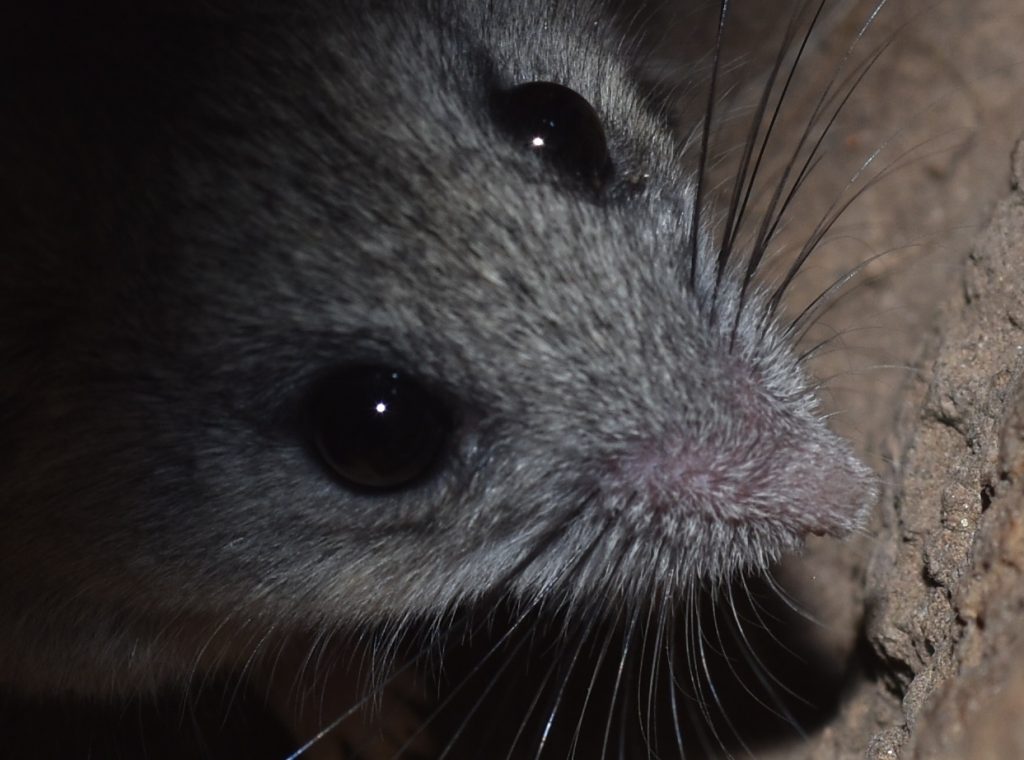
Same mouse…

Like the house mouse, deer mice are easy to breed in captivity. They look very similar to house mice, but have much larger eyes (as evidenced in the above photos). They’re also more energetic and tend to jump and run around more, which makes them a bit more difficult to handle. This skittishness is what inspired their common name.

Deer mice have a number of characteristics that make them better suited for some types of research. For one thing, they tend to live longer than house mice. Peromyscus spp. have a maximum lifespan of 5-7 years, as compared to just 3 years for M. musculus. This makes them more useful in aging research. Deer mice are also the main carriers of the Sin Nombre hantavirus, a virus which can cause a fatal disease in humans. Obviously, this makes them of great interest to medical researchers.

As I mentioned earlier, deer mice are native to North America. This makes them a convenient subject for North American researchers who want to study the evolution and distribution of species. The fact that deer mice don’t normally live in close association with humans is also helpful in this regard, since it means we haven’t been spreading them all over the place like we have with M. musculus.

A House for the Mouse 6

In the 1910s and 20s, Francis B. Sumner began investigating the use of deer mice as laboratory animals. He built the first Peromyscus “mouse house” at Scripts Institute in California. When his work was discontinued, Sumner passed the torch (and his mouse stock) to Lee R Dice at the University of Michigan. In the 30s and 40s, Dice continued refining methods for maintaining deer mouse colonies. He also began cataloging mutations in the mice, which he gave names like gray, dilute, and epilepsy.

In 1985, Wallace Dawson at the University of South Carolina used Dice’s deer mouse strains to start the Peromyscus Genetic Stock Center (PGSC). PGSC maintains six Peromyscus species, as well as numerous mutant strains, which it makes available to researchers. You can click here to visit their website.

The above video shows a California mouse (P. californicus). It was filmed on 4/17/17 in Mission Trails Regional Park in San Diego County.

The Mouse Leaves Its Mark 7

In the course of his pioneering work with Peromyscus, Sumner investigated its geographic variation. At the time, this represented one of the few major studies of evolution in wild populations. Important figures in evolutionary thought like Sewall Wright, Theodosius Dobzhansky and JBS Haldane closely followed his work and drew conclusions from it. Wright, for instance, used Sumner’s observations as evidence that quantitative traits in organisms were subject to the same rules of inheritance as discrete traits. (A discrete trait is a trait that an organism either has or doesn’t have like blue eyes. A quantitative trait is one that can vary in quantity like pigmentation.)

Dobzhansky used Peromyscus data in his studies of reproductive isolation. Some subspecies of P. maniculatus live in overlapping geographical areas, but are separated by habitat. For instance, one subspecies lives only in forests, while another lives only in open habitats like prairies. These subspecies are able to mate and produce viable offspring in the lab, but because they inhabit different environments in the wild, the subspecies remain isolated from each other.

Peromyscus has been attracting the attention of these and other evolutionary biologists for about a century now. It’s become one of the best studied mammals and there are many ongoing research projects devoted to it.

Glaciation and an Ancient River Drive the Evolution of New Species8

The map below shows Northwestern Mexico and the Southwestern United States. The thin blue line is the Colorado River feeding into the Gulf of California. (The area highlighted in pink shows the distribution of the desert iguana, which has nothing to do with this article. I just needed an image that was in the public domain and this was the best I could find.)
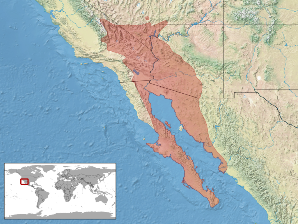
The Colorado River shown emptying into the Gulf of California
Author: compiled by rbrausse with Quantum GIS
This file is licensed under the Creative Commons Attribution-Share Alike 3.0 Unported license.

The Gulf of California began forming around 12 million years ago as a result of the movement of the underlying tectonic plates. Before that, the Colorado emptied directly into the Pacific. Eventually, it was diverted to empty into the Gulf. The important take-away from this is that for millions of years, the Colorado River has acted as a semi-permeable boundary, isolating land-bound animals living north of the river from those living south of it.9

When a group of organisms is isolated from other members of their species for a long period of time, that group will begin to diverge from the rest of the species. If this goes on for long enough, they may form a new species. This is what has happened to deer mice living around the Colorado.

The river is not the only thing that’s been shaping the evolution of these mice, though. The Earth has experienced a number of ice ages. During an ice age, a cooling climate causes glaciers to extend farther south in the northern hemisphere (and, of course, farther north in the southern). The last ice age, which started about 2.58 million years ago,10 is technically still going on, although we’re slowly coming out of it as the Earth warms. (Well, not so slowly anymore, thanks to human activity, but that’s another story.) The Last Glacial Maximum or LGM is defined as the most recent period when glaciers were at their thickest and the seas were at their lowest (due to all that water being locked up in ice). The LGM occurred around 24,000 to 18,000 calendar years ago.

As the glaciers melted and sea level rose, new islands formed as a result of the surrounding land being flooded by sea water. Many of these islands formed in the newly opened Gulf of California. A few also formed off the western coast of Baja, as well. Deer mouse populations trapped on these islands experienced further isolation from their confreres, resulting in more speciation. The formation of deserts and arid regions as the climate grew hotter and dryer resulted in new niches and pushed speciation still more. Undoubtedly, other changes related to climate and geology have played a role, as well.

A scientific paper published in the Journal of Mammalogy (click here to read it) describes research done on this process of speciation in deer mice in the areas bordering the Colorado. This paper looks at a subgenus of Peromyscus called Haplomylomys, which includes two species groups, californicus and eremicus. The californicus group is made up of just the one species, P. californicus. Eremicus is comprised of 10 species, including the one from which it takes its name, P. eremicus. Californicus lives in habitats with moderate amounts of water, like chaparral and woodlands. The eremicus group is restricted to arid and semi-arid regions.

Haplomylomys has been studied extensively, starting in the early 20th century. This new paper extends that research, concentrating on the species that developed on the various islands around Baja California. The researchers extracted genetic data from a great many species on both the islands and the mainland. They also extracted ancient DNA from skeletal specimens of P. guardia and P. pembertoni. I won’t go into all the details of what they found. Suffice it to say that they were able to find evidence of the effects of all the previously mentioned natural forces on the development of the various species of deer mouse in the region.

Of Mice and Monogamy 11 12 13

The vast majority of mammals are promiscuous. This makes sense from an evolutionary perspective. The more females a male mates with, the better the chances that he will succeed in passing on his genes to future generations. Nevertheless, there are cases where monogamy is the better strategy. If the young of a species are slow to develop, taking a long time to become independent, then it might make sense for the male to stick around and help care for his progeny. This is why humans are at least semi-monogamous.

Monogamy has evolved independently in two species of deer mouse — P. californicus and P. polionotus — which makes them of special interest to scientists who study social behavior in mammals. P. polionotus is also called the oldfield or beach mouse.14 Oldfield mice are found in the southeastern United States.15 In a study published in the Journal Nature,16 17 researchers took oldfield mouse pups and moved them into the nests of P. maniculatus, a polygamous species of deer mouse. They also took pups from P. maniculatus and put them into oldfield mouse nests. When the oldfield mouse pups grew up, they exhibited the same monogamous behavior as other members of their species, despite being raised by polygamous parents. Likewise, the P. maniculatus pups exhibited polygamous behavior as adults, despite being raised by monogamous parents. What this tells us is that, in deer mice at least, it’s genetics that determines whether or not a mouse is monogamous.

The scientists went on to try to identify some of the genes associated with monogamous behavior. Oldfield mice exhibit a number of behaviors associated with monogamy. For example, they build more elaborate nests than their polygamous cousins. The males spend more time licking their pups, huddling with them, and handling them. The researchers mated P. polionotus mice with P. maniculatus mice and found that the offspring exhibited varying mixes of the aforementioned behaviors.

By examining the DNA of the hybrid offspring, the scientists were able to identify stretches of DNA (called loci) that were associated with certain behaviors. In a follow up study, they focused on one of the genes in one of those loci — a gene that controls the production of a hormone called vasopressin. Some of this hormone is produced in the hypothalamus of the brain. P. maniculatus, it turns out, produces three times as much vasopressin in their hypothalamus than oldfield mice. The researchers tried injecting vasopressin into the brains of oldfield mice and found that the mice started making simple nests like the nests of P. maniculatus. The injected vasopressin had no other observable effects on the parental behavior of the mice. So it’s likely that the gene controlling vasopressin is just one of a number of genes that control behaviors associated with monogamy or polygamy.

This was just a brief overview of the paper in Nature. There are a lot of details that I glossed over, so you might want to read the New York Times article for a more in-depth treatment. Click here for that article. You can view the original Nature article by clicking here.

So what about the other species of monogamous mouse? We met P. californicus, the California mouse, earlier. This mouse is found in California and Mexico, on the other side of the continent from P. poloniotus. Lots of research has been done on P. californicus and it’s social behaviors. I won’t bore you with any specifics here, except to mention one article that briefly discusses a possible reason for the evolution of monogamy in this mouse. The article, entitled Tracking the Monogamous Mouse, was published in the Baltimore Sun. (Click here to read it). The article discusses a study by Doctors David Ribble and David Gubernick.

In the article, Dr. Ribble explains that the California mouse is “about twice the size of a normal field mouse”. (Field mouse is a generic term for any mouse that commonly inhabits fields.) Having to power a larger body means that you have less energy left over to devote to other things like making babies, so, other things being equal, larger animals tend to have smaller litter sizes. Most deer mice average four pups per litter, whereas, the much larger California mice produce only two. It turns out that four pups can share enough body heat to keep themselves warm, but two pups need the additional warmth from their parents. So in California mice, the male sticks around to huddle with the pups when the female is off foraging for food.

This neatly explains why P. californicus evolved monogamy, but why did they evolve a larger size? I don’t know. I wasn’t able to find any articles that answered that question. Maybe nobody has studied it. There’s far more stuff out there on deer mice than I’ve had time to read, though, so it’s quite possible that I just haven’t found the right article.

A Note In Closing 18 19

Walt Disney is said to have been in the habit of feeding one of the field mice that would wander into his Kansas City animation studio. This mouse was likely P. leucopus. In 1925, another animator, Hugh Harman, poked fun at Disney by drawing sketches of mice around a photograph of him. This inspired Disney to direct one of his employees to sketch a new mouse character. The rest, as they say, is history. So it seems that our model mouse may have been the model for Mickey.
(More or Less) Carnivorous Plants

What is a carnivorous plant? Seems like a simple question, right? Of course, nature is rarely simple. Suppose an animal dies next to a rosebush. Suppose also that, instead of being scavenged, its body decomposes where it fell. The rosebush will happily take up the nitrogen and other nutrients released by the decomposing body. Does that make the rosebush carnivorous? If so, that would mean that almost all plants are carnivores. So just being capable of absorbing nutrients from decomposing flesh is probably not a good definition of carnivory.

Various botanists have tried to come up with a more satisfactory definition. None of them is universally accepted, but the following, rather strict, definition is often used. According to this definition, a plant must meet the following criteria to be carnivorous: 7

  1. It has to do something to attract prey.
  2. It has to capture the prey.
  3. It has to digest the prey, using digestive enzymes that it secretes itself.
  4. It has to absorb the products of digestion. (I guess that one’s pretty obvious, but it’ll be important later.)

In this article, I’m going to go through most of the different kinds of (more or less) carnivorous plants and point out how they do or don’t meet these criteria.

Venus FlyTrap (Dionaea muscipula) 11

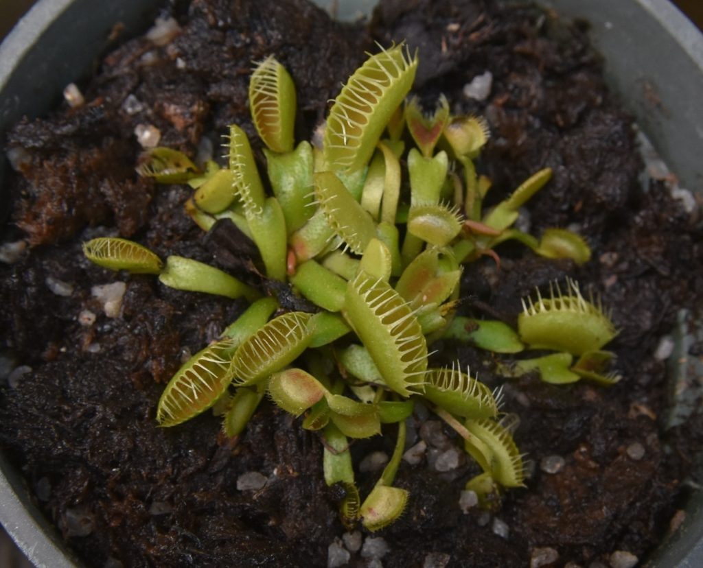
We may as well start with the most famous carnivorous plant, the Venus flytrap. Venus flytraps are native to the east coast of North and South Carolina. Each trap is a modified leaf, consisting of two lobes, hinged at the midrib. The interior of the lobes are a reddish color and there are small nectar-secreting glands along the rims. So any insects that happen by think they’ve found a flower and blithely crawl into the trap.

Venus FlyTrap (Dionaea muscipula)
The Huntington Botanical Garden, San Marino, CA 4/22/18
Interesting that all the traps seem to be sprung. I suspect that’s because the plant is in a botanical garden. A trap probably isn’t open for long before some kid comes along…

On the upper surface of each lobe are three trigger hairs. To trigger the trap, two of these hairs much be touched within 20 seconds of each other or one of the hairs must be touched twice in quick succession. The calibration of the triggering mechanism is so finely tuned that it will not normally be triggered by non-prey stimuli, such as raindrops. After snapping shut, the trap only begins digestion if it receives at least five more triggers. This ensures that it’s caught a live bug, not just some piece of detritus.

The trap doesn’t close completely right away, though. The edges of the leaf lobes are fringed by stiff hairs called cilia. When the trap closes part-way, the cilia mesh together blocking the escape of larger prey, but very small bugs that aren’t worth eating can get out through the gaps in the cilia. If the trap fails to capture a suitable bug, it will reopen after around 12 hours.

Once the trap closes all the way, it forms a hermetic seal and essentially becomes a stomach for digesting the prey. Glands on the leaf lobes release digestive enzymes that break down the body of the prey into small components that can be absorbed through the leaf surface. Digestion takes about ten days, after which, the trap reopens, releasing the empty husk. The flytrap catches mostly crawling arthropods, such as ants, beetles and spiders.
Is it carnivorous? D. muscipula fits all the criteria listed above. By any definition, it’s a carnivore.

Waterwheel Plant (Aldrovanda vesiculosa) 12

The only extant (non-extinct) plant with a trapping mechanism similar to the Venus flytrap is the waterwheel plant, which is the only living species in the genus Aldrovanda. The waterwheel is a free-floating aquatic plant with no roots. The traps are arranged in whorls around a central stem. They’re similar to the traps of the Venus flytrap, but much smaller — the lobes are only 2-3 mm long. They trap very small prey like nematodes and Daphnia (water fleas).

Waterwheel plant (Aldrovanda vesiculosa)
This file is licensed under the Creative Commons Attribution-Share Alike 3.0 Unported license.

A. vesiculosa is the most widely distributed of all the carnivorous plants — it’s native to Europe, Africa, Asia, and Australia. This is due to its mechanism of distribution — the small plants stick to the feet of migrating waterfowl which drop them off at their next stop. Despite its wide distribution, the waterwheel is not doing well. There are only 50 confirmed populations left in the world.

Is it carnivorous? The waterwheel fits all the criteria of carnivory except one. It doesn’t appear to do anything to attract prey. Maybe its prey is so common that it doesn’t need to. Seems like nit-picking, though. I think most botanists would still consider it a carnivorous plant.
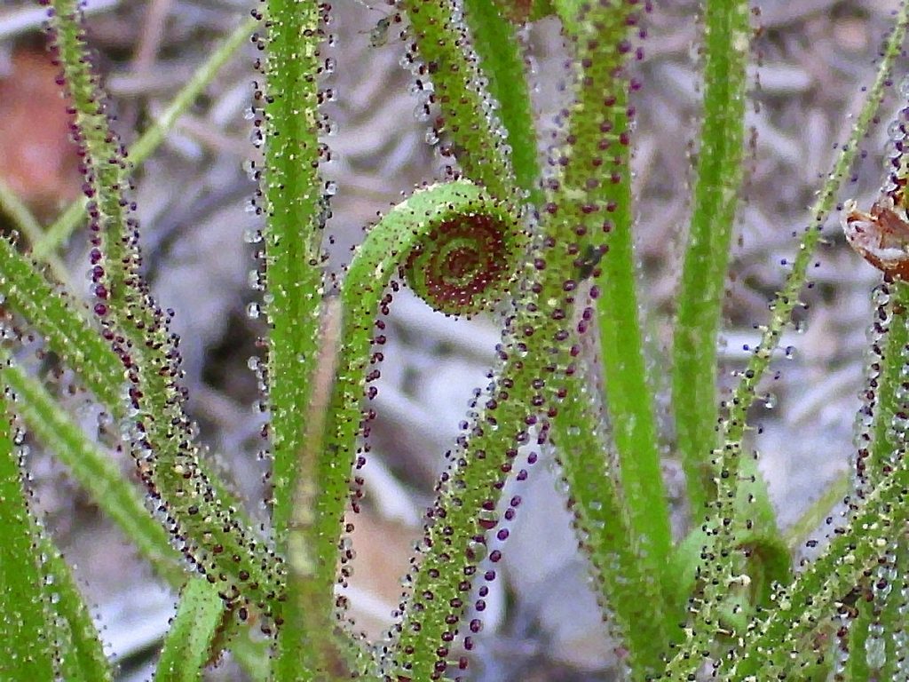
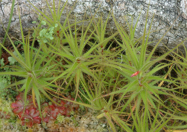
Sundews (genus Drosera) 17

The sundews make up the genus Drosera, which contains at least 194 species, making it one of the largest genera of carnivorous plants. It’s also very widespread — there are native sundews on every continent except Antarctica. Here in California, there are at least two native species — the great sundew (D. anglica) in the central and northern part of the state and the round-leaved sundew (D. rotundifolia), which seems to have a slightly larger distribution, extending a bit farther south. There are also a number of introduced species.20

Sundew (Drosera sp.)
The Huntington Botanical Garden, San Marino, CA 4/22/18

In most species of sundew, the leaves are covered with two types of glands. The first type takes the form of long tentacles, topped with sticky globs of sweet, glistening mucilage. These globs attract insects, snare them, and then secrete the enzymes that digest them. (Talk about multi-purpose!) The second type are sessile (stalkless) glands that absorb the products of digestion.

A fly trapped by a sundew
(Closeup from the last photo)

As soon as an insect is snared by the mucilage, the surrounding tentacles begin to bend toward the center of the leaf, bringing more globs of mucilage in contact with the bug. The mucilage clogs the spiracles (breathing pores) of the insect, causing it to suffocate. In some species, the leaf itself will bend around the insect to bring even more tentacles in contact with it. (You can see this behavior in the above photos.)

Are they carnivorous? Sundews easily meet the criteria for carnivory.

An Evolutionary Aside 21

The Venus flytrap (Dionaea), the waterwheel plant (Aldrovanda), and the sundews (Drosera) are grouped together into the family Droceraceae. Molecular and morphological studies suggest that Dionaea and Aldrovanda are more closely related to each other than to any other genera. Studies also suggest that Dionaea and Aldrovanda share a common ancestor with the sundews. The fossil record for carnivorous plants is far too sketchy to allow us to trace the evolution of these groups, but genetics and morphology can give us some clues. According to current thinking, Dionaea and Aldrovanda probably evolved from a sundew ancestor.

We’ve already seen that the leaves of many types of sundew will curl around a trapped insect and that this reflex is triggered by the insect touching the glandular tentacles of the leaf. The leaves of the various species of Drosera vary greatly in form. In the particular species shown above, the leaves are long and strap-like, but in many species they’re round. It’s not too hard to imagine how a round leaf with a propensity for closing around prey might have evolved into the snap-trap of a Venus flytrap or waterwheel. In this scenario, the glandular tentacles would have evolved into the trigger hairs of the trap, losing in the process their ability to secrete mucilage, which would no longer be needed.

Currently, there’s no evidence as to whether Dionaea evolved before Aldrovanda or vice versa. It could have happened either way.

Portuguese Sundew (Drosophyllum lusitanicum) 22

The Portuguese sundew is native to the Iberian Peninsula and Northern Africa. It’s not closely related to the true sundews (genus Drosera), but it does resemble them, having long glandular tentacles that trap insects. Unlike the tentacles of the sundews, however, these tentacles do not move in response to stimuli.

Portuguese sundew (Drosophyllum lusitanicum)
Author: Javier martin
Released into the public domain on Wikipedia Commons. Link to source page.

Is it carnivorous? D. lusitanicum excretes it’s own enzymes to digest trapped insects. So, although it’s traps are completely passive, it fits all the criteria for strict carnivory.

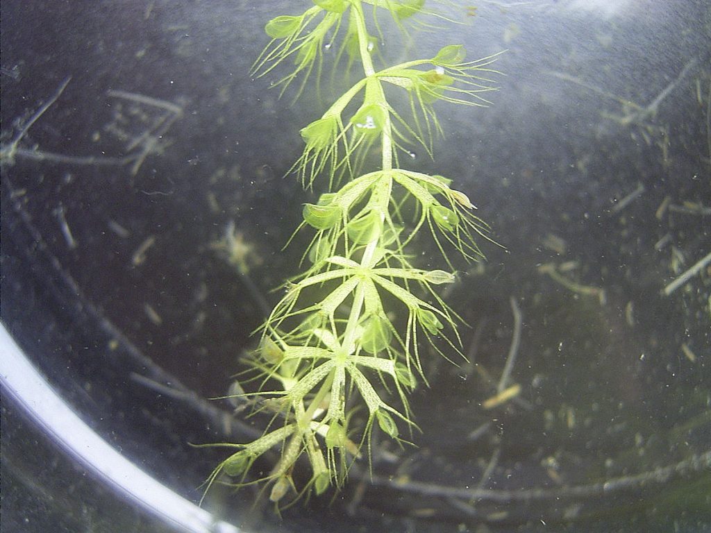
Bladderworts (genus Utricularia) 23 24 25

Bladderworts are found all over the world, except, of course, Antarctica. Quite a few species are native to California. Utricularia includes both terrestrial and aquatic species. In both cases, the main part of the plant grows beneath the surface.

Bladderwort (Utricularia dimorphantha) in Japan
Author: fischermans
This file is licensed under the Creative Commons Attribution-Share Alike 3.0 Unported license.

All bladderworts are carnivorous, trapping small prey using bladder-like traps. These traps are quite sophisticated. In fact, Utricularia may be the among the most highly developed plants in the world. Each trap has a trapdoor with trigger hairs. When a prey animal touches a trigger, the trapdoor opens and the animal is sucked into the bladder. The trapdoor quickly closes, trapping the animal inside. This process takes only ten to fifteen milliseconds. The bladder secretes digestive enzymes into the trap and the plant absorbs the products of digestion.

Water is constantly being pumped out of the trap. This creates a negative pressure inside the trap, due to the elasticity of the bladder walls. That is, the walls of the bladder are sucked inwards as water is pumped out. When the trapdoor opens, the walls spring apart, creating the suction that pulls in the prey.

Terrestrial species of bladderwort tend to feed on very small prey such as protozoa and rotifers swimming in the water-saturated soil. Aquatic species usually have larger traps and can feed on larger prey such as Daphnia, nematodes, mosquitoes, young tadpoles, and even fish fry.

Are they carnivorous? Yes.

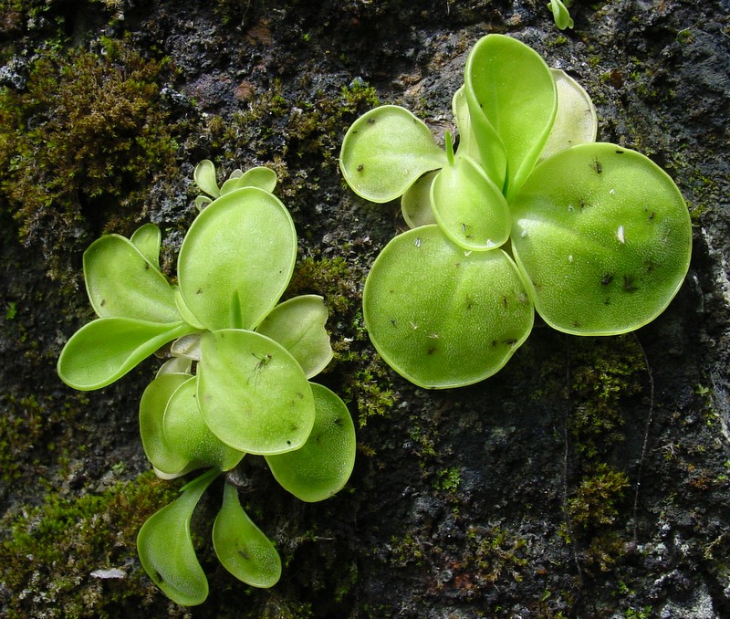
Butterworts (genus Pinguicula) 26 27 28

Butterworts are found throughout the Northern Hemisphere, as well as Central and South America. Mexico has the most numerous species. The common name comes from the greasy feel of the plant’s leaves.

Butterwort (Pinguicula conzattii) in Oaxaca, Mexico
Author: NoahElhardt
This file is licensed under the Creative Commons Attribution 2.5 Generic license.

Pinquicula tend to be small plants with flat leaves. They trap prey by means of tiny glandular hairs that cover the leaves. There are two types of these glands. The peduncular glands consist a of single stalk cell with a few secretory cells at the tip. These cells produce mucilage, which forms visible droplets on the leaf surface. This gives the leaf a wet appearance, which probably helps to attract insects. The peduncular glands secrete a small amount of digestive enzymes, but are mostly used for luring and trapping small insects. Some species are able to bend the leaf edges slightly, in order to bring more of the glands into contact with the prey.

The second kind of gland found on the leaves are sessile glands, which lie flat on the leaf surface. When the peduncular glands trap a bug and start the process of digestion, the flow of nitrogen stimulates the sessile glands to release additional enzymes that complete the process. The products of digestion are absorbed through the leave surface.

Butterworts will also digest pollen grains that land on their leaves.

Are they carnivorous? Butterworts fulfill all the criteria for carnivory.

Corkscrew Plants (genus Genlisea) 29 30 31

Corkscrew plants are small plants native to Africa and Central and South America. Genlisea have no roots. Instead, they have two types of leaves — above-ground leaves that carry out photosynthesis and highly modified subterranean leaves that attract, trap, and digest tiny invertebrates, especially protozoa. Each of these underground leaves forms a hollow tube which forks into a pair of capillary tubes. Spiral grooves along the length of each capillary tube allow the prey to enter and inwardly-pointing hairs prevent them from escaping. At the fork, the prey enters the main tube, which also has inwardly-pointing hairs. Eventually, the animal reaches the digestive chamber — a vesicle containing glands which secrete digestive enzymes.

Corkscrew plant (Genlisea lobata). Native to Brazil.
Author: Denis Barthel
This file is licensed under the Creative Commons Attribution-Share Alike 3.0 Unported license.

Besides trapping prey, the subterranean leaves of the corkscrew plant serve many of the functions of roots, including anchoring the plant and absorbing water and nutrients.

Are they carnivorous? Several of the articles I read mentioned that corkscrew plants attract prey, but I couldn’t find any that explained how they do this. So, assuming this is true, Genlisea does fit the strict definition of carnivory.

Another Evolutionary Aside 32 33

The bladderworts, butterworts, and corkscrew plants are grouped together into the family Lentibulariaceae. The corkscrews branched off first from the common ancestor of Lentibulariaceae and the other two genera branched off later. Scientists believe that the most recent common ancestor of all these plants was terrestrial and had a basal rosette of flat leaves and a primary root.

The carnivorous technique of the butterworts is pretty similar to that of the sundews, minus any movement of the glands. The bladderworts and corkscrews, on the other hand, seem to be totally unique in the carnivorous plant world. Unfortunately, I haven’t been able to find any information about the evolution of carnivory in these two genera. If I find anything in the future, I’ll update this page.

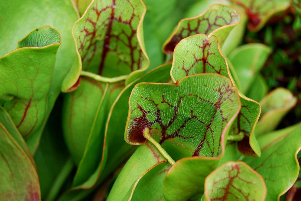
American Pitcher Plants (family Sarraceniaceae) 34 35

The pitcher plants in the Sarraceniaceae family may be the most well-known of the carnivorous plants after the Venus flytrap. The family contains three extant genera — Sarracenia, Darlingtonia, and Heliamphora.

Sarracenia composes the North American pitcher plants (excluding Darlingtonia). The pitchers are formed by long leaves that curl up to form tall, water-tight tubes. Usually, many pitchers radiate out from a central point. The tubes are partially filled with rainwater mixed with digestive enzymes.

Purple pitcher plant (Sarracenia purpurea)
Author: Geoff Gallice from Gainesville, FL, USA
This file is licensed under the Creative Commons Attribution 2.0 Generic license.

The pitcher attracts insects with secretions from nectaries located around the lip. In most species, a waxy coating on the lip causes insects to fall in and more wax on the inside of the pitcher makes it difficult for them to climb out. Many species also have downward-pointing hairs on the inside the pitcher that make climbing out even more difficult. Further down the tube, the insects fall into the water where they drown and are digested.

At least some members of the Sarracenia genus produce their own digestive enzymes. However, most, if not all, species also use symbiotic bacteria and protozoa to supplement their supply of enzymes and some may rely on them entirely.

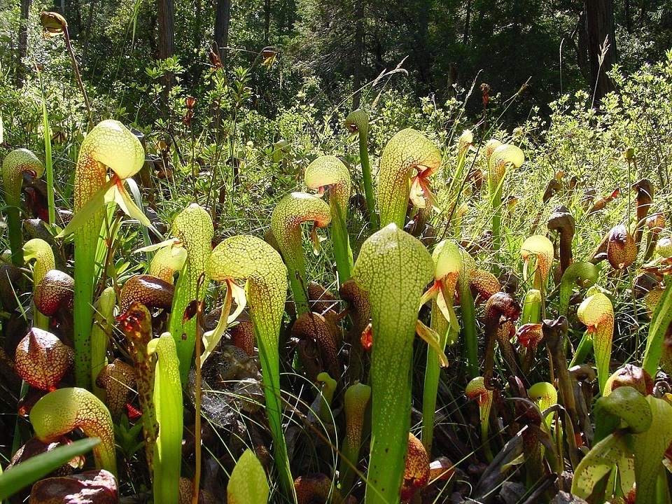
Members of the genus Darlingtonia are often called cobra lilies because of the their resemblance to the snake. Darlingtonia californica is the only species in the genus and it’s native to Northern California and Oregon. D. california is unusual in that it doesn’t trap rainwater in its pitchers. The water is drawn up from the roots. The hood prevents rainwater from diluting the contents of the pitcher.

California pitcher plant (Darlingtonia californica) a.k.a. cobra lily, Botanical Trail, Butterfly Valley Botanical Area, Plumas County, California
Author: NoahElhardt (talk · contribs)
This file is licensed under the Creative Commons Attribution-Share Alike 3.0 Unported license.

It used to be believed that the cobra lily did not produce any of its own digestive enzymes and, instead, relied entirely on symbiotic microorganisms to digest its prey. However, recent studies have shown that it does secrete at least one such enzyme.

Heliamphora is the genus of South American pitcher plants, which are called sun pitchers in common parlance. They’re similar to the other plants in Sarraceniaceae with the exception that most of the species in the genus do not produce their own digestive enzymes. Instead, they rely on symbiotic bacteria.

Are they carnivorous? As already mentioned, many of the species in Saraceniaceae do secrete at least some of their own digestive enzymes. Those species, at least, fit the strict definition of carnivory. Most of the members of the genus Heliamphora, however, rely entirely on symbiotic bacteria for digestion and, so, would only fit a looser definition.

Tropical Pitcher Plants (genus Nepenthes, family Nepenthaceae) 36

The pitchers of Nepenthes are remarkably similar in form to the pitchers of Sarraceniaceae, even though the the two groups are not closely related. They’re a wonderful example of convergent evolution, which is where two unrelated organisms evolve very similar features simply because they are under similar evolutionary pressures. In other words, they find similar solutions to a common problem. In this case, the common problem is how to supplement their diets. One thing all carnivorous plants have in common is that they grow in areas that are nutrient poor. Carnivory is a way of getting the nutrients that they can’t get from the soil.

Upper pitcher of Nepenthes alata photographed on Mt Ambucao, near Sagada and Banaue, Luzon in The Philippines
Author: Attenboroughii
This file is licensed under the Creative Commons Attribution 2.0 Generic license.

Unlike the pitcher plants of the Americas, most species of Nepenthes grow as woody vines (lianas). Most of them produce two types of pitchers. The lower pitchers typically sit on the ground. The upper or aerial pitchers are usually smaller and have different features. In both cases, the pitchers grow from tendrils of the liana. The tendril of an upper pitcher will often loop around a branch to steady the pitcher. In some cases, the upper and lower pitchers are specialized to attract different types of prey.

The prey of Nepenthes usually consist of insects, but the species with the largest pitchers sometimes catch vertebrates such as lizards and even rats! A plant that eats small mammals… that may be the closest we’ll ever get to Audrey II! 37

Are they carnivorous? The members of Nepenthes are believed to secrete at least some of the digestive enzymes found in their pitchers, although these are supplemented with enzymes produced by symbiotic microorganisms. So Nepenthes seems to fit the strict definition of carnivory.

A Special Mention for Nepenthes lowii 38 39

We’re not quite finished with Nepenthes yet, because now we come to the one carnivorous plant that I absolutely had to write about. Nepenthes lowii is a tropical pitcher plant that grows in Borneo. Lower pitchers are rare, because the plant quickly enters the climbing stage in which the upper pitchers are produced. And the interesting thing about these upper pitchers is that they look a lot like toilet bowls. With lids. But the really interesting thing is that they are toilet bowls.

Nepenthes lowii
Author: JeremiahsCPs
This file is licensed under the Creative Commons Attribution-Share Alike 3.0 Unported license.

The underside of the lid produces a white secretion that looks a lot like snail eggs, but is actually produced by the plant itself. This secretion attracts birds, tree shrews, and the occasional rat. The opening of the pitcher and the placement of the lid have evolved in such a way that a tree shrew or rat feasting on the secretion will naturally straddle the opening of the pitcher. In fact, the pitcher appears to have evolved to be a perfect fit for tree shews. Any excrement produced by the rodent will drop into the pitcher. Poop is high in nitrogen — just what the plant is looking for. So, to put it bluntly, N. lowii eats poop.

Oh, and I almost forgot… the white secretion seems to contain a fast-acting laxative.

Is it carnivorous? Nepenthes lowii meets all the criteria for carnivory except one: it doesn’t eat the animals — just their excrement. Actually, that’s not entirely true. Insects do fall into N. lowii‘s pitchers and they do get eaten, so technically I guess it is carnivorous. However, the plant eats mostly feces and studies have shown that it is quite capable of subsisting entirely on a diet of poop. So N. lowii seems be a case of a carnivorous plant that is evolving away from carnivory.

Bromeliads (family Bromeliaceae) 40

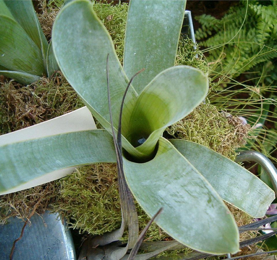
The bromeliads are one of the few groups of monocots that includes carnivorous plants. (Monocots and dicots are two of the major groups into which the flowering plants are divided. The seeds of monocots contain only one embryonic leaf. Those of dicots contain two.) Bromeliads include both terrestrial species and epiphytes. An epiphyte is a plant that grows on another plant and gets its water and nutrients from air, rain, water (in aquatic environments) or debris that accumulates around them. Epiphytes are not parasites. They just use other plants for support.

In many bromeliads, a rosette of leaves forms a tank that catches rainwater. A pool forms in the tank and the leaves absorb water from the pool. Entire ecosystems often form in these pools, including insects and tree frogs. Animals defecate and die in the pools. Other plants, such as algae, grow in the pools and detritus collects in them. All this supplies the bromeliad with a rich source of nutrients to be absorbed by the leaves.

Brocchinia reducta. Native to South America.
Author: BotBln
This file is licensed under the Creative Commons Attribution-Share Alike 3.0 Unported license.

A number of bromeliad species have been found to attract insects, which drown in the pools and are digested at least partly by enzymes produced by the plants.

Are they carnivorous? The bromeliads that attract, kill, and digest insects appear to fit the definition.

Another Special Mention: The Sheep-Eating Plant 41

Puya chilensis is a tree-like bromeliad that grows in the Andes. Its leaf rosettes are used by several bird species as nests. I’m not sure if P. chilensis absorbs any nutrients through its leaves, but run-off from the nests may fertilize the roots.

P. chilensis has very sharp, outwardly-pointing spines, which it probably uses to discourage predators. Birds are often trapped and killed by these spines and run-off from their decomposing bodies might also fertilize the roots of the plant. Where it gets crazy, though, is that sheep sometimes get their wool entangled by the spines of these plants and, if not rescued, they may die and decompose at the base of the plant. The roots of the plant would, of course, absorb the nutrients released by that decomposition and a rotting sheep carcass releases a lot of nutrients. For this reason, P. chilensis is sometimes called the “sheep-eating plant”.

P. chilensis does’t produce digestive enzymes, so it doesn’t fit the strict definition of a carnivorous plant. It’s still pretty amazing, though. It makes you wonder kinds of bizarre, now-extinct carnivorous plants might have existed in the past.

Click here to watch a video of some sheep caught by P. chilensis.

Dewstick or Fly Bush (genus Roridula, family Roridulaceae) 42

Roridula is a genus of shrubs native to South Africa. The leaves and sepals are covered with sticky tentacles, which trap insects. The dewstick does not digest the insects directly, however. Instead, there are two species of insect, both in the genus Pameridia, that prey on the captured bugs. These insects deposit their droppings on the leaves, which absorb nutrients from them.

Dewstick (Roridula gorgonias) in Kew Gardens
Author: Polypompholyx
This file is licensed under the Creative Commons Attribution-Share Alike 3.0 Unported license.

Are they carnivorous? The members of Roridula don’t excrete their own digestive juices. They don’t even eat the trapped insects — just the feces of the symbiotic Pameridia. So they certainly don’t fit the strict definition of carnivory. And yet, they do attract and capture insects and, eventually, the nutrients from those insects get into the plant by way of the Pameridia. So maybe the strict definition is too strict.

Protocarnivorous Plants 43 44

Occasionally, we encounter a plant that seems to meet all the criteria for carnivory except one: they don’t actually eat their prey. For instance, there are plants that attract insects, capture the insects, and even secrete digestive enzymes to digest the insects, but they don’t absorb the products of digestion. What could possibly be going on here?

In fact, the solution to this conundrum is pretty simple. Plants don’t just kill insects in order to eat them. They also kill them to protect themselves from being eaten. It’s not hard for plants to attract insects. They do it all the time without meaning to. So plants have had to evolve defenses against being eaten. Often this means producing toxic or bad-tasting chemicals to discourage the bugs. Another approach is to secrete a sticky substance that traps them and clogs up their pores.

Suppose a plant has taken this second approach and evolved sticky glands to trap insects. How could it take the next step and evolve a way to eat them? Actually, this isn’t as hard as it sounds. It turns out that regular, non-carnivorous plants are able to absorb nutrients through their leaves. They do it through their stomata (breathing pores), but also directly through the epidermis — the outer layer of the leaf. Some horticulturalists and other plant people have taken advantage of this by applying liquid fertilizer directly to the leaves — a process called foliar feeding.45

So some plants that trap and kill insects, but don’t eat them, may be on their way to becoming fully carnivorous. This kind of plant is sometimes called a protocarnivorous plant. The term is also used more broadly to refer to any plant that comes close to being fully carnivorous, but doesn’t quite fit the bill. Many of the plants discussed above could be classified as protocarnivorous. (You may also encounter terms like paracarnivorous, subcarnivorous, and borderline carnivorous. They’re all used pretty much interchangeably.)

A great many groups of plants have been examined for possible carnivory or protocarnivory — many more than I care to cover in this post. But if you’re interested, you can click here to read a paper that goes through various plant families, examining each one for evidence for carnivory. It’s actually a pretty good read and I used it as a reference for much of the information presented here. For the purposes of this article, though, I’ll just mention one of the possibly protocarnivorous plants that I’ve encountered in Southern California…

The Catchfly, a Possible Protocarnivore

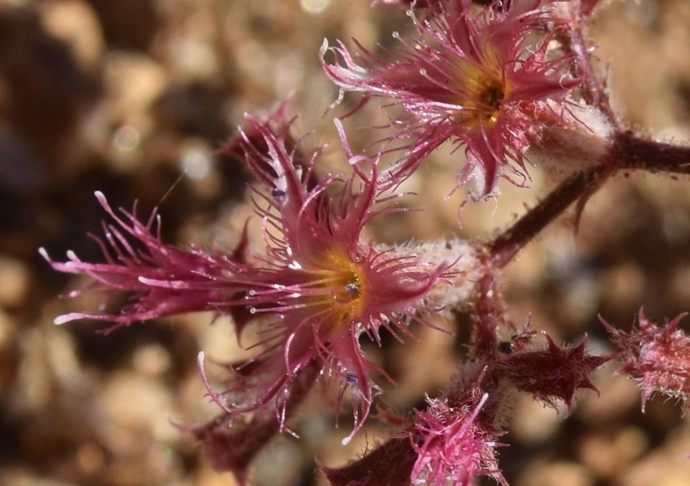
Plants in the genus Silene (family Caryophylaceae) are called catchflies because many species have sticky hairs on their stems and calyxes that trap and kill insects. They don’t seem to digest the insects, though, so they’re probably doing it as a defense against predation.

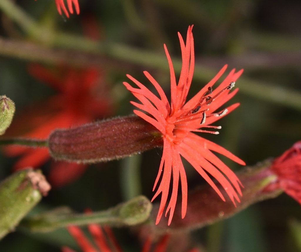
I’ve seen a couple of species of Silene in San Diego County. The beautiful scarlet catchfly (Silene laciniata angustifolia) is a California native:

Scarlet catchfly (Silene laciniata angustifolia)
Mission Trails Regional Park, San Diego County 5/30/19

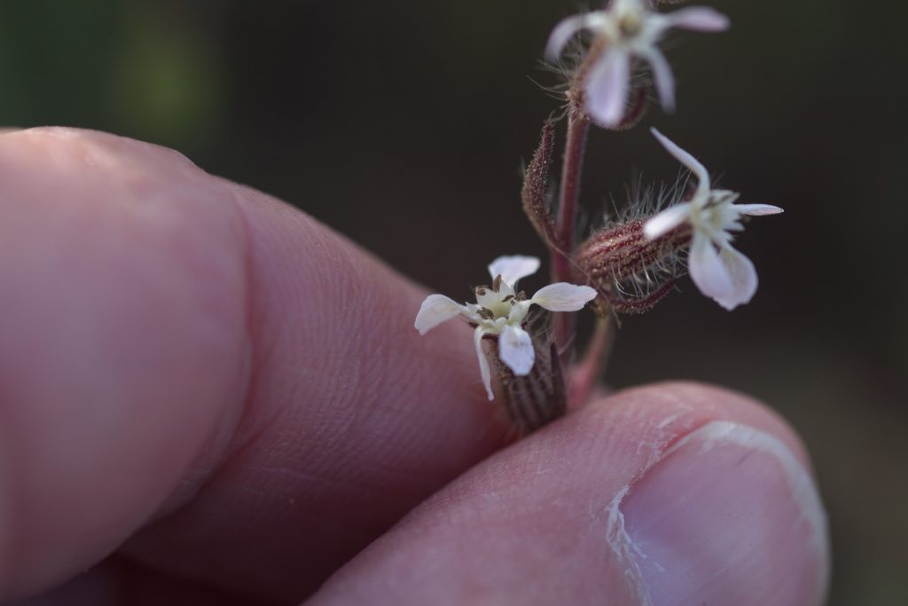
Small-flowered catchfly or common catchfly (Silene gallica) is another commonly seen catchfly in my area. A non-native, unfortunately.

Small-flowered catchfly (Silene gallica)
Mission Trails Regional Park, San Diego County 4/15/19
Small-flowered catchfly (Silene gallica)
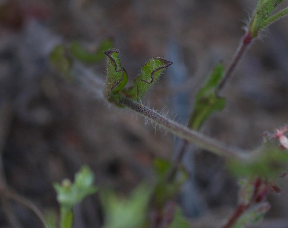
Note the hairs on the stem and leaves.
Small-flowered catchfly (Silene gallica)
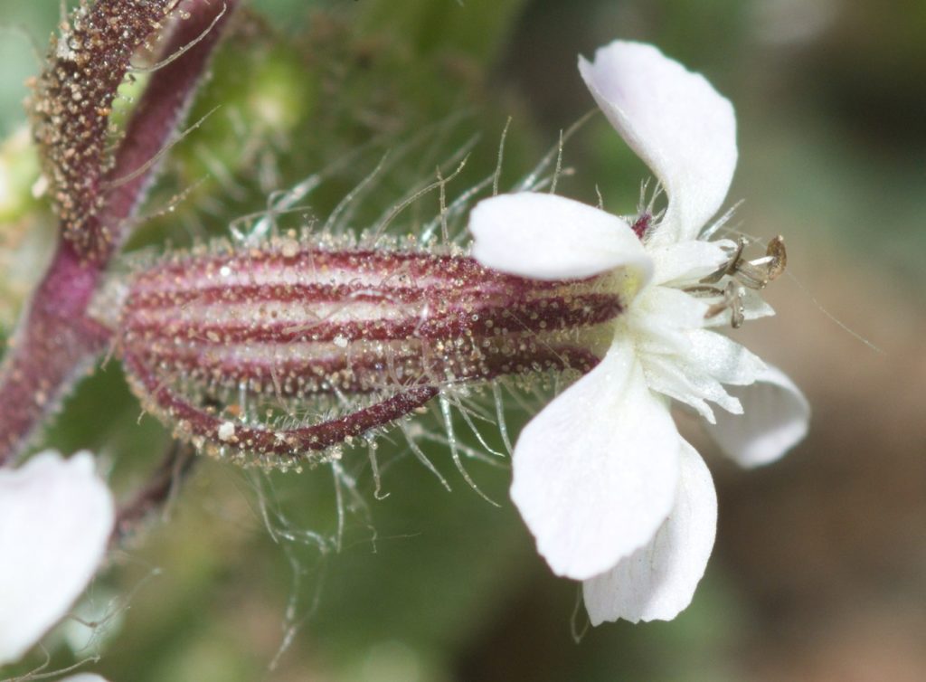
Note the glands on the calyx of the flower.

Carnivorous Seeds? 46 47

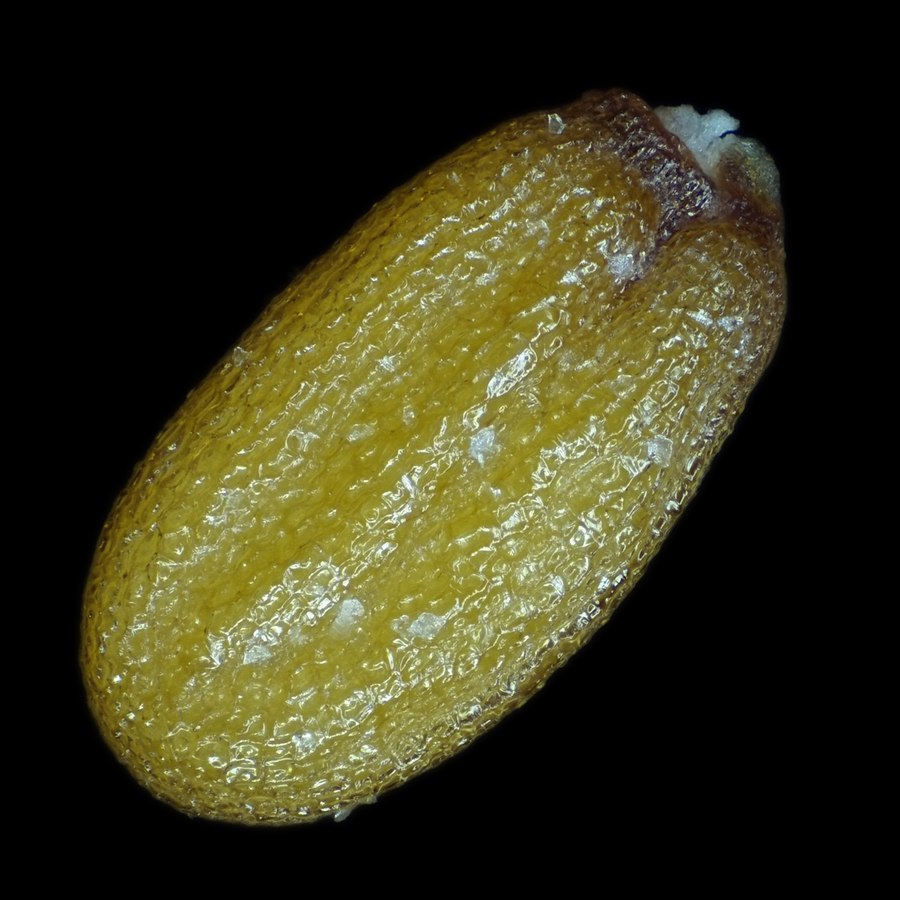
Because… why not? Shepherd’s purse (Capsella bursa-pastoris) is a small plant in the mustard family (Brassicaceae). According to findings presented in Nature,48 nematodes — very tiny roundworms that are super-abundant in soil — are attracted to the seeds of this plant, get stuck in the sticky mucilage coating the seeds, and eventually die, fertilizing the soil around the plant. The researchers found that plants germinated in soil containing nematodes did better than those germinated in soil without nematodes, especially if the soil was nutrient-deficient.

Capsella bursa-pastoris seed
Author: Alexander Klepnev
This file is licensed under the Creative Commons Attribution 4.0 International license.

Is it carnivorous? It’s not known whether or not enzymes produced by the seeds play a role in the digestion of captured nematodes. It’s possible they may enlist the aid of symbiotic microorganisms to do the job, but there’s no conclusive evidence for that, either. So, at present, C. bursa-pastoris cannot be said to fit the strict definition of a carnivorous plant. It certainly counts as a protocarnivorous plant, though. And, in any case, a seed that eats worms is pretty freaking awesome!

Crayfish Journeys

When Charles Darwin published On the Origin of Species in 1859, he argued the case for evolution by presenting what he hoped would be an overwhelming amount of evidence. I think most scientists would agree that he succeeded admirably. Nevertheless, Darwin had to contend with the fact that much of the scientific knowledge we take for granted today didn’t exist at the time. Obviously, he knew nothing about the genetic code. Even the basics of heredity were almost unknown. (Gregor Mendel had made a great start, but his work was pretty much ignored until decades later.) So the mechanism underlying natural selection was a black box to him. This was a problem, but not an insurmountable one. Darwin simply had to present evidence that there must be some such mechanism; he didn’t actually need to find it.

A more serious problem was continental drift, a.k.a plate tectonics. There were many cases of identical fossils found on widely separated bodies of land with no obvious way that the organisms could have gotten from one body to the other. For instance, there were fossils of animals found on the west coast of Africa that were identical to fossils found on the east coast of South America. Today the explanation is obvious. We know that all the continents used to be joined together into one vast super-continent called Pangaea. Pangaea eventually broke up and the fragments drifted apart to form separate land masses. The animals found in both South America and Africa evolved there when the two continents were connected.

Darwin knew nothing about continental drift. He didn’t even know about radioactive elements (whose decay heats up the Earth’s mantle and produces the massive convection currents that move the continents.) No one at the time seriously believed that the continents could be moving. So Darwin was forced to come up with alternative explanations for such fossils. When I read On the Origin of Species as a young man, I remember thinking that the suggestions he put forth were not very convincing.23 27

So what does all this have to do with crayfish? Crayfish are basically small, freshwater lobsters, but, unlike their marine relatives, they can’t survive in the open ocean. Nevertheless, there are native crayfish found on all the continents except Africa and Antarctica. So crayfish are a textbook example of a population of organisms that evolved when all the continents were joined together and were broken up into separate populations when Pangaea broke up. Those populations continued to evolve after the breakup. I’ll be discussing this in more detail later, but, first, let’s get to know our subject a little better.

Red swamp crayfish (Procambarus clarkii)
Sweetwater River near Jamul, CA 8/19/2017
Claw (chela) of red swamp crayfish
Sweetwater River near Jamul, CA 8/19/2017

Crayfish, Crawfish, or Crawdad?

The word crayfish probably comes from the Frankish word krebitja, which is a diminutive of the word for crab.31 This evolved into the Old French word for crayfish — “escreviss” — and eventually into our version of the word. Crayfish are also called crawfish or crawdads in some localities. “Crawfish” is obviously just a variation on the pronunciation. The origin of “Crawdad” is a bit more mysterious. It could be related to the use of “dad” in “doodad”, but no one seems to know for sure.

Crayfish are crustaceans, which means that they are related to such animals as crabs, shrimps, prawns, and woodlice. Crustaceans are, in turn, a subgroup of Arthropods. Like other arthropods, such as insects and spiders, crustaceans have exoskeletons. Unlike other arthropods, they have biramous limbs. A biramous limb is an apendage that branches into two parts. The bottom part is often (but not always) used for locomotion, while the upper part is used for other purposes — gills, for instance.

Crayfish evolved from lobsters. Some authors treat them as a type of lobster; some don’t. It’s not important from a scientific point of view, since “crayfish” and “lobster” are both common names. Historically, crayfish have generally been classified as belonging to three families — Astacidae, Cambaridae, and Parastacidae. Together, these three families can be referred to as Astacida. So Astacida is the scientific name for crayfish as a group.33 The study of crayfish is called Astacology. 40

Crayfish in California

There is only one extant (not extinct) species of crayfish that’s native to California. That’s the Shasta crayfish (Pacifastacus fortis). It lives only in Shasta County in northeast California. It’s threatened by habitat loss and also by competition from an invasive species, the signal crayfish (Pacifastacus leniusculus).43 47

Another invasive species that’s found in California, including here in San Diego County, is the red swamp crayfish (Procambarus clarkii), which is in the Cambaridae family. P. clarkii is native to the southern and southeastern United States and northern Mexico. Its native range doesn’t extend much farther west than Texas, but it’s been introduced to many other places in the U.S. and around the world. In some places, it’s become a serious pest.49

The red swamp crayfish is the most widely farmed species of crayfish in the world. It’s used as food, as bait for fishermen, in the aquarium trade, and as research animals in schools and laboratories. All this accounts for its wide dispersal. In some cases, P. clarkii has been deliberately introduced into an area in hopes of an economic return. In other cases, schools or individuals have released them into the wild just because they didn’t know any better.

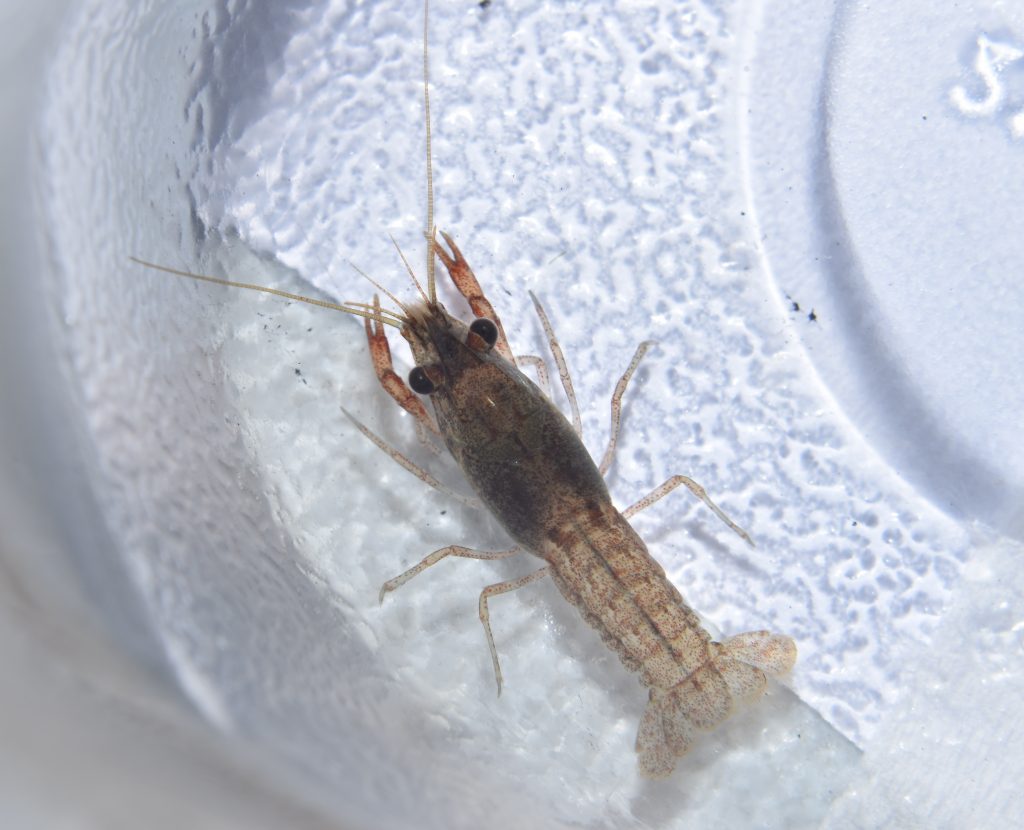
P. clarkii is the only crayfish that I’ve encountered in southern California. All the photos that I’m including with this post, unless otherwise noted, are photos of this species and all were taken of individuals that I found in the Sweetwater River near Jamul, CA.

Crayfish Anatomy50

Crayfish are a popular subject for dissection in high school and college biology classes. As a result, there are some excellent resource videos online. The easiest way to learn crayfish anatomy is to watch some of these videos. Click here for a good anatomy video. And here’s a couple of links to videos that actually show the dissection process: Crayfish Dissection 1, Crayfish Dissection 2

Just in case you don’t feel like watching a bunch of videos, I’ll give you a quick lesson. Figure 1 shows the basics of crayfish external anatomy.

Figure 1 – This image is in the public domain. It was downloaded from Wiki Commons at this url.

For the internal anatomy, refer to figure 2 below:

Figure 2 – This image is in the public domain. It was downloaded from Wiki Commons at this url.

Figure 1 shows a female crayfish in the genus Cambarus. Other crayfish species should have similar anatomies. I don’t know what species was used for Figure 2.

Crayfish are decapods, a word which means “ten-footed”.51 In the case of crayfish, this refers to the five pairs of walking legs or pereiopods. The first pair — the pair with the big claws — are also called chelipeds.

On the sides of the thorax, hidden by the carapace, are the gills. Each gill looks like a feathery appendage, but it’s actually the upper branch of one of the walking legs. Unfortunately, the gills are not shown in the illustrations, but if you watch the dissection videos, you’ll get a good look at them.

The head and thorax are joined and together make up the cephalothorax. This is covered on the top and sides by a shell called the carapace. The carapace includes the rostrum — the pointed “nose” or beak at the front of the head.

Crayfish have two pairs of antennae — one long pair and another much shorter pair. The shorter pair are called antennules. Each antennule is branched. That is, they are biramous appendages like the walking legs. Arthropods are believed to have evolved from annelids — segmented worms with non-branched (uniramous) appendages on each segment. As new species evolved from this ancestor, segments were added, lost, or fused, and became specialized in various ways. The appendages went on to differentiate into new forms that served different purposes. Some, for instance, evolved into mouth-parts and others became antennae.

In the crustaceans (or maybe in an earlier ancestor), the appendages evolved to be biramous. It’s likely that two segments were fused into one segment with two pairs of appendages and, later, the two pairs of appendages fused to become one biramous pair.52

Note the “pleopods” on the “tail” (really the abdomen) of the crayfish. These are also called swimmerets. These are thin, flimsy-looking, biramous appendages that are used by the female to hold her eggs to the bottom of her abdomen after she lays them. Despite the name, they’re not actually used for swimming, although they may help to keep water flowing over the gills. They may also help in steering.

In the male crayfish, the first pair of pleopods are enlarged and are used for transferring sperm during copulation. These more specialized pleopods are called gonopods.

The tail is made up of the telson (which is actually the last segment of the body) and four paddle-like structures called uropods, two on each side of the telson. When frightened, a crayfish will often propel itself rapidly backwards by flipping its tail in an arc underneath its body.

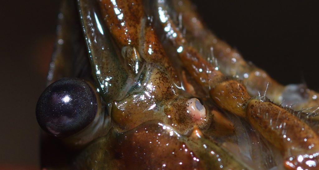
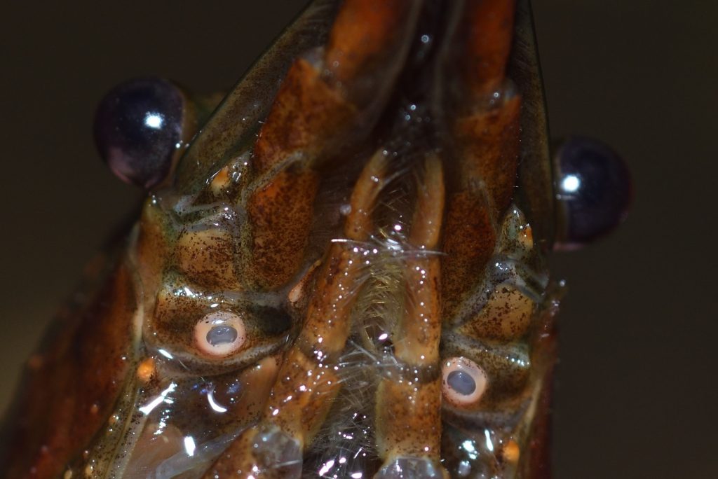
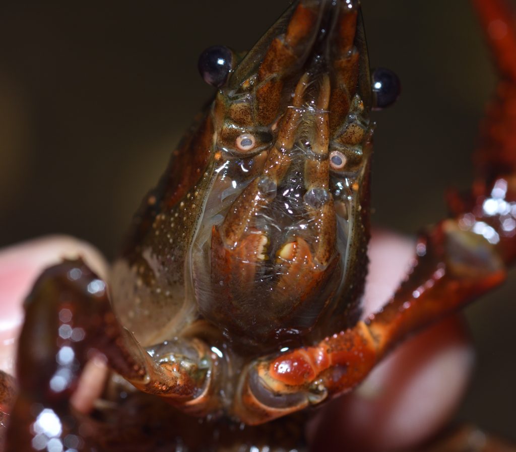
On the underside of the head, just a little farther back than the eyes, are the openings to the nephridia. The nephridia (there are two of them) are glands that are present in many invertebrates that function like kidneys — they eliminate toxic substances and waste products from the urinary and circulatory systems.53 In crustaceans, the nephridia are also called green glands, antennal glands, or maxillary glands. The following two photos clearly show the openings of the green glands. The actual glands are not visible in the above illustrations, but one of the openings is labeled in each figure.

Red swamp crayfish (Procambarus clarkii)
The pink oriface is the opening of the green gland.
Sweetwater River near Jamul, CA 10/4/2019
Red swamp crayfish (Procambarus clarkii)
Showing the openings of the green glands.

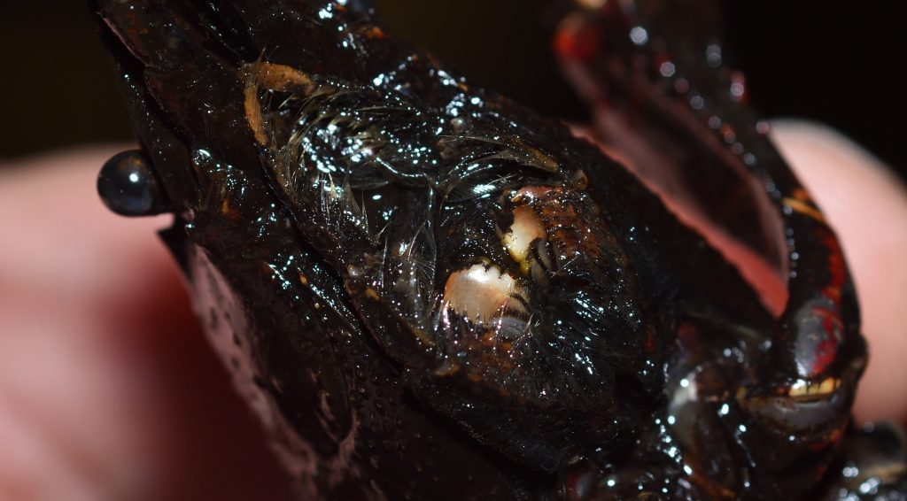
In the above photos, you can see the largest of the three pairs of maxillipeds. These are appendages used to manipulate food. They’re also labeled in figures 1 and 2. The photo below gives you a more complete view.

Red swamp crayfish (Procambarus clarkii)

Also visible in the above photo are the mandibles. Those are the small, light tan structures between the maxillipeds. The mandibles are used for grinding up food.54 Here’s a better view of them:

In the following shot, you can see two of the pairs of maxillipeds from the side. (The third pair is hidden underneath the body.)

Red swamp crayfish, showing two of the three pairs of maxillipeds

The following video shows a crayfish using its maxillipeds to search the sediment for food.

In the next photo, you can see the rostrum, the antennae, the antennules, and the compound eyes. The eyes are mounted on short stalks called pedicels.

Red swamp crayfish (Procambarus clarkii)

Like the compound eyes of many insects (which probably evolved from crustaceans), a crayfish eye consists of thousands of ommatidia — thin, columnar structures that run from the surface of the eye to the retina. The ommatidium of a crayfish consists of a cornea, a lens, and photoreceptor cells. (Figure 3 shows a generic drawing of an ommatidium.) Each of the tiny, individual facets visible in the following photos is the cornea of an ommatidium. When excited by incoming light, the photoreceptor cells produce electrical impulses representing the intensity and color of the light. These impulses are processed by the nervous system and brain of the crayfish to form an image.

Crayfish eye
Figure 3 – A generic ommatidium
A and B represent the cornea and lens, respectively.
This image is in the public domain. It was downloaded from Wiki Commons at this url.

I mentioned earlier that arthropods are believed to have descended from a segmented worm with a pair of appendages on each segment. Originally, all the segments were probably identical externally, except for the head and tail, where the orifices for the mouth and anus would have been located. The body of the crayfish is still divided into segments, although these segments have been greatly modified, and each of the segments still has a pair of appendages.

To summarize, here’s a chart that lists the segments of the crayfish body and their associated appendages:

Head: 5 segments
Segment 1: Antennas
Segment 2: Antennules
Segments 3-5: Mouth-parts, consisting of 3 pairs of modified appendages (Only the mandibles are visible in the photos.)

Thorax: 8 segments
Segments 1-3: Three pairs of maxillipeds used for manipulating food
Segment 4: Chelipeds (the 1st pair of walking legs with the large claws)
Segments 5-8: The other 3 pairs of walking legs

Abdomen: 5 segments
Segments 1-5: Pleopods or swimmerets

Tail: 1 segment
Segment 1: Telson and uropods

Daily Life

Most crayfish live in bodies of freshwater, such as lakes, rivers, streams and ponds, although a few species can live in brackish water. They are mostly nocturnal, hiding under rocks or logs or in burrows during the day and coming out to forage at night. There are cave-dwelling species that have lost eyes and pigmentation. Most crayfish are omnivores, eating a variety of plants and small animals, both living and dead.

Many, but not all, species of crayfish dig burrows. Those that do often dig them all the way down to the water table, which insures that they will have access to water even if the surface water body dries up. Some species spend their entire lives in their burrows, some come out to forage at night and return to their burrows during the day, and some spend most of their time outside their burrows, returning to them only for certain activities, such as mating or egg laying or for protection when environmental factors become a threat.55

The Life Cycle 56 57 58 59 60

The exact details of the crayfish life cycle varies greatly from species to species and genus to genus. The following description focuses on the red swamp crayfish (P. clarkii).

P. clarkii becomes sexually mature in as little as two months. The male crayfish comes in two forms, officially named form 1 and form 2. Form 1 is the mating form. In form 2, the male is not capable of mating and may attack a female if it comes near. The crayfish changes from one form to the other by molting. Mating usually takes place in the fall, but there may be a second period of mating in the spring in warmer, wetter climates.

After the female lays her eggs, she gathers them up and glues them in a mass to the underside of her abdomen and tail. An adhesive called glair is used to glue the eggs. The swimmerets also help to hold the egg mass to the body. A crayfish lays hundreds of eggs at a time, but only 20 to 40% of them produce young. Many fail to hatch because they didn’t get fertilized. Others come loose from the egg mass and, without the mother’s protection, will almost certainly get eaten. P. clarkii females carrying eggs or young can be found year-round, which certainly contributes to the invasive capacity of the species.

The exoskeleton of a crayfish doesn’t grow, so the crayfish has to shed it periodically. After a molt, it takes a couple of days for the new exoskeleton to harden. During that time, the crayfish is very soft and vulnerable, so it will stay hidden (usually in a burrow). You can click here to see a sped up video of a crayfish molting. Click here for another video that shows the process at normal speed.

P. clarkii has a life span of around 2 to 5 years in the wild. The life span of other crayfish species varies widely. It’s usually from 1 to 20 years, but there’s a genus of spiny crayfish (Euasticus) in Australia that can live for 50 years or more.61

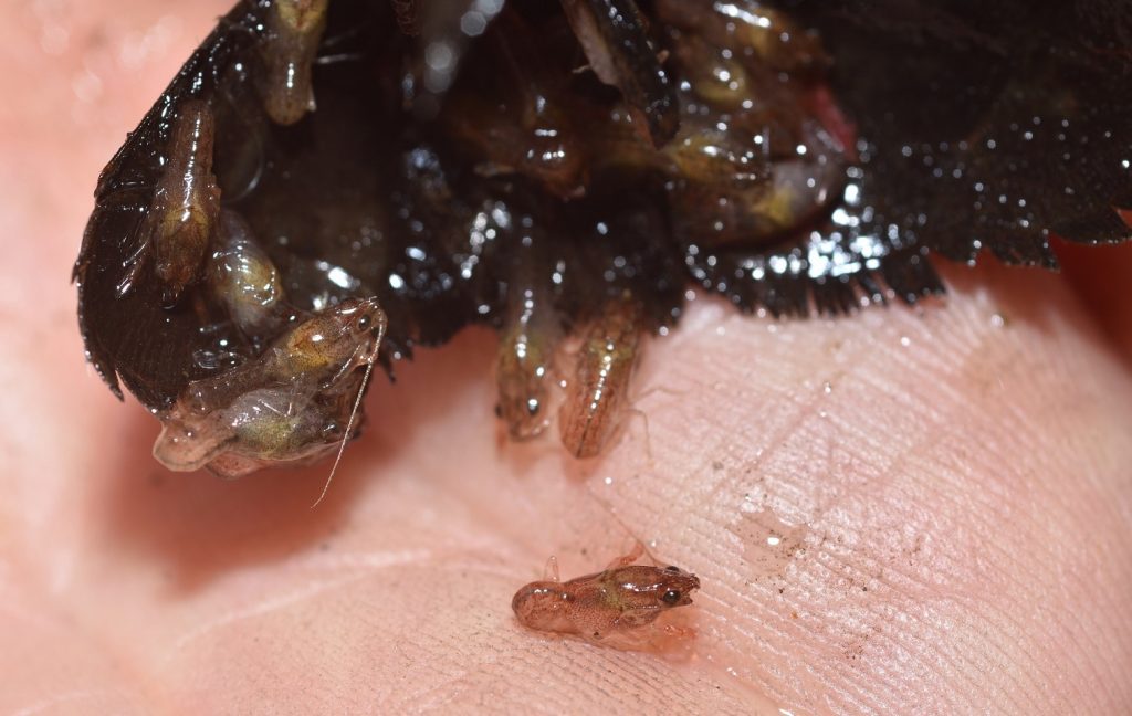
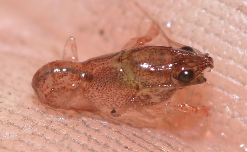
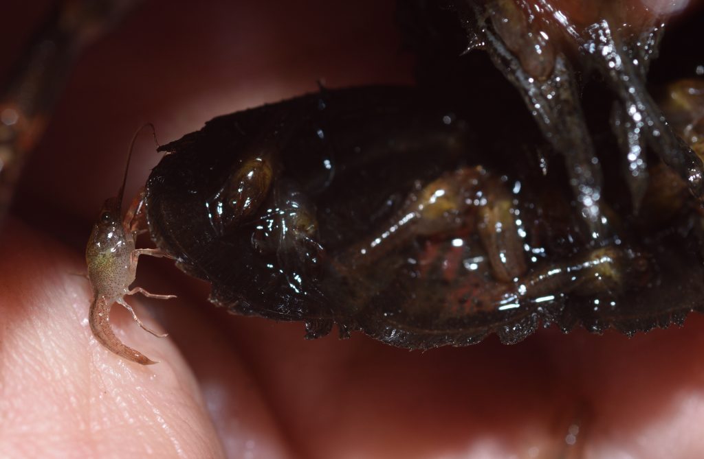
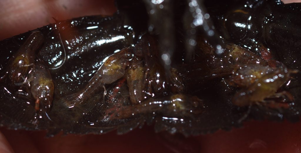
One of the crayfish that I caught in the Sweetwater River had babies. The following photo shows the underside of the tail with a bunch of babies still clinging to it (and falling off onto my palm).

Red swamp crayfish (Procambarus clarkii) with babies
Sweetwater River near Jamul, CA 10/4/2019
Another view of the babies on the tail

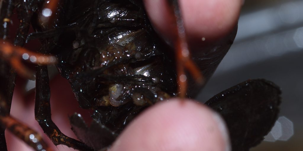
The next two photos show a few babies clinging to the underside of the abdomen. The pleopods or swimmerets help hold them to the mother.

I released the mother and babies back into the river next to the rock where I found them. Hopefully, all the babies found their way back to the mother, but, honestly, most of them aren’t going to make it to adulthood anyway. Young crayfish are much more susceptible to predation than the adults.

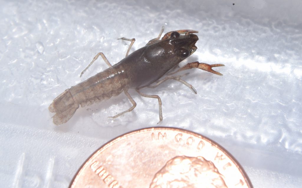
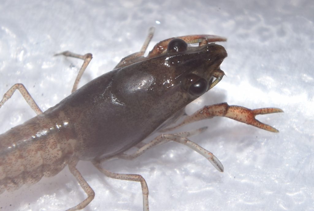
The next few photographs show a very young crayfish. I’m not sure how old it was, but it was no longer with its mother.

A very young red swamp crayfish (Procambarus clarkii)
Sweetwater River near Jamul, CA 9/9/2017
A very young red swamp crayfish (Procambarus clarkii)

And finally, here’s some shots of a juvenile. Crayfish don’t go through extensive metamorphosis. Aside from their chelae (main claws) being much smaller, the juveniles and even the babies look very much like the adults. This is one thing that differentiates them from lobsters, which have a distinct larval stage. It would be interesting to know why this is. My guess is that it’s because lobster larvae are part of the plankton, drifting near the surface. When they grow too large for this, they sink to the bottom and become benthic (bottom-dwelling) organisms. It probably makes more sense for freshwater crayfish, which often live in small streams, to start out living on the bottom.

A Personal Anecdote

Before I moved to California, I lived in Virginia. One day I was walking through the woods and, as I crossed a small stream, I caught sight of a crayfish in the water. I reached down and scooped it up. It immediately pinched my hand with one of its claws. It was quite painful, but I managed to hold on to it and pry its claw loose. I took it home and put it in an aquarium.

After a week or two, the crayfish molted. In nature, the empty exoskeleton usually gets broken up by rocks and currents. In this case, however, since there were no currents in the aquarium, the exoskeleton was almost perfectly intact. It was missing a few legs, but otherwise, it was in pristine condition. Even the remarkably long antennae were still attached.

I took the exoskeleton out of the aquarium and let it dry. Then I bent a paperclip into a mount for it. I glued one end of the mount to the bottom of the exoskeleton and the other to the bottom of a small plastic display case. The result was a cool little display of a crayfish exoskeleton. Sadly, a couple of years later, I dropped the case and the fragile exoskeleton broke apart. I haven’t been able to find any photos of the exoskeleton in its display case, but I was able to find a drawing that I did of the exoskeleton. That’s posted below.

Drawing of the exoskeleton of a Virginia crayfish. Probably Cambarus sp.

Meanwhile, Back in Pangaea…

I mentioned at the beginning of this article that crayfish are an example of an organism that evolved when all the continents were joined together into one super-continent called Pangaea. When that continent broke up into separate land masses, the population of crayfish was broken up into various isolated pockets, which then went on their separate evolutionary ways. Now, I’m going to try to describe that population breakup and subsequent differentiation in a little more detail. Most of the following is taken from a paper in the Journal of Crustacean Biology entitled Phylogeny of Fossil and Extant Freshwater Crayfish and Some Closely Related Nephropid Lobsters.62

In this paper, the authors examined both modern and fossil crayfish, applying molecular and morphological analyses to determine the relationships and likely evolutionary history of crayfish groups.

The paper concludes that “The freshwater crayfish are a monophyletic group consisting of three families (and possibly a fourth grouping to include only [the genus] Gramastacus, pending further investigation).” What this means, first of all, is that the traditional division of crayfish into three families is probably correct. The “monophyletic” part means that crayfish are all evolved from one common ancestor. This may seem obvious, but there have been many cases where organisms that seemed very similar turned out to be only distantly related. This is not the case with crayfish.

The authors also found evidence of a single freshwater invasion by crayfish. It could have been possible that crayfish evolved in saltwater and that two or more subgroups in different locations independently adapted to freshwater. Again, this does not seem to be the case. This is important because it means that crayfish radiated from a single freshwater location in Pangaea.

The reference paper contains a map showing the current distribution of the three extant crayfish families. Click here to go directly to that map. (The legend will appear at the bottom of the page.) You can see from the map that the family Parastacidae is limited to the Southern Hemisphere, including South America, Australia, Madagascar, New Zealand, and New Guinea. All these land masses were originally part of Gondwana, the southern-most of the two super-continents that formed when Pangaea broke up.

Pangaea began its breakup about 175 mya during the Triassic. It initially broke up into the southern super-continent of Gondwana and a northern super-continent called Laurentia. Gondwana would go on to break up into the land masses of the Southern Hemisphere, while Laurentia would break up into North America and Eurasia. (South America was initially part of Gondwana. North and South America would not come together and be joined by the Isthmus of Panama until many millions of years later.)

So the members of Parastacidae are descended from the crayfish that were isolated on Gondwana after the initial breakup of Pangaea. The other two families are both limited to the Northern Hemisphere, so these were the crayfish that were isolated in Laurentia. As Laurentia and Gondwana proceeded to break apart in their turn, further populations of crayfish found themselves isolated from one another; however, the breakup of these two super-continents didn’t happen all at once. There were a series of breakups over a long span of time. So the populations within families are more closely related than the families are to each other.

The paper came to a number of other conclusions. For instance, crayfish appear to have evolved from the Chilenophoberidae family of lobsters, rather than the Erymid lobsters, as it was previously thought. A minor detail, perhaps of interest only to specialists, but it’s another small piece in the great puzzle of evolution.

Rattlesnake!

“Everybody knew that you should never provoke a rattlesnake, much less tie it into a bow. But that didn’t stop Judd. What did stop him was the rattlesnake.” -Demetri Martin

It’s easy to forget how remarkable the rattlesnake is. We all learn about rattlesnakes as kids, so we’re used to them, even if we’ve never actually seen one in the wild. And, of course, many people are too busy being terrified of rattlers to think about them in any other way. But stop for a minute and really think about it — a snake with a rattle on its tail! If it didn’t actually exist, it would be a ridiculous idea. The evolution of the rattle will be one of the things I’ll be covering in this article, but first, we need to get the basics out of the way…

Rattlesnakes are native to North and South America with the greatest concentration in the southwestern United States and northern Mexico. There are two genera of rattlesnakes, Crotalus and Sistrurus. Sistrurus is the more basal genus, which is to say, Sistrurus evolved first and Crotalus branched off from it. The larger, more well-known of the rattlers are in Crotalus. Those in Sistrurus are smaller and have smaller, higher pitched rattles. There are 33 species of rattlesnakes altogether, only 2 of which are in Sistrurus.60

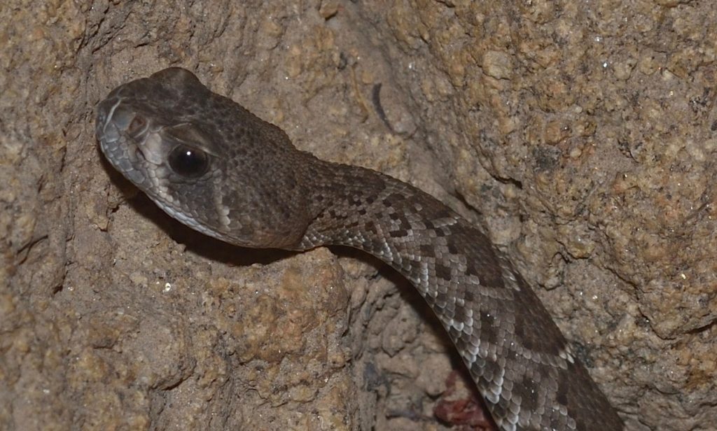
Rattlers are pit vipers (subfamily Crotalinae of family Viperidae). The “pit” part of the name refers to the pit organs, small heat-sensing pits located on each side of the head. These act like eyes, allowing a rattlesnake to detect warm-blooded prey even in pitch dark.63

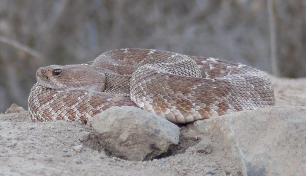
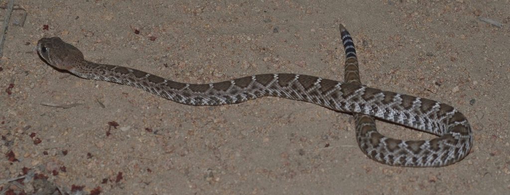
There are a number of Crotalus species here in southern California, but I’ve only had the uh… good fortune to encounter a couple of them. One is the red diamondback or C. ruber, which has a very pale, reddish coloration and a diamond pattern on its back. The other is the western diamondback or C. oreganus. This has a similar diamond pattern, but is usually dark gray or brown. Most of the western diamondbacks that I’ve seen have been in the subspecies C. oreganus helleri, which is also called the southern Pacific rattlesnake.64 65 66 (A third species, C. atrox, also goes by the common name western diamondback. Their range is supposed to include the southern tip of California, but, as far as I know, I haven’t encountered any.)67

I’ve run into rattlesnakes (almost literally a couple of times) in various places around San Diego County, but most of the encounters were in Mission Trails Regional Park, just west of San Diego. I don’t suppose they are any more common there than in most of the other places I hike; I just hike in Mission Trails a lot more often, since it’s close to where I live.

That said, I saw around 17 rattlers in 2018, almost all of them in that park. I came close to stepping on one twice. The second time was the scariest. I had just turned onto a trail. Overhanging branches of broom baccharis were blocking my view of the rattler, a huge southern Pacific. My foot landed right in front of the snake, which started rattling very loudly. Needless to say, I jumped back. I don’t know if the snake had already been coiled up when I almost stepped on it, but it was coiled up by the time I saw it. The snake partially uncoiled and, using some kind of beautiful, sinuous motion that I can’t begin to describe, propelled itself several yards away, deeper into the bushes, hissing and rattling the whole time. It stopped there and continued to rattle angrily as we took photos of it. Fortunately, there wasn’t much undergrowth, so I was able to get some great shots and a nice, long video. Those are posted below.

Southern Pacific Rattlsnake (C. oreganus helleri)
Mission Trails Regional Park, San Diego County 8/16/2018
This is the snake from the above video.
A closer look

In the video, you can hear one of the people I was hiking with calling to some women who were walking by on the main road, asking them if they wanted to see a rattlesnake. They declined. You can hear my friend responding “You’ll catch the movie…” Lol.

It was a fairly unnerving experience, since I’d already come close to stepping on a smaller rattler earlier that year. After that second encounter, I bought some snake chaps and started wearing them whenever I hiked in Mission Trails. I kept wearing them until the weather turned cooler, making rattlesnake encounters less likely. For whatever reason, I haven’t encountered as many rattlers this year (2019) and I haven’t felt the need to wear the chaps. You can bet I keep to the middle of the trail whenever I pass that particular overhanging bush, though.

Red diamondback (Cotalus ruber)
Mission Trails Regional Park, San Diego County 8/7/2018
A closer view

As hair-raising as that last story may have sounded, rattlesnake bites are actually very uncommon and, when they do occur, they’re rarely fatal. On average, there are 7000 to 8,000 venomous snake bites in the U.S. per year — that’s all types of venomous snakes, not just rattlers. And, on average, only 5 of those 7000 to 8000 snakebite victims die. Rattlesnakes don’t want to bite you. They will do so only if they feel threatened. If you step on one or try to catch it, then, sure, it’s going to bite. According to Wikipedia, “A majority of victims (about 72%) are males, often young and intoxicated”.68

One reason rattlesnake bites are rarely fatal is that the snakes have voluntary control over how much venom they inject. It takes energy for a snake to make venom, so it doesn’t want to waste it on something that it can’t eat. When a rattlesnake bites a person, it will often inject a smaller amount of venom than it would if it was attacking prey. In fact, around 20% of rattlesnake bites are dry bites — no venom at all!69

I do know one person who was bitten by a rattlesnake. A woman that I often hike with had just finished a hike. She went into the bushes for a call of nature and, as she was pulling her pants up, her foot broke through the ground and into a rattlesnake den. The snake started rattling and my friend felt a light tap on her calf. She said it felt like a tap on the shoulder. She thought maybe the snake’s rattle had hit her. After pulling her foot out (and presumably putting some distance between herself and the rattler), she examined her leg. She didn’t see any marks at first, but when she squeezed her leg, two tiny beads of blood oozed up from where the fangs had penetrated.

As I mentioned, this was at the end of the hike, so one of the other hikers was able to drive her directly to a hospital. This hiker was driving a Corvette and, in his words, “I’d been waiting for this my whole life!” To make a long story short, my friend managed to survive the drive to the hospital and the bite turned out to be dry. She never felt any effects from it at all. The poor snake was probably too surprised to even think about injecting venom.

Note: As soon as my friend was bitten, they called 911 and the ambulance met them as they were driving to the hospital. The paramedics checked her over quickly, but she wasn’t exhibiting any symptoms and, since the sports car was able to handle the twists and turns in the road better than the ambulance, the paramedics let her continue the trip to the hospital in that.

Rattlesnake bite
copyright © 2019 Hazell Belvin
Used with permission

So, to summarize, you’re unlikely to ever get bitten by a rattlesnake and, if you are, there’s a good chance that the rattlesnake will not inject a full dose of venom. But if you do get bitten, what should you do?

What To Do If Bitten

I was going to include list of things to do in case of snake bite, but I’m not a medical professional, so it’s probably better if I just direct you to a few websites that seem trustworthy. Click here for the Mayo Clinic’s snake bite page. Click here for a site that’s specific to rattlesnake bites. And finally, click here for a link to Johns Hopkins’s page.

Now that we’ve covered the basics, let’s take a closer look at the various things that go into the making of a rattlesnake.

The Pit Organ 70 71 72

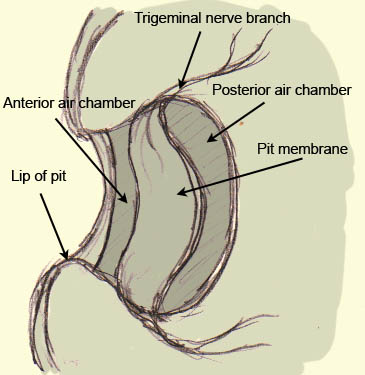
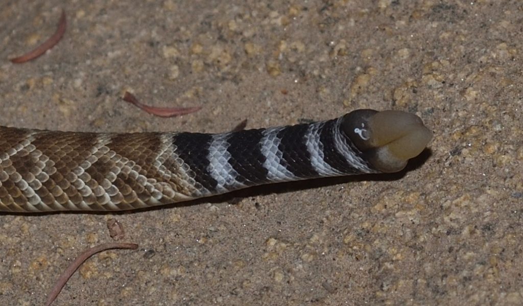
As mentioned earlier, the pit organs are small, heat-sensing pits on each side of the rattlesnake’s head. In the photo below, you can see one of the pits. It’s positioned between the eye and nostril, a little lower on the snout. The silvery oval inside the pit is the pit membrane. It may look like it’s swelling outward, but I think that’s an optical illusion. It’s actually curved inward, as depicted in the diagram below the photo.

Probably another southern Pacific rattler
The silvery oval is the pit membrane.
Mission Trails Regional Park, San Diego County 8/14/2017
A diagram of the pit organ
This image is in the public domain. It was downloaded from Wikipedia Commons at this url.

The pit organs function as crude eyes for detecting infrared light, a.k.a heat. For those of you who need a quick physics refresher, heat has several ways of traveling from place to place. One way is in the from of light waves in the infrared portion of the spectrum. The only difference between infrared light and red light is the wavelength. The wavelength of infrared light is longer than that of red light and cannot be detected by our eyes. As it happens, it can’t be detected by the rattlesnake’s eyes either. That’s why it needs pit organs.

In its regular eyes, the rattlesnake has special chemicals that are sensitive to light in the visible spectrum. The pit organ works a little differently. When infrared light from a warm object like a mouse hits a spot on the pit membrane, it heats up that spot. Infrared (IR) receptors in the form of heat-sensitive proteins detect the change in temperature and generate an electrical impulse that travels down nerves to the snake’s brain. The pit membrane is rich with blood vessels which quickly cool the spot back down to ambient temperature once it’s no longer being heated up.

Note that the opening that lets infrared light into the pit is smaller than the diameter of the pit. This means that the pit acts like an infrared version of a pinhole camera. The infrared light entering the pit forms a crude image in the form of a heat map on the surface of the membrane. So the pit membrane serves the same function as the retina in a regular eye.

The snake’s brain integrates the heat map from its pit organs with the visual images from its eyes. It’s kind of like how our brain integrates the 2-d images from each eye to form one 3-d image. So, what does this look like to the snake? I have no idea. I’m not a snake (despite what you may have heard). It’s like a person who’s been blind from birth trying to imagine what eyesight is like.

The heat image formed by the pits must be rather crude, but it’s good enough for the snake to detect and strike prey in total darkness. A BBC video that I found on YouTube shows some infrared images from a heat-sensitive camera developed by the military. According to the narrator, the rattlesnake’s pit organs are “30 times more powerful” than the camera. I think this probably means that the pit organs are able to detect a heat signature that is 30 times weaker; I doubt they form as sharp an image. It’s a great video, though. You can watch it here.

There is also evidence that the snake uses the pit organs for avoiding predators and for thermal regulation. In fact, it’s possible that the pit organs originally evolved for prey avoidance and/or thermal regulation and that rattlesnakes (or their ancestors) only later started using them for hunting.

So how could pit organs have evolved? I wasn’t able to find much about this, but one article mentioned that some snakes in the Boidae family have IR receptors in their labial scales (scales on the sides of the snout), without any specialized structures. Other snakes in the same family have IR receptors in the bottom of simple labial pits.73 These would seem to be examples of possible intermediate steps in the formation of a pit organ.

Those Eyes 74

Humans and many other animals have a small, central pit in the retina called a fovea consisting of tightly packed cone cells (the cells responsible for color vision). The fovea is where our vision is sharpest and it makes it possible for us to do things like reading and driving. A rattlesnake eye doesn’t have a fovea, so rattlesnakes are not able to see sharply defined images. (Never let a rattlesnake drive!) They rely mostly on the perception of movement and don’t appear to follow objects with their eyes.

Red diamondback (C. ruber)
Same one as in the earlier photos

Rattlesnakes are adapted for hunting at night, so they have lots of rod cells (the cells that are sensitive to dim light, but not color). They also have cone cells, though, so they have some color vision.75

Rattlesnakes have vertical pupils. That is, when the pupil is contracted, it forms a vertical slit. Not all snakes have vertical pupils. Studies have shown that it’s mostly ambush predators that have them. Computer simulations have shown that a vertical pupil allows a predator to better judge distance without moving the head — an obvious advantage, since moving the head could alert prey. (For a fascinating article about pupil shape, click here.)

Those Ears? 76

Yes, rattlesnakes have ears. They just don’t have ear openings. Or eardrums. What they do have are middle ears, but even those are not as specialized as those of mammals. In fact, mammals in general have much better hearing than reptiles because a couple of bones that form part of the jaw in reptiles (which are the ancestors of mammals) migrated into the middle ear and now form part of a mammal’s hearing apparatus — a remarkable and bizarre fact that I definitely need to do a blog post about one of these days.77

So rattlesnakes don’t have very good hearing. They do seem to be very sensitive to vibrations in the ground, though. These are transmitted to their ears through their bodies, especially their bones. So I suppose they do have pretty good hearing in that sense.

That Flicking Tongue 78

Snakes use their forked tongues for smelling. The forked tongue is not unique to rattlesnakes, of course — many reptiles have them — but it certainly adds to their mystique.

Snakes have a great sense of smell. In fact, it’s probably their most important sense. When a snake senses a change in its environment (through vibrations in the ground, for instance), it immediately starts flicking its tongue in and out, sampling the air. A groove, called a rostral groove, in the snake’s upper lip allows the tongue to move in and out without the snake having to open its mouth. When the tongue darts out, chemicals in the air stick to it. When the tongue is withdrawn into the mouth, the tines are inserted into two tubes in the roof of the mouth. These tubes form the Jacobson’s organ, also called the vomeronasal organ (VMN). This organ is stimulated by chemicals on the tongue to send nerve signals to the part of the brain that processes smell.

So why do snakes have forked tongues? This allows them to sense the direction from which a smell is coming. For instance, if more of a particular chemical sticks to the left tine than to the right, then the smell is probably wafting in from the left.

Snakes are also able to smell through their nostrils. I’m not sure how sensitive their noses are, but I get the impression that their tongues are far more sensitive. According to one article, a snake may pick up a scent through its nostrils and then start flicking its tongue to get a better sniff.79

I can’t resist mentioning that humans, as well as many other mammals, also have a Jacobson’s organ. It also consists of two tubes in the roof of the mouth. There’s a lot of debate about what purpose the Jacobson’s organ serves in humans. Some people think that it’s stimulated by sexual pheromones. Others disagree. There doesn’t seem to be any definitive evidence either way.

Those Venomous Fangs80

Rattlesnakes, in case you haven’t noticed, have two thin, curved fangs in the front of their mouths. These fangs are very much like hypodermic needles. They have a hollow core with an opening near the top where the venom enters the fang and a narrow slit on the front of the fang near the tip, through which the venom is ejected. Each fang is supplied with venom through a duct that connects to a large venom gland located near the back of the head. So there’s a separate venom gland for each fang. These glands are what give rattlesnake heads a triangular shape.81 When the snake bites, muscles on the sides of the glands squeeze the venom through the ducts into the fangs.

When the rattlesnake closes its mouth, the fangs fold back into the roof of the mouth, where they rest in a sheath of soft skin.82 Click here to see a video of a rattlesnake skull with the jaws rigged up to keep opening and closing. If you spend a little time watching this video, you can get a pretty good idea how the system works. Fangs are modified teeth, of course. They grow from the maxilla bone of the upper jaw. The other teeth, which are used for gripping the prey while the snake swallows it, grow from a separate bone, the pterygoid, that connects to the end of the maxilla and extends backwards toward the rear of the skull. The maxilla is connected at the other end to the rest of the upper jaw. The connections between all these bones are flexible, allowing them to rotate with respect to each other. When the rattlesnake opens its mouth, the pterygoid bones are pushed forward and the maxillas rotate, swinging the fangs into biting position.

Rattlesnakes shed their fangs periodically (once every 60 days or so) and they always have replacement fangs waiting in reserve. In fact, they have a series of 7 pairs of fangs in various stages of development behind their functional fangs! So even if a snake unexpectedly breaks a fang, it won’t have to wait long before it can deploy a new one.

So how did this whole remarkable system evolve? Let’s start with the venom. Venom is basically very strong saliva. This may raise a few eyebrows. How does saliva get magically transformed into venom? Well, it may surprise you to learn that ordinary human saliva injected under the skin of the hand can cause pain, redness, swelling, and partial loss of function.83

Obviously, human saliva did not evolve to be toxic. It just happens to contain some substances that don’t have very nice effects when injected under the skin. Actually, this isn’t too surprising given that the purpose of saliva is to aid in digestion by beginning to break down food in the mouth. So it makes sense that if it’s injected into the hand, it would begin to break down tissue. It’s also not hard to imagine evolution working with snake saliva to slowly increase its toxicity.

Venom — and this is true for almost all venomous creatures — is not a single substance. It’s a complex cocktail of chemicals and the different chemicals may serve very different purposes. For instance, some chemicals may act to actually kill the prey, while other chemicals only paralyze it. Still other chemicals may start the process of digestion. Different components of venom may also act on different systems. One chemical might break down muscle while another attacks the nervous system.

Different species of rattlesnake have evolved different mixtures of chemicals in their venoms to match their preferred prey. For instance, the venoms of C. ruber and C. oreganus are mostly necrotic — they break down tissue. The Mojave rattlesnake (C. scutulatus), on the other hand, has more neurotoxic components in its venom. Research has shown that the common ancestor of these three species had a more general purpose venom that contained both necrotic and neurotoxic elements. As this species differentiated into multiple species, its descendants modified the mix of chemicals in their venoms to reflect their presumably more specialized diets.84

That takes care of the venom, but how in the world did these snakes manage to evolve hypodermic needles with which to inject it? The first venomous snakes didn’t have fangs, but they did have teeth. These snakes probably caught their prey and held it in their mouths while the venom slowly seeped into the puncture marks made by the teeth. Chewing probably sped up the process. Eventually, some of the teeth evolved to be longer than the others and developed grooves that helped to channel the poison into the wounds. We still have these kinds of snakes today. These are the opisthoglyphous or “rear-fanged” snakes which make up the Colubridae family. (Technically, opisthoglyphous refers to the type of fang, not the group of snakes, but it’s often used informally to refer to the group.)

When I first read about rear-fanged snakes, I found the idea very confusing. How can a snake use fangs that are in the rear of its mouth? A picture of a snake called a boomslang on Andrew Durso’s excellent (and excellently-named) blog “Life Is Short, But Snakes Are Long” cleared things up for me. Click here to see the photo I’m talking about. It’s around the middle of the page. In this photo, you can see that the fangs aren’t actually in the rear of the mouth. They’re just at the rear of the maxilla bones. The pterygoid bones continue on beyond the fangs. (In these snakes, the maxilla and pterygoid bones are not able to rotate relative to each other.) So the fangs are really more toward the middle of the mouth. These snakes bite down on their prey and “chew” the venom into it.

The fangs of rattlesnakes — the kind that are in the front of the mouth and fold back when not in use — are called solenoglyphous fangs. These types of fangs evolved to inject large amounts of venom quickly and deeply. When a rattlesnake strikes a prey animal, it doesn’t hold on to it. After injecting venom into the prey, the snake immediately releases it. It then uses its excellent sense of smell to track the prey to where it finally collapses. This way, the snake doesn’t risk being injured by a struggling animal.

There’s another group of snakes that have what’s called proteroglyphous fangs. These fangs are also in the front of the mouth, but they don’t fold back. They tend to be about a third as long as rattlesnake fangs and they are also hollow. Snakes that have this type of fang are all in the Elapidae family and they include such infamous serpents as cobras, mambas, and death adders. Elapids bite their prey and hold on while they wait for the venom to take effect.

Scientists believe that solenoglyphous and proteroglyphous snakes both evolved from opisthoglyphous ancestors. So the distant ancestors of rattlesnakes would have had grooved fangs near the middle of the mouth. Over time, the ridges of the grooves folded over to form tubes and the fangs migrated to the front of the mouth. Evidence for this last part can be found in embryology. In the rattlesnake embryo, the developing fangs are located in the middle of the mouth in the same place as in rear-fanged snakes. As the embryo grows, the short piece the maxilla behind the fang grows much faster than the rest of the maxilla, pushing the fang forward in the mouth. You can find a nice set of photos illustrating this on the same blog page that I mentioned earlier, right under the picture of the boomslang. Click here to go back to that web page.

Those Insanely Flexible Jaws

Click here to see a video that explains how a snake swallows its prey. (It’s not a rattlesnake, but the principal is the same.) Snakes have more bones in their jaws than mammals do and these bones are not as tightly bound together. They have ligaments that stretch, allowing the bones of the jaw to spread apart, making room to swallow the prey whole.

You may be wondering how a snake goes about pulling a prey animal down its throat. I haven’t found any explicit description of this, but I’m guessing the snake clamps down harder with one side of the jaw than the other (remember how much more flexible those jaws are than ours!) and then twists its head so that the other side of the jaw moves a little farther up on the animal. It then quickly opens its mouth and snaps down again to get a new grip and repeats the process, this time clamping down with the other side of the jaw and twisting the head in the other direction.

It’s also possible, and this is entirely speculation on my part, that the pterygoid bone can be drawn back a little farther into the mouth to pull the prey a little farther in. Then the snake releases its grip long enough for the pterygoid to spring forward a little before it clamps down again. As always, if anyone out there has more info, please leave a comment. Once the prey gets down into the throat, the muscles of the throat and body can take over.

And Finally, That Rattle 85

The rattle consists of a series of hollow, interlocking segments. The segments are made of keratin — the same material that makes up our hair and fingernails and the scales of reptiles. In fact, the rattle segments are modified scales. Rattlesnakes are not born with rattles. Instead, the new-born rattler has a pre-button or first button on the end of its tail, which is a single segment of keratin. The pre-button is lost after a few days when the baby rattlesnake sheds its skin for the first time. The pre-button is replaced with the first rattle segment (labeled d in the diagram below). Obviously, this segment was developing before the snake molted, so it must have contained living tissue. By the time it’s ready to replace the pre-button, though, it has become a hollow shell of dead material and it has a second, living segment forming behind and partially within it. By the time the snake is ready to molt again, the living material in this second segment will have died, leaving a hollow keratin shell, and a third segment will have started growing behind it. In this way, the snake adds a new segment to its rattle every time it sheds its skin.

The parts of a rattle
This photo is in the public domain. It was downloaded from here.

The series of segments at the bottom of the above diagram represents a disassembled rattle. Notice how each segment bulges out in two places. The front bulge of a segment fits loosely inside the rear bulge of the segment in front of it. So the segments remain connected to each other, but are loose enough to rattle against each other when the snake shakes its tail. 86

The pre-button on the tail of a baby red diamondback (C. ruber)
Mission Trails Regional Park, San Diego County 10/14/19

The YouTube channel What’s Inside? has an entertaining video in which the creator of the channel cuts open a rattle to see, well… what’s inside. As you can hopefully guess by now, he found that there was basically nothing inside — just the interlocking walls of the segments. You can watch that video by clicking here.

This is the baby rattlesnake from the previous photo.
Close up

The muscles that shake the rattle are some of the fastest muscles known — comparable to those that power a hummingbird’s wings. They fire an average of 50 times per second and can keep firing for hours.

A rattlesnake sheds its skin several times a year, depending on the availability of food, so the number of segments in the rattle cannot be used to determine the age of a snake. Also, a rattlesnake will often lose segments from the end of the rattle in battles with other rattlers, when they are bitten off by predators, and just from general wear and tear. Usually, the rattle of a full-grown snake will have no more than 7 or 8 segments. (Note, the rattlesnake shown in the video near the beginning of this article — the one I almost stepped on — had a rattle with 12 segments! That was one big, healthy snake!)

So how did this amazing device evolve? Obviously, the rattle of modern rattlesnakes is a warning device (an aposematic device if you want to be technical). A rattle could not have just appeared full-blown in the rattlesnake lineage, though — it had to have evolved gradually. But it’s hard to imagine how a very early, proto-rattle could have been of much use as a warning device. So how could it have come about?

Actually, many modern snakes that don’t have rattles still shake their tails. They do this for a variety of reasons. Some snakes appear to shake their tails as a distraction. If a predator is trying to eat it, a snake may shake it’s tail to try to attract the predator to that end of its body. Obviously, the snake has a better chance of surviving the encounter if the predator is attacking its tail rather than its head.

Other snakes shake their tails to attract prey. A small, writhing tail may look like a worm or insect larva to a hungry bird. Some snakes actually have tails that have evolved to resemble specific kinds of insect larvae or other animals.87 Without a doubt, the most remarkable example of this is the Iranian spider-tailed horned viper which, as the name suggests, has evolved a structure on the end of its tail that resembles a spider!88 Click here to watch a video of this stunning creature.

Finally, a rapidly shaking tail, even without a rattle, may serve as a warning to predators that have learned to avoid such snakes.

In one study, researchers compared the tail shaking of 56 species of snake that didn’t have rattles to the tail shaking of rattlesnakes. They found that the more closely a species was related to rattlesnakes, the more similar their tail shaking was to that of rattlers.89

So the rattle probably started with a shaking tail. Over time, various modifications were added for various reasons. Eventually, in one lineage of snakes, these modifications led to the evolution of the rattles we see today.

In Conclusion

There’s so much more I could write about rattlesnakes. I should at least mention that rattlesnakes are ovoviviparous, meaning that the mother snake incubates her eggs inside her body and the babies are born live. Then there are rattlesnake roundups and the story of antivenin and dens full of hibernating rattlesnakes and on and on and on… but this is supposed to be an article, not a book, and it’s already much longer than I planned. If you made it this far, I hope you enjoyed reading it. And, as always, if you have any interesting facts or stories to add, feel free to put them in a comment. I’ll leave you with a few photos of rattlesnake skulls that I took at the San Diego Natural History Museum:

Red diamondback (C. rubers) 9/13/2019
It looks like a replacement fang is coming in for the right fang. It’s not unusual for the original fang and the replacement to both be present for a time before the original finally works loose.
Red diamondback (C. rubers) 9/13/2019
Front view of previous skull
In this photo, you can see that the replacement fang is coming in to the outside of the function fang. It will shift into the inner position, displacing the old fang.
Western diamondback (C. oreganus) 9/13/2019
This snake also appears to have had a replacement fang coming in.
Western diamondback (C. oreganus) 9/13/2019
Front view of previous skull
Note the orifices in the bases of the fangs where the venom would enter them.
Western diamondback (C. oreganus) 9/13/2019
Top view of previous skull

Diving Deep Into Dodder

It’s been called witch’s hair, devil’s hair, devil’s guts, devil’s ringlets, scaldweed, beggarweed, fireweed, strangleweed, strangle tare, hairweed, pull-down, hailweed, and hellbine — the parasitic plant dodder doesn’t get a whole lot of love.69 If you do much hiking in southern California, you’re probably used to seeing huge, yellow-orange masses of dodder draping vegetation. Many people assume it’s invasive, but it’s actually native to California.

A buckwheat plant almost completely smothered in dodder
Noble Canyon, Pine Valley, CA 07/07/19

Dodder is in the genus Cuscuta, which contains between 100 and 170 species, all of them parasites. It has a world-wide distribution. Cuscuta used to be treated as the only genus in the family Cuscutaceae, but it’s now been incorporated into the morning glory family (Convolvulaceae). The various species can be yellow, orange, or red. At least one species, Cuscuta reflexa or giant dodder, is green. (That one’s found in India, so I probably won’t be posting any photos of it here.)

Dodder (Cuscuta sp.)
Cowles Mountain, San Diego County 05/31/19

Cuscuta is classified as a stem parasite, because it invades the stems of other plants (as opposed to their roots). Like all parasitic plants, dodder evolved from photosynthetic ancestors. At least some species still retain traces of chlorophyll, although not nearly enough to survive without a host. Generally, if a plant is parasitic, but still does some of its own photosynthesis, it’s called a hemiparasite. If, on the other hand, it has totally lost the ability to photosynthesize, it’s called a holoparasite. Dodder seems to be an edge case. Some of the articles I’ve read confidently state that it’s a hemiparasite. Others just as confidently call it a holoparasite. To some extent, it probably depends on the species. As mentioned earlier, the Indian species — C. reflexa, is green, so it retains more chlorophyll than the other species and probably photosynthesizes at least slightly. When it comes to the other dodders, the various articles can’t seem to agree on whether they photosynthesize just a little bit or none at all.

C. subinclusa growing on laurel sumac. The leaves have been reduced to tiny scales.
Cowles Mountain, San Diego County 07/23/19

When a dodder stem contacts a plant, it wraps itself around the plant. (It even wraps itself around itself, as you can see from the photos in this post). If the plant is a potential host, the dodder will produce very thin, modified roots called haustoria (singular haustorium), which burrow into the stems of the host plant and invade its vascular system, tapping into both xylem and phloem.76 The dodder then proceeds to extract all the water, sugar, and nutrients it needs from the other plant.

A Closer Look at Haustoria

The following description is based on an article published in the online journal “Frontiers of Plant Science“. You can read that article here.

When a dodder stem comes into contact with an appropriate host, it goes into an “attachment phase”, in which the cells at the contact point develop into a secondary meristem.89 This secondary meristem forms an adhesive disk or “prehaustorium”, which adheres to the stem of the host plant. Cuscuta even induces the host plant to produce sticky substances of its own to reinforce the connection.

This photo shows a couple of dodder stems wrapped around the green stem of a buckwheat plant. The swellings where the dodder contacts the host stem are the prehaustoria or adhesive disks.
San Diego County 05/31/19

Next comes the “penetration phase”, in which a haustorium grows from the adhesive disk and penetrates the stem of the host. Cuscuta secretes enzymes that work to degrade the cell walls of the host and the developing haustorium forces its way through the weakened or ruptured walls. Meanwhile, the dodder stem continues to grow longer, attaching a second prehaustorium, then a third, and so on.

Cells at the tips of the haustoria form “searching hyphae”, which search for xylem and phloem. When hyphae come into contact with one of the sieve cells of the phloem, they grow around the cell “like the fingers of a hand”. The hyphae then fuse with the cell so completely that it’s hard to tell where the parasitic hyphae end and the host cell begins.

This is a section of dodder stem that I pulled loose from the host plant. You can see the undersides of the prehaustoria. Haustoria would have been growing from the prehaustoria into the stem of the host plant. They were too integrated with the host tissue to be pulled free.

Similarly, when the searching hyphae find xylem tissue, the dodder stimulates the host xylem to develop synchronously with its own xylem tissue (formed within the hyphae), resulting in a continuous tube of xylem connecting the two plants.

Another view of adhesive disks pulled loose from a host stem
Here you can see the wounds left by the prehaustoria ripping loose from the buckwheat stem. The haustoria are still imbedded in the host stem.

To quote from the referenced article:

“The haustorium […] is the defining feature of all parasitic plants, representing the interface where nutrients, solutes, and carbohydrates are exchanged between host and parasite. […] This interface also allows the exchange of macromolecules between the host plant and the parasite.”

And:

“… the impressive extent of exchanged macromolecules such as RNAs confirms the haustorium as an ‘open door’ between host and parasite that seems not especially guarded.”

Cuscuta can reproduce asexually from bits of stem spread by birds or other animals. Dodder is a flowering plant, however, so it can also grow from seed. Most, if not all, species are annuals, so I suppose it has to regrow from seeds each year. A newly germinated seedling has roots, but little or no chlorophyll, so it has to reach a host as soon as possible. The endosperm of the seed, which is much like the yolk of an egg, contains enough food to keep the young plant alive for 5 to 10 days after germination, but if it fails to find a host within that time period, it dies. Experiments have shown that dodder locates potential hosts by sensing airborne chemicals released by the plants. In other words, it smells them. Dodder has been shown to be sensitive to a number of different volatile compounds and to be repelled by others. Once a seedling attaches itself to a host, its roots are no longer needed, so they wither away.

Dodder seeds don’t have any obvious dispersal mechanism — they simply drop to the ground. The plants probably rely on the dispersal of bits of stem by animals to spread to new areas.

The white flowers belong to California buckwheat. Remarkably, even loaded down with dodder, buckwheat still manages to produce flowers.
Cowles Mountain, San Diego County 05/31/19
Cuscuta flowers (C. californica)
Cowles Mountain, San Diego County 07/12/19

Many people who are familiar with dodder don’t realize that it’s a flowering plant. That’s probably because they’ve never bothered to take a close look. Dodder produces flowers in abundance, but they’re very tiny — around 3 mm wide. In most species, they’re white or cream-colored, so they don’t stand out against the pale yellow-orange of the stems.

There are a number of species of dodder in southern California, but the two most common ones are Chaparral or California dodder (C. californica) and canyon dodder (C. subinclusa). I’ve seen both species growing on Cowles Mountain in Mission Trails Regional Park, just west of San Diego. California dodder grows at or near the bottom of the mountain. It infests mostly buckwheat. The flowers are short, white, and urn-shaped.

The canyon dodder in the park grows mostly on laurel sumac. Its stems are noticeably thicker than those of California dodder. The flowers are also white, but longer and more tube-shaped. The petals are joined for maybe two-thirds of their length.

A dodder flower has five petals. The petals are joined together near the base, but peel back at the ends to expose five stamens90 and two pistils.91 The pistil of a dodder consists mostly of a style — a long, thin stalk — sporting a very small, round stigma on the end. When a pollen grain sticks to a style, it grows a thin tube that penetrates the style and follows it down to the ovary, which it fertilizes. As a flower matures, the styles grow longer. They may not grow equally, though. In many species, one style will grow noticeably longer than the other.

Cuscuta flower (C. californica)
Cowles Mountain, San Diego County 07/12/19
Cuscuta flower (C. californica). Note the unequal length of the styles.
The reddish plant matter to the left of the flower is part of the buckwheat host.
Cowles Mountain, San Diego County 07/12/19
Cuscuta flower (C. californica)
Cowles Mountain, San Diego County 07/12/19

Why would a flower have two unequal styles? This represents two different methods of assuring reproductive success. An insect flying from flower to flower will pick up pollen from one flower and transport it to the next. The long pistil stands a better chance of getting pollen from the visiting insect stuck to it, so it has a better chance of picking up pollen from a different plant. The short pistil, on the other hand, has a better chance of receiving pollen from the stamen growing next to it. In other words, one pistil favors cross-pollination and the other favors self-pollination.

The whole point of sexual reproduction is to give a species a better chance of coping with changes in the environment by producing offspring with many different characteristics. If the environment changes in such a way that one individual has less chance of survival, maybe another individual with different characteristics will stand a better chance. So, all things being equal, cross-pollination is better than self-pollination. On the other hand, what if the pollinators that a flower usually relies on are in short supply? In that case, the plant can still reproduce by self-pollination.

Which is better — cross-pollination or self-pollination — can vary with circumstances. For instance, many plants have evolved to take advantage of disturbed ground. When a large tree falls, clearing a space in the forest, or a fire kills all the vegetation in an area, some plants have evolved to quickly colonize the newly vacated plot of ground, squeezing in a few generations before other plants crowd them out. For this kind of plant, self-pollination may be the more successful strategy, simply because it’s quicker — the plant doesn’t have to wait for a bug to come by or for the wind to blow a few pollen grains of the right sort in its direction. Believe it or not, some flowers have evolved to grow flower buds that never open! They still have stamens and styles, but the pollen from the stamens always ends up on the styles in the same bud.

As a plant species encounters different environments, it will often evolve different pollination strategies that allow it to adapt to those environments. If some members of a species evolve a new strategy that is so different from the old strategy that they are no longer able to breed with the plants that use the old strategy, then those members have formed a new species.

In his Master’s thesis, which you can read online here, Michael Wright discusses the evolution of sexual reproduction in Cuscuta. He comes to the conclusion that the evolution of two styles of unequal length was crucial to the success of the genus. To quote Dr. Wright, “The more flexible floral design enabled Cuscuta to evolve different mechanisms of reproductive assurance in coordination with their exploitation of novel host species and new pollination environments.”

Some of the examples he gives are fascinating. In one subspecies, a style tends to collect pollen from it’s own flower near its tip, but, as it grows longer and the stamens begin to wither and die, the style bends over, so that its lower part is exposed to any visiting insects. In this way, if it collects a pollen grain from an insect, that grain will likely be closer to the bottom of the style than any pollen that it picked up from its own stamens. So the tube that grows from the foreign pollen may be able to reach the ovule first. Thus, preference is given to the potentially more beneficial foreign pollen.

This springtail (order Collembola) was crawling around inside the flower of a California dodder. I don’t know if springtails are an effective pollinator of dodders, though. Just because an animal visits a flower, it doesn’t follow that it acts as a pollinator. For this reason, pollination studies

The Consequences of Intimacy

A single Cuscuta plant will routinely infect multiple host plants and multiple Cuscuta plants will routinely infect the same host. And, as already noted, the haustoria of Cuscuta form a very intimate connection with the vascular tissue of the host plant, allowing all sorts of things to pass between them. These several facts have some important consequences:

The transfer of viruses. Viruses seem to pass freely from an infected host plant to Cuscuta and from there to an uninfected host. Sometimes a virus is able to infect the dodder, as well as the host plant. In this situation, the dodder can go on to pass on the virus to other host plants even if all the original hosts have died. In other cases, Cuscuta may be immune to infection by the virus. This does not prevent it from transferring the virus between hosts, however.

Horizontal gene transfer (HGT). HGT is most familiar in microbes. Bacteria, for instance, routinely exchange DNA. This is one reason they are able to evolve resistance to antibiotics so quickly. What scientists have only recently begun to appreciate is that HGT can also occur in plants and animals. The mechanisms are not fully understood and it probably doesn’t happen very often, but there is good evidence that parasitic plants, such as Cuscuta, can exchange genes with their hosts. It’s likely that much of Cuscuta‘s success as a parasite is a result of its ability to incorporate useful genes from host plants and express them in its haustoria.

Communication between hosts. The fact that Cuscuta steals resources from its host plant is obviously detrimental to the host. There’s no getting around that. However, it doesn’t benefit a parasite if its host dies. So, perhaps it’s not so surprising that a parasite will sometimes do things that actually benefit the host. There is experimental evidence that when a dodder connects multiple host plants, it forms a communication network between them, passing useful info from plant to plant. See the section below for more details.

How Dodder Helps Its Hosts

A study published in PNAS92 showed that when two host plants are connected by a Cuscuta bridge and one of the hosts is attacked by an insect, the dodder will pass signals to the other host that cause that host to react in a way that makes it more resistant to attack. (Click here to read the original paper.)

Multiple buckwheat plants connected by dodder bridges

The study was done on Cuscuta australis. The insect pest in the study was tobacco cutworm (Spodoptera litura). Several host plants were used.

A number of experiments were done in the study. In a typical experiment, C. australis was allowed to infect two host plants growing close together, but not in contact. S. litura was allowed to feed on one of the host plants. I’ll call this plant A and I’ll call the other plant (which was not being fed on) plant B. After a period of time (say 48 hours), leaves from both host plants and vines from the dodder were collected and subjected to RNA-sequencing analysis to determine what changes had occurred in gene expression (up-regulation or down-regulation of the genes).

When an insect feeds on a leaf of a plant, the other leaves will typically start to produce chemicals that make them less attractive to the insect. The results of this study showed that the same thing happened in plant B. Apparently, when an insect feeds on a leaf of a host plant, the leaf produces signals that cause its other leaves to ramp up their defenses. These same signals are passed along by the dodder to any other host plants that it is attached to, causing them to follow suit.

The same experiment was done on plants that were close together, but not connected by dodder. In this case, plant B did not ramp up its defenses. This shows that whatever signals were being produced by plant A, they were only passed on to plant B if A and B were connected by dodder. Surprisingly, the results were similar even when plants A and B were in different families.

After collecting the leaves and vines for analysis, S. litura was allowed to feed for a time on plant B. Data was then collected on the amount of damage caused by the insects. This data showed that when a Cuscuta bridge was present, plant B experienced less insect damage, lending support to the conclusions drawn from the RNA-sequencing analysis.

Some Clues About How Dodder Became Dodder

A genetic analysis done on C. australis found that dodder split from its common ancestor with Ipomoea (the largest genus in the morning glory family) around 33 million years ago. This analysis produced a number of other interesting results. Apparently, the common ancestor of Cuscuta and Ipomoea experienced a whole-genome triplication event before the split. This is not too uncommon in plants. Such an event in an animal would almost certainly be fatal, but plants tend to be less affected by such things. In fact, having multiple copies of a gene often allows the extra copies to go on to evolve new functions, thus helping to drive evolution. Chances are, it was this triplication event that led to the split between Cuscuta and Ipomoea.

Two other results of this analysis were that Cuscuta experienced accelerated molecular evolution and large-scale gene loss. Both of these findings are typical of parasitic plants, especially holoparasites. Since a holoparasite has no need to photosynthesize and no need to extract resources from the soil, it can safely rid itself of the genes that control those functions. The parasite also has less need to produce substances to defend itself from infection and predation, since it can often use substances produced by its host plant for that purpose.

As for the accelerated molecular evolution, I haven’t been able to find any explicit explanation for that in my reading. My guess would be that the switch to a parasitic lifestyle introduces novel selective pressures that other plants in an area would not experience.

Click here to read this study.

Mysteries of the Third Eye

Back when we were kids, my sister gave me this book for my birthday. The title was “Creatures from the Inner Sphere” and the premise, as far as I could tell, was that the Lock Ness Monster is the Devil and flying saucers are angels. (My sister knew I liked science, so I guess she just grabbed the first book that looked “sciency” to her.)

You may be wondering if this article is of a similar nature. It’s not. It’s about the parietal eye of lizards, which is often informally referred to as the third eye. The parietal eye is located on the top of the head at the mid-line. The other two eyes are called lateral eyes to distinguish them. Not all lizards have a parietal eye, but most do. The parietal eye is sometimes called a pineal eye, because of its connection to the pineal gland. (We’ll get to that later.)

You’re probably wondering why you’ve never noticed that lizards have three eyes. That’s because the parietal eye is much smaller than the lateral eyes, so it’s easy to miss. You may also be wondering if the parietal eye is a real eye. That depends on who you ask. The parietal eye is sensitive to the intensity, wavelength, and direction of the light hitting it, but it doesn’t actually form an image. On the other hand, it does contain a cornea, a lens, and a retina, although these are less developed than in the lateral eyes. Also, it doesn’t contain rods and cones like the lateral eyes — it uses a different biochemical method of sensing light.93 So I guess it’s a matter of opinion whether or not it’s a real eye. It’s definitely “eye-like”, though.

Home Invasion

I recently came home from a camping trip only to find that my apartment had been broken into. The culprit was a baby western fence lizard (Sceloporus occidentalis). The little scamp had made herself 94 at home — eating my food, drinking my beer… (okay, I made up that last part). I caught her and put her in a Tupperware container. I figured since she snuck into my home uninvited, the least she could do was pose for a few pictures.

Lizzy the Lizard
A baby western fence lizard (Sceloporus occidentalis)
La Mesa, CA 8/11/2019

The first thing I noticed was that she had a very prominent parietal eye. There’re some good shots of it in the photos below. This inspired me to learn a bit more about the organ and that led to this post.

The parietal eye is is the white spot on top of the head
A closer view
Closer still
And a lateral eye

So what does the third eye do? Well, when a lizard goes into a trance and drifts out of its body… sorry. The function of the third eye is not completely understood, but it’s connected to the pineal gland and is known to be involved in circadian rhythms and the regulation of body temperature. One article describes it as acting like a “dosimeter” in the sense that it keeps track of changes in light intensity and wavelength. In fact, it’s probably worth quoting the article, since I think it sums it up pretty well:

“The parietal eye is thought to play a role in both hormone production and thermoregulation, by acting as a dosimeter, with an ability to sense changes in intensity and wavelength of light then convert photic stimuli into neuroendocrine messages, allowing optimal timing for reproduction and other activities.” 95

One of the things the parietal eye controls is the length of time a lizard suns itself. Experiments have shown that if a lizard’s third eye is blocked or removed, it will stay out in the sun longer, apparently no longer being able to tell when it’s had enough exposure.96

In another experiment, researchers at the Università di Ferrara in Italy showed that at least some lizards use their parietal eyes like compasses. The lizards in the experiment were ruin lizards (Podarcis sicula). The researchers released the lizards into an outside, hexagonal pool of water and trained them to swim to a platform on the periphery of the pool. After the lizards learned to find the platform, the researchers lowered the platform to just below the surface of the water and released the lizards again. The lizards were still able to find the platform. Then the researchers removed the platform altogether. When the lizards were released into the pool, they swam to the part of the pool where the platform had been located.

In the second part of the experiment, the researchers shifted the lizards’ internal clocks either 6 hours forward or 6 hours backward. (Presumably, they did this by keeping them in artificially lit rooms where the light cycle was out of step with the sun.) This time, when the lizards were released into the pool, the ones whose internal clocks were shifted back looked for the platform further to the right, while the ones whose clocks were shifted forward looked further to the left.

In the final phase of the experiment, the researchers covered the lizards’ parietal eyes with paint. This time, the lizards swam around at random looking for the platform. The researchers also tried surgically removing the parietal eye from some of the lizards. Again, they just swam around at random. Click here to read the original article in the “Journal of Experimental Biology”.

The Long, Confusing Evolution of the Third Eye 97

Note: If you don’t feel like delving into all the messy details of third eye evolution, feel free to skip down to the Summary section at the end. I won’t blame you.

The parietal eye is an ancient structure, predating the vertebrates. Evidence for this is found in a group of living animals, the sea squirts or tunicates. The sea squirt is a chordate — a member of a group of animals, the Chordata, that possess, at some point in their lives, a notochord — a cartilaginous rod which is the precursor to the vertebral column. The adult sea squirt looks nothing like a chordate, showing no evidence of a notochord (and no third eye), however, the larval form is a recognizable chordate, possessing brain, spinal cord, gill slits, fins, a notochord, and a single eye on the top of the head.

This eye is a relatively simple structure consisting of a one-celled, pigmented cup containing a lens composed of three cells and surrounded by a layer of around 15-20 sensory cells. It’s sensitive to the presence and direction of light, but does not form an image.

All this suggests that the parietal eye evolved at least as far back as the chordate ancestors of vertebrates.

The first fossil evidence of parietal eyes is found in fish. Many fossil fish from the Devonian period (350 to 400 million years ago) display openings in their skulls where the parietal eyes would have been. In some, the openings were paired or partly subdivided, suggesting that they had a partially subdivided third eye or even two third eyes!

Actually, this isn’t too surprising because the modern lamprey also possesses two parietal eyes. Lampreys are a type of jaw-less fish. Jawed fish evolved from jaw-less fish, so the presence of paired parietal eyes in lampreys suggests that they were present in the jaw-less ancestors of modern fish more than 430 million years ago. In addition to lampreys, parietal eyes are found today in sharks and some bony fish.

The parietal eyes of the lamprey are superimposed on each other in the mid-line and “they show right (pineal) and left (parietal) connections to the brain.” 98 Apparently, even when an animal only has one parietal eye, the underlying brain structure develops as two symmetrical parts. The left part becomes the parietal eye and the right part becomes the pineal sac. In reptiles, the pineal sac secretes melatonin and the parietal eye senses light.99

In lampreys, fishes, and many other vertebrates, the parietal eye is associated with an accessory organ called the parapineal organ. I haven’t been able to find any info at all about what the parapineal organ does. Maybe not much is known about it. The parietal eye itself develops from the pineal organ in most vertebrates. However, an extinct lizard, called Saniwa, has been found that had both a pineal and a parapineal eye and, in this case, the parietal eye developed from the parapineal gland. This implies that lizards re-evolved a parietal eye. That is, they originally had a parietal eye that was evolved from the pineal gland, lost it, then re-evolved it from the parapineal gland.

Fossil amphibians also show evidence of third eyes. Even in the stem amphibians, the labyrinthodonts, the parietal foramen was almost always present. The parietal eyes of fish and amphibians are smaller than those of lizards. This makes sense in view of the fact that water absorbs much of the light, making a parietal eye less useful to aquatic animals. Apparently, fish and amphibians do get some use out of them, however, since so many still have them. The following photo shows the tiny parietal eye on the head of a bullfrog.

American bullfrog (Lithobates catesbeianus)

We’ve already noted that most lizards have parietal eyes. What about other reptiles? The tuatara (genus Sphenodon) is a reptile that lives in New Zealand. It’s the only surviving member of the order Rhynchocephalia, a group that flourished 200 million years ago. Tuataras are not lizards, but they are the closest living relatives of the squamates (lizards and snakes). As a hatchling, it has a pronounced parietal eye, although it loses it as an adult.100

Snakes are very closely related to lizards. In fact, many authors describe them as legless lizards.101 Nevertheless, snakes do not have parietal eyes. Snakes are believed to have evolved underground. This may explain why they lost their third eye.102 Many burrowing lizards still have their third eyes, though. Is it possible that they took up the burrowing lifestyle much later in evolutionary time than the snakes and have just not have time to lose the extra eye? I don’t know.

The parietal eye is absent in turtles and archosaurs, which includes birds and crocodilians. It’s also absent in the ancestors of archosaurs, which includes the non-avian dinosaurs and the pterosaurs. Reptiles evolved from amphibians, which have third eyes, so the ancestors of turtles and archosaurs must have had third eyes, too. So they lost them at some point. Why is this? All animals that have parietal eyes are cold-blooded. Presumably, once an organism evolves the ability to maintain its body temperature through metabolism, it no longer has as much use for a parietal eye. Birds are warm-blooded, so it makes sense that they would lose the third eye. Crocodilians are cold-blooded, but they evolved in the tropics, close to the equator, where the length of the day doesn’t vary much and the temperature is usually warm, so a parietal eye wouldn’t be very useful to them, either.

Turtles are more of a mystery. The evolutionary origin of turtles is not very well understood. The latest research tends to favor the view that turtles evolved their shells as an adaptation to a burying lifestyle. So, like the ancestors of snakes, the early turtles may not have had much use for a parietal eye.

And that brings us to mammals. I already mentioned that no warm-blooded animals have third eyes and mammals are warm-blooded, so it’s no surprise that mammals don’t have parietal eyes. Mammals evolved from reptiles, though. Specifically, they evolved from a group of reptiles called therapsids and fossil evidence indicates that most of these had parietal eyes.

This brings up an interesting point. It stands to reason that early mammals would have lost their parietal eyes at around the same time that they became warm-blooded. A group of researchers at the University of Witwatersrand in South Africa decided to use this to see if they could figure out when mammals evolved warm-bloodedness. The best fossils of therapsids are found in the Karoo region of South Africa, so the researches examined fossils from this region looking for the presence or absence of the parietal foramen, which is indicative of a third eye. What they found was that the shift from cold-bloodedness to warm-bloodedness must have occurred around 246 million years ago, about 10 million years before the first dinosaurs.

Finally, what remnants of the third eye are there in modern mammals, including us? Well, we still have a pineal gland. However, it’s no longer at the top of the head. Instead, it has migrated deep inside the brain to a point just behind and below the thalamus. Its primary function is to produce melatonin, which helps control sleep/wake patterns, but studies also suggest that it influences the pituitary’s production of sex hormones. The pineal gland itself also puts out many other hormones, including serotonin. It no longer contains photosensitive cells, so, presumably, it gets its info about the outside world from the lateral eyes or maybe indirectly from other parts of the brain.103

Summary

Now I’m going to attempt to summarize all of this, based on my understanding of it, which, admittedly, is not very good. If any of you readers have a better understanding, then, by all means, please enlighten me in the comments.

The pineal gland originally evolved as a sensory organ at the top of the head in chordates. It contained photosensitive cells, so it actually formed the parietal eye. It was often associated with an accessory organ called the parapineal gland. In some organisms, both glands were visible at the top of the skull, sometimes separated by a short space. Presumably, both were photosensitive. Maybe they had closely related, but different functions. In many vertebrates, the parapineal gland was eventually lost, leaving only the parietal eye (the one that was developed from the pineal gland). In mammals, the pineal gland migrated away from the top of the skull and lost its photosensitive properties. So modern mammals do not have a parietal eye.

In lizards, the pineal gland also lost its photosensitive properties, but, at some point in their evolution, these properties became useful again, so lizards re-evolved them. But this time, they used the parapineal gland instead of the pineal gland to do so.

Told you it was confusing.

Parasitic Plants

We expect plants to be green. Sure, flowers come in pretty much every color of the rainbow and even leaves may contain pigments that mask the underlying green, but, because plants contain chlorophyll and chlorophyll is green, we know the green must be there somewhere.

Except when it isn’t. About 1% of angiosperms (flowering plants) have either partly or wholly given up photosynthesizing in favor of becoming parasites. If you’re no longer using sunlight to make your own food, you no longer need chlorophyll. If you no longer need chlorophyll, then, over the course of millions of generations, you’re going to slowly lose the genes that allow you to make it. And so, you slowly lose the green.

A plant that is just trying out the parasitic lifestyle will still be green. Maybe the only difference you’ll notice is that its leaves are unusually small. Eventually, though, as it commits more and more to the life of a thief, its leaves will be reduced to scales or disappear altogether and its stems will turn a pasty yellow or a ghostly white. The stems may even disappear altogether as the parasite moves underground or into the body of its host. In that case, you’ll never see it at all until it flowers.

I said that 1% of flowering plants are parasites. So what about non-flowering plants? For some reason, parasitism is much rarer outside the angiosperms, but there are a few examples. Some people argue that, in bryophytes (mosses, liverworts, and hornworts), the sporophyte is parasitic on the gametophyte. (See my earlier post, In Among the Mosses – An Introduction to Liverworts.) That seems like a bit of a stretch to me, since the gametophyte depends on the sporophyte to reproduce itself and vice versa, but I guess that’s just quibbling over terminology.

There’s only one known parasitic species of conifer — a plant named Parasitaxus usta, which grows in New Caledonia. (Or maybe two. We’ll get to that later.) Other than that, the only non-flowering parasitic plants that I know of are some types of algae. Helicosporidium is a genus of algae that parasitizes the guts of insects103. As a general rule, parasitic plants tend to have greatly reduced genomes104, simply because they rely on their host to perform many of the functions that they would otherwise have to handle themselves. Interestingly, scientists have found that Helicosporium has retained most of its functions that are not directly related to photosynthesis. This suggests that Helicosporium may be fairly new to the parasite game. Given time, the species in this genus will probably lose many more genes that they no longer use or use very little. Another genus of algae, Cephaleuros, is parasitic on higher plants105. There are probably many others.

Getting back to angiosperms, parasitic flowering plants can be broadly divided into stem parasites and root parasites. In both cases, the plants have evolved thin, root-like filaments called haustoria (singular haustorium) that penetrate into the tissues of their host plants (stems, in the case of stem parasites, or roots, in the case of root parasites) and steal resources from them. Parasitic plants can also be classified by the degree of parasitism. A holoparasite is a parasite that has completely lost the ability to photosynthesize and relies entirely on its host to survive. A hemiparasite, on the other hand, still retains some ability to photosynthesize and only gets some of its resources from its host. In many cases, a hemiparasite may be on its way to becoming a holoparasite. It just hasn’t gotten there yet.

There are about 4,500 known species of parasitic flowering plants in approximately 20 families. Most of these families contain both parasites and non-parasites. The last common ancestor of the plants in such a family must have been photosynthetic (since it’s very unlikely that a plant will re-evolve photosynthesis once it has lost it.) It follows that plants have independently evolved parasitism many times. It also follows that haustoria (the thin, root-like organs) have evolved independently many times.

Now that we’ve gone over a few of the general characteristics of parasitic plants, let’s take a closer look at some of them. Each of the groups of parasitic plants that I discuss here is certainly deserving of its own article and I plan to devote future articles to some of them. For the moment, though, I’ll just give each of them a brief mention.

The one parasitic plant that most people have heard of is mistletoe. Mistletoe is actually the common name for a great many hemiparasites in the order Santalales. In fact, molecular studies have shown that plants in this order evolved parasitism independently five different times. So the different kinds of plant that we call mistletoe are not necessarily very closely related. As such, they have widely varied lifestyles.

Phainopepla poop on a mesquite branch. It looks like it’s entirely mistletoe seeds.
Lyceum Wash in Anza Borrego State Park 03/24/19

The mistletoe that I see most commonly in southern California is desert mistletoe (Phoradendron californicum), also known as mesquite mistletoe. It’s found in the Sonoron and Mojave Deserts, where it grows as a stem parasite mostly on trees in the pea family, such as mesquite and catclaw. P. californicum extracts water and nutrients from its host plant through its haustoria, but it does its own photosynthesis. (This despite having no leaves — I guess it has enough chlorophyll in its stems.) Mistletoe is spread by birds that eat the berries and poop out the indigestible seeds. A song bird called the Phainopepla is the main disperser of mistletoe seeds in western North American. It’s not uncommon when hiking in the Sonoron Desert to see a limb of a mesquite tree covered in seed-ridden droppings from this bird.

Dodder (Cuscuta sp.) on California buckwheat
Cowles Mountain, San Diego County 05/31/19

Another common parasitic plant in southern California is dodder. Dodder is any member of the genus Cuscuta. The most common dodder in my area is Cuscuta californica, but there are a number of other species found here. All of them take the form of huge masses of stringy, orange-yellow tangles of thin stems and tendrils draping host plants such as buckwheat and laurel sumac. Dodders still retain traces of chlorophyll and may photosynthesize to varying degrees, so they should probably be classified as a hemiparasites. They are well on their way to being holoparasites, however, extracting water, nutrients, secondary metabolites, proteins, and even mRNAs106 from their hosts.

Update 8/25/19: Actually, the articles I’ve been reading can’t seem to agree on whether Cuscuta is a hemi- or holoparasite. Some of them say it’s totally parasitic, others say it still does a bit of photosynthesis. Maybe it varies according to the species. I would guess that most species are pretty close to being holoparasitic, but, hey, I’m not even a professional botanist.

Dodder (Cuscuta sp.)
Cowles Mountain, San Diego County 05/31/19

Dodders are stem parasites. When a dodder first germinates from a seed, it has a small, rudimentary root that serves to anchor it, but it absorbs no nutrients from the soil and this root starts to degenerate once the plant acquires a host. The plant soon becomes entirely rootless. Dodder produces tiny white or cream flowers in abundance, but they usually go unnoticed by the casual observer, since they are only a few millimeters wide.

A buckwheat stem completely swathed in dodder
Cowles Mountain, San Diego County 05/31/19

The broomrape family, Orobanchaceae, is a large family of mostly root parasites. It includes both hemi- and holoparasites. Of the hemiparasites, some are obligate parasites (i.e. they can’t survive without a host). Others are facultative parasites — they can get along on their own, if necessary.

A common genus of broomrapes found in southern California is Castilleja. This includes coast Indian paintbrush (C. affinis), a perennial herb with beautiful red flowers that is well-known to hikers. Indian paintbrush is a hemiparasite. It can still photosynthesize, but its leaves are noticeably reduced. C. affinis is never found in nature without a host plant. It can survive and reproduce in a greenhouse without a host, but the plants will be less vigorous than they would be with a host. So does this make C. affinis an obligate or a facultative parasite? It’s not clear. This is another case where it’s just a matter of how you define your terms.

Coast Indian paintbrush (Castilleja affinis)
A root hemiparasite in the broomrape family
Mission Trails Regional Park, San Diego County 04/16/19

Another very common member of the genus Catilleja is the oddly named purple owl’s clover, Catilleja exserta. (Apparently, it’s also called exserted Indian paintbrush, although not by anyone I know.) C. exserta is also a hemiparasite. It’s somewhat similar in form to coast Indian paintbrush, growing in erect stems with small, thin leaves, but its flowers are a striking purple or lavender color with large spots of white. Purple owl’s clover is an annual plant. It seems to be much pickier about when it blooms than its relative, C. affinis, but, in the Spring, when the time is right, it can be seen blooming in profusion.

Purple owl’s clover (Castilleja exserta)
Other common names include escobita and exserted Indian paintbrush.
Mission Trails Regional Park, San Diego County 04/16/19

Yet another hemiparasitic broomrape in my area — one that I just recently learned about — is bird’s beak, specifically stiffbranch bird’s beak (Cordylanthus rigidus). It’s an odd-looking plant, again with small, much reduced leaves on thin stems. The flowers are strange white blossoms that apparently remind some people of bird’s beaks. The dark-tipped bracts107 that surround the blossoms are what really give the plant its weirdness.

Stiffbranch bird’s beak (Cordylanthus rigidus)
A root hemiparasite in the broomrape family
Otay Mountain, San Diego County 06/22/19

Two more species of broomrape found in southern California are chaparral broomrape (Orobranche bulbosa), which I saw on Otay Mountain near the Mexican border, and Parish’s broomrape (Orobanche parishii), which I encountered a bit farther North near Pine Valley, CA. Both are holoparasites, depending entirely on resources extracted from the roots of their host plants. Lacking both leaves and chlorophyll, the only parts of these plants visible above ground are the thick clusters of flowers growing from single stems.

Chaparral broomrape (Orobanche bulbosa)
A weird, root holoparasite in the broomrape family
Otay Mountain, San Diego County 06/22/19
More chaparral broomrape (Orobanche bulbosa)
Otay Mountain, San Diego County 06/22/19
Parish’s broomrape (Orobanche parishii)
Another weird, beautiful member of the genus Orobanche
Noble Canyon, Pine Valley, CA 07/07/19
Parish’s broomrape (Orobanche parishii)
Noble Canyon, Pine Valley, CA 07/07/19

That about covers the parasitic plants I’ve come across on my hikes, but there are a few others I want to mention. One plant that I’ve looked for unsuccessfully for years is Thurber’s stemsucker (Pilostyles thurberi). P. thurberi is only a few millimeters long. It has no roots or leaves and lives entirely inside the stem of its host plant. The only time the plant is visible is when it blooms, sending out a tiny flower that protrudes from the stem of the host.

P. thurberi parasitizes shrubs in the pea family, especially Emory’s indigo bush or dyebush (Psorothamnus emoryi). This plant is very common in parts of the Sonoran Desert. Every time I see dyebush, I look for the flowers of the parasite, but, so far, I haven’t had any luck.

I mentioned earlier that there was only one known species of parasitic conifer — Parasitaxus usta, which grows in New Caledonia. This plant always grows attached to the roots of another conifer, Falcatifolium taxoides. It has no roots of its own, but also, no haustoria, so it’s not entirely clear how it gets its sustenance. Scientists think P. usta is a myco-heterotrope — a parasite on the mycorrhizal fungi that form symbiotic relationships with many plants. Studies have shown that it gets carbohydrates from the fungus. However, it also appears to get nitrogen and water directly from the host tree. (I’m not sure how it does this without haustoria. None of the articles I read were clear about it.) Click here to read an interesting article about this unique parasite.

This would not be the first time a plant evolved to parasitize fungi. Ghost plant or ghost pipe (Monotropa uniflora), which grows in a small part of northern California (and other parts of the world), is a known parasite of mycorrhizal fungi. Lacking chlorophyll, it sprouts a single stem that looks like a frilly white candy cane without the stripes.

I hinted earlier that there may be one more example of a parasitic conifer. I say may be, because this one’s a little different. Ever heard of an albino redwood? I hadn’t either until I started researching this article. An albino redwood is a mutant coast redwood (Sequoia sempervirens) that grows from the base of a normal redwood and lives on sugars extracted (usually) from the parent tree. It has no chlorophyll and so, its needles are a ghostly white. It’s also much smaller than a normal redwood, which makes sense, considering that it has to live off what it can get from its parent. Although it’s usually regarded as a parasite, in 2016 one scientist suggested that normal redwoods may actually support the albinos because they store toxic heavy metals. The albinos have defective stomata (breathing pores) that cause them to accumulate the metals in their tissues. If this is the case, then, far from being a parasite, the albino redwood could be benefiting the surrounding trees by absorbing poisons from the soil. There are only around 400 albino redwoods known and their locations are secret.

I’ll end this post with another plant that I’ve never seen in the wild (and probably never will, since it’s native to Sumatra and, possibly, Borneo). The corpse lily or corpse flower (Rafflesia arnoldii) is a parasite of a few species of vines in Tetrastigma, a genus in the grape family. R. arnoldii is most famous for producing the largest single flower in the world. This flower grows to a diameter of around one meter (3.3 feet), which is even more remarkable considering that, as with Pilostyles thurberi, the rest of the plant grows entirely inside its host. The name corpse flower comes from its scent, which mimics the smell of rotting flesh to attract its pollinators, carrion flies.

Horned Lizards

Show the average person a photo of a lizard — any lizard at all — and chances are the first words out of their mouth will be “Is that an iguana?” The horned lizard may be the one exception to that rule. Most people have heard of horned lizards and may even have a pretty good idea what they look like, although they may know them as horned toads or horny toads.

The horned lizard is in the genus Phrynosoma, which means “toad-bodied” 108 — an obvious reference to its short, fat, toad-like body. There are currently 14 recognized species, all native to the western United States and Mexico.

Blainville’s horned lizard (Phrynosoma blainvillii)
Noble Canyon, San Diego County 07/07/19

The most common species here in San Diego County is Blainville’s horned lizard (Phrynosoma blainvillii). P. blainvillii was formerly a subspecies of P. coronatum, the coastal horned lizard. This species has since been broken up, but some of the literature still uses the older classification.

Horned lizards eat mostly ants, especially harvester ants. Some species eat almost nothing else. Blainville’s horned lizard will eat other types of bugs, including beetles, wasps, grasshoppers, and caterpillars, but it’s been estimated that ants make up 90% or more of its diet. They eat ants by lapping them up with long, sticky tongues. Ants are not very nutritious, so a horned toad has to eat a lot of them to meet its nutritional needs. Hence, the large, toad-like belly. Their short skulls, wide jaws, and fine teeth are also adaptations to a diet of mostly ants.

Blainville’s horned lizard (Phrynosoma blainvillii)
Noble Canyon, San Diego County 07/07/19

Horned toads can live in a wide variety of arid and semi-arid environments, ranging from desert to woodland, but those environments do need to meet certain requirements. They need to live in an area that has native ants (they can’t or won’t eat the invasive Argentine ants). They hunt by burying themselves in sand near ant hills and ambushing passing ants, so they need to live in an area with fine, loose sand. They also need shade for when it gets too hot. This usually means living where there’s lots of shrubbery.

I read about one exception to this last requirement in a paper about horned toads living on Table Mountain in southern California. (I’ve actually hiked this mountain a couple of times, so it was kind of cool to read a scientific paper about it.) The plateau of Table Mountain is mostly devoid of shrubbery and larger vegetation. Nevertheless, a population of P. blainvillii has managed to establish itself there by using the burrows of a species of kangaroo rat (Dipodomys californicus) as refuges from the heat. The rodents presumably receive no benefit from the lizards using their burrows, but they aren’t harmed by it either and the lizards probably could not survive in the area without the shelter of the burrows. This kind of relationship, where one partner benefits and the other is neither benefited nor harmed, is called commensalism. Here’s a link to the paper, if you’re interested.

Blainville’s horned lizard (Phrynosoma blainvillii)
Laguna to Cuyamaca hike, San Diego County 05/13/18
I think this is probably a juvenile, judging by the size of the horns.

Horned lizards get a lot of the water they need by extracting it from the insects they eat, but they are known to take advantage of the small amount of rain that falls in their areas using an unusual technique called rain-harvesting. When it rains, a horned lizard will raise its rear end above its head, spread it’s limbs, and flatten its back, so that the rainwater runs down its back toward its mouth. It’s evolved a network of tiny channels between its scales that channel the rainwater by capillary action. The lizard even works it jaws in a way that appears to pump the water toward its mouth! Click here to read an article about rain-harvesting. The linked article talks about research done on Texas horned lizards (Phrynosoma cornutum), but I’ve seen another paper on rain-harvesting in desert horned lizards (Phrynosoma platyrhinos) 109. I would guess that P. blainvillii exhibits similar behavior, but I don’t know if anyone’s look into it.

Horned lizards are slow moving compared to most lizards. Their main defense is their protective coloration, which allows them to blend in to their environment. They can even change their color from dark to light or vice versa to better match their surroundings. This takes them just a few minutes. If blending in doesn’t work, they’ve got some impressive armor to fall back on. The rows of spikes on their bodies are modified scales, but the horns on their heads have bony cores, which are extensions of the skull.

Horned lizards are pretty good at recognizing the type of predator they’re dealing with and reacting accordingly. When encountering a predator that swallows its prey whole, like the Greater Roadrunner, a horned lizard will puff itself up to make itself harder to swallow. Rattlesnakes are ambush predators and don’t chase their prey, so a horned lizard will actually run from them. A whip snake, on the other hand, is a fast, active hunter, so a horned toad encountering one of these snakes will hold very still and rely on its camouflage.

When a horned lizard does run, it just goes a short distance and then freezes. This is an effective strategy because most predators focus on motion. When the lizard suddenly stops moving, the predator will often lose sight of it.

The horned lizard reserves its most impressive defense mechanism — its superpower, if you will — for canids like dogs, coyotes, and foxes. If a coyote has you in its claws, you aren’t going to be running anywhere and puffing up isn’t going to do you any good either. So, what’s a horny toad to do in that predicament?

Well, you could always squirt a stream of foul-tasting blood out of your eyes… By restricting the blood flow from its head, a horned lizard increases the pressure, rupturing thin blood vessels around its eyes and sending a stream of blood shooting out, often directly into the predator’s mouth. The blood contains chemicals that make it repellent to canids. (It’s been suggested that it gets these chemicals from the harvester ants it eats, which are known for their painful stings.) Click here to watch a great National Geographic video of a horned toad squirting blood.

Blainville’s horned lizard (Phrynosoma blainvillii)
Laguna to Cuymaca hike, San Diego County 05/13/18

Blood-squirting must be a rather expensive form of self-defense, because horned lizards only use it as a last resort. They don’t normally do it with people. When a human catches them, they usually just hold still and hope for the best. Maybe they recognize humans as low-risk. (I suppose if you tried to eat one it might work, but, well… let’s just not try that.) I’d be interested to know if horned lizards have any control over the direction in which they squirt blood. If so, none of the articles I read mentioned it.

A baby horned lizard
Rattlesnake BM hike, San Diego County 08/27/17
Here’s one more from Noble Canyon. This is different individual from the one shown earlier.
Noble Canyon, San Diego County 07/07/19

The Spineflowers

One of my favorite plants in southern California is fringed spineflower (Chorizanthe fimbriata). It’s a wonderfully strange-looking plant. The leaves form a tiny rosette around the base of the stem and are usually not visible unless you push the inflorescence out of the way. Even then, they’re often dry and shriveled up. The flower petals are narrow and pointed and, as the name suggests, fringed. At the base of each flower is a number of tiny spines. Closeup, the flowers are really spectacular, as you can see from the following photos. Fringed spineflower is found in a variety of habitats from chaparral to woodlands.

Fringed spineflower (Chorizanthe fimbriata var. laciniata)

The spineflower in the photo above and the one in the next photo are different varieties of the same species. The first one is Chorizanthe fimbriata var. laciniata and the one below is Chorizanthe fimbriata var. fimbriata. When members of a species routinely exhibit different characteristics, but those characteristics don’t prevent them from interbreeding, they’re sometimes classified as different varieties.

Fringed spineflower (Chorizanthe fimbriata var. fimbriata)
Mission Trails Regional Park, San Diego County. 05/31/19

Just to give you an idea how small we’re talking here…


Fringed spineflower (Chorizanthe fimbriata)
Mission Trails Regional Park, San Diego County. 06/11/19

The plant shows a bit more green before it’s in full bloom.

HereFringed spineflower (Chorizanthe fimbriata)
Mission Trails Regional Park, San Diego County. 06/11/19

And here’s a photo of the rosette.

Fringed spineflower (Chorizanthe fimbriata)
Mission Trails Regional Park, San Diego County. 06/11/19

There are many other species of spineflower in Chorizanthe. A common one in chaparral and scrub environments is Chorizanthe staticoides or Turkish rugging. (Is that a cool name or what?) It’s very similar to C. fimbriata, but the petals look more like regular petals, without the elaborate fringes.

Turkish rugging (Chorizanthe staticoides)
Sunshine Mountain, near Descanso, CA. 06/02/19
Turkish rugging (Chorizanthe staticoides)
Sunshine Mountain, near Descanso, CA. 06/02/19

A common species of Chorizanthe in the Sonoran desert is devil’s spineflower (C. rigida). It looks quite a bit different from C. fimbriata and C. staticoides. It can be single-stalked or multi-stalked, but it doesn’t form mats and the spikes are much more pronounced. The leaves are also more visible. They form an interesting contrast to the spikiness of the stems and add to the overall strangeness of the plant. Devil’s spineflower grows to around 4 or 5 inches tall. Its flowers are yellow, but very tiny and hard to see. After flowering, the plant dies and the leaves and stems dry up, but the stiff skeleton retains its shape, turning grey, brown, or almost black.

Devil’s Spineflower (Chorizanthe rigida)
Anza Borrego State Park. 03/24/19

In desert flats, debris washed downstream often collects around the plant’s skeleton, forming a small hillock which can obscure the plant in the center. This debris may serve as fertilizer for the next generation. Although small and easy to overlook, the evil-looking spines of devil’s spineflower makes it easy to see how it got its common name.

Brittle spineflower (Chorizanthe brevicornu) is a widely distributed plant, but it’s most common in the desert. It has a similar form to C. fimbriata and C. staticoides, but, unlike them, the whole plant is usually bright green. It has very tiny white or greenish-yellow flowers.

Brittle spineflower (Chorizanthe brevicornu)
Anza Borrego State Park. Probably March, 2019

Last, but not least, we have prostrate spineflower (Chorizanthe procumbens). It’s often yellowish-green, as in these photos, but can be gray or reddish. The tiny flowers are white or yellow. You can see a white one near the center of the following photo.

Prostrate spineflower (Chorizanthe procumbens)
Los Penasquitos Canyon, San Diego County. 06/09/19
Prostrate spineflower (Chorizanthe procumbens)
Los Penasquitos Canyon, San Diego County. 06/09/19

All the spineflowers mentioned above are annuals. To find perennial species, you’ll have to go down to Chile or Argentina, which I haven’t gotten around to doing yet. Maybe someday.

Chorizanthe and Eriogonum

Spineflowers are in the buckwheat family, Polygonaceae. In particular, they’re very closely related to plants in the genus Eriogonum. If you do any hiking at all in southern California, you’ve seen lots of California buckwheat (Eriogonum fasciculatum). The following photos show E. fasciculatum in various stages of development. Can you see similarities to any of the spineflowers?


California buckwheat (Eriogonum fasciculatum)
San Diego County. 07/04/19
California buckwheat (Eriogonum fasciculatum)
San Diego County. 07/04/19
California buckwheat (Eriogonum fasciculatum)
San Diego County. 07/04/19

A common species of Eriogonum in the Sonoran and Mohave deserts of southern California is E. inflatum or desert trumpet. Here’s a photo I took of this weird plant in the badlands of the Sonoran desert. You can just see the dried remains of a rosette of leaves at the bottom of the plant.

Desert trumpet (Eriogonum inflatum)
Anza Borrego State Park. 12/09/17

According to a molecular study done by A. S. Lamb Frye and referenced in an article on the Flora of North America website, Chorizanthe is actually embedded within Eriogonum. This means that either all the species in Chorizanthe should be moved into Eriogonum or Eriogonum should be broken up into multiple genera. You can check out the Flora of North America article here.

So it looks like any botanists out there that happen to specialize in Chorizanthe or Eriogonum can expect a bit of an upheaval in the near future as taxons are reorganized. As always, I’m amazed by the extraordinary variety of forms exhibited by organisms that are, nevertheless, very closely related.

Vegan Wasps and Flying Mice


There is a family of wasps, the Sphecidae or thread-waisted wasps, that practice mass-provisioning. These wasps construct brood chambers which they fill with dead or paralysed insects or spiders. Different species of wasp specialize in different prey. Some collect mostly spiders, some collect mostly beetles, and so on. Once a chamber is fully-provisioned, the wasp lays a single egg in it, seals it, and begins work on the next chamber. Adult wasps feed on pollen and nectar from flowers, so it’s likely that pollen sometimes gets brushed off and falls into the brooding chamber. A lot would be stuck to the bodies of prey insects, as well. Pollen is an excellent source of protein, so it’s likely that the wasp larvae often eat the pollen.

At some point, around 130 million years ago, some unidentified species of Sphecid wasp must have started routinely supplementing its prey collection with pollen. Perhaps this came in handy when prey was in short supply. Over time, this wasp evolved to rely more and more on pollen and less and less on insects until, eventually, it was collecting only pollen. The first bee was born.

“[Bees] are simply wasps that have gone vegan” 110

Sphecid wasps are almost exclusively solitary, so the first bees would also have been solitary. Most of them still are. The average person can recognize only two types of bee — honey bees and bumblebees. This is a bit surprising, since there are about 20,000 bee species worldwide. Just in San Diego County, where I live, we have several hundred native species. To be honest, though, I’ve only seen a tiny fraction of those and I only recognize a few of them on sight. So maybe I’ll learn as much writing this post as you’ll learn reading it.

We may as well start with honey bees. Honey bees are members of the genus Apis. Currently, Apis includes seven species, only two of which — Apis mellifera and Apis cerana indica — have been domesticated. Apis mellifera, the western or European honey bee, is the only one of the two that has been spread widely beyond it’s native range. This is the honey bee most people are familiar with. Some people may be surprised to learn that honey bees are not native to North America. In fact, they’re believed to have originated in either Africa or Asia. It’s hard to tell for sure because humans have been spreading them around for thousands of years.

Honey bee (Apis mellifera) on nightshade blossom (Solanum umbelliferum)
Los Penasquitos Canyon, San Diego County. 06/09/19

Honey bees are famously social insects. Technically, they are eusocial. Eusociality is the highest level of sociality. To be classified as eusocial, a species must meet the following criteria:

  1. Cooperative brood care. Members of a colony care for offspring that are not their own.
  2. Overlapping of generations within colonies. Multiple generations live within a colony at the same time.
  3. Division of labor into reproductive and non-reproductive castes. Only one or a few individuals reproduce. The rest perform brood care and other tasks.
Honey bee (Apis Mellifera)
San Diego County. June 2017

Honey bees and other eusocial creatures are sometimes thought of as superorganisms. This is the idea that the entire colony functions as a single organism in which the individual members act more like cells than independent beings. Experiments have shown that in some ants — another eusocial organism — the colony as a whole exhibits more intelligence than the individual ants.

Honey bee (Apis Mellifera)
San Diego County. June 2017

An Very Close Look at a Honey Bee

I found this honey bee clinging to a rock. It was very sluggish and barely responded at all as I filmed it with my usb microscope. I’ve seen this sort of thing before in bees. Sometimes you’ll find one that’s so slow and unresponsive that you can pick it up in your hand without it reacting. Bees have very short lifespans. I suspect that bees get like this when they’re nearing the end of their days.

At any rate, this video gives us a chance to really examine the anatomy of the bee. Notice the “simple” eye on the top of the head between the two compound eyes. This is called an ocellus (plural ocelli). An ocellus has a single lens and just a few sensory cells. It’s used to detect movement.

I could easily devote the rest of this article to honey bees. (Heck, I could devote a whole blog to them.) But we’ve got a lot more bees I want to get to, so let’s move on to bumblebees…

Bumblebees are members of the genus Bombus. Prior to World War II, they were sometimes called “humblebees”, but that term has pretty much disappeared, except in some of the older literature. (Darwin called them that.) And, in times past, “dumbledor” was sometimes used to refer to bumblebees and other buzzing insects. (Now where have I heard that name before?)

A bumblebee (Bombus sp.) expelling waste
San Diego County
Do bumblebees really pee? Click here to learn the answer.

Most bumblebee species are also eusocial; however, they have not taken it quite as far as the honey bees. For one thing, bumblebee colonies are much smaller — usually between 50 and 400 bees. Honey bee colonies, by comparison, generally consist of around 50,000 individuals! Also, in temperate regions, bumblebee nests last for only one season. When the weather turns cold, they die off. The young queens go into hibernation for the winter and search for a place to start a new colony the following spring. Honey bee colonies, on the other hand, are perennial — they can last for many years.

There are many other ways in which bumblebees fall short of the level of social organization that honey bees have achieved. They don’t make combs of hexagonal cells — their brood cells are much more roughly made; they don’t make honey (honey bees make it to use during the winter when they can’t forage); their workers are not as specialized — a single bee will perform many different tasks; and they don’t do a “dance” to tell other bees where to find the good flowers.

One distinctive thing about bumblebees is that they do tend to be rather large insects. The largest is believed to be a South American species named Bombus dahlbomii. Adult queens of this species are about 1.6 inches (40 mm) long. They’re also quite fluffy, which has led to the popular name “flying mice”. Author Dave Goulson, perhaps most memorably, described the insect as “a monstrous fluffy ginger beast”. 111

A solitary bee that I often see in San Diego County is the mining bee or digger bee (genus Andrena). There are hundreds of species in the genus, but the ones I’ve seen are medium- sized, hairy bees with a tan or golden color. They’re called mining bees because the female digs a tunnel in the ground with branches leading off to individual brooding cells.

Mining bees (Andrena sp.) in hedgehog cactus bloom
Anza Borrego State Park, San Diego County. 03/30/19
Closeup
Well, I guess they’re not always solitary…
Note: This video is not sped up — it’s actual speed.
Mining bee (Andrena sp.) in four-o’clock blossom (Mirabilis sp.)

Another group of bees that are sometimes called mining bees are bees in the genus Perdita. These bees are small and often brightly colored. As you can see, the specimen in the photos below has a metallic sheen to the head and thorax. Perdita is one of the few types of bees that don’t have stingers.

Mining bee (Perdita sp.) in weed mariposa lily blossom
Mission Trails Regional Park, San Diego County. 06/04/19

Another group of solitary bees found in southern California is the long-horned bees (tribe Eucerini, family Apidae). These are small, hairy bees. The males in most species have unusually long antennae; hence, the “long-horned” in the common name. Presumably, this helps them locate potential mates. Although technically solitary, many of them nest in large groups and males are sometimes found in large aggregations. All long-horned bees nest in the ground.

Long-horned bees (tribe Eucerini) on splendid mariposa lily
Los Penansquitos Canyon, San Diego County. 06/09/19
Long-horned bee (tribe Eucerini)

I think the bee in the following photo may also be in Eucerini, but I haven’t been able to confirm that.

Bee in hooked pincushion plant (Navarretia hamata)
San Diego County. 06/04/19

The following series of photos are of an evening bee in the genus Hesperapis, family Mellitidae. I haven’t been able to find out much else about it. One of the species in this genus, H. regularis, which is native to California, constructs its brooding chambers underground. I would guess the other species in the genus do this, too, but I don’t know for sure. I saw this bee in the Sonoran Desert near East Butte.

Evening bee (Hesperapis sp.)
Sonoran Desert. 02/03/19
Evening bee (Hesperapis sp.)
Sonoran Desert. 02/03/19

Yet another solitary bee is the chimney bee or sunflower bee (genus Diadasia). Its host plants include asters, bindweeds, willowherbs, cacti, and mallows. Species that frequent cacti are sometimes called cactus bees. The most common host plants are mallows, which suggests that this was likely the ancestral host plant for the genus.

Diadasia constructs its brood chambers underground. The common name “chimney bee” comes from the fact that they built small turrets above the entrances to their tunnels. It’s not known what purpose these turrets serve. It’s been suggested that they help to prevent flooding, keep debris from falling into the tunnels, and/or protect the nest from predatory insects. At least sometimes, after egg-laying, a bee will disassemble the turret and use the mud to plug the tunnel entrance.

Chimney bee (Diadasia sp.) on a cholla cactus
Los Penasquitos Canyon, San Diego County. 06/09/19
Chimney bee (Diadasia sp.)

One last bee I want to talk about is the carpenter bee (genus Xylocopa). Unfortunately, I don’t have any good photos of them, but they’re so common and distinctive that I felt I should mention them anyway. These are large bees that look a lot like bumblebees, but they’re mostly black. Some species are entirely black. One way to tell for sure whether a bee is a bumblebee or a carpenter bee is to examine its abdomen. Carpenter bees generally have shiny abdomens; whereas, bumblebee abdomens are extremely hairy. They’re called carpenter bees because most species construct their nests by burrowing into wood or other hard plant material like bamboo.

Bee Impostors

Many harmless insects have evolved to resemble bees or other harmful insects (harmful as in stinging or biting or just tasting really bad), thus decreasing the chances that a predator will try to eat them. This is called Batesian mimicry. What looks like a bee in the following photos is actually a type of hoverfly or Syrphid fly.

Hoverfly (Eristalis sp.) in splendid mariposa lily
San Diego County. 06/02/19
A closer look…